|
Case Report
Development of a Marjolin’s ulcer within a non-healing laparotomy wound
1 MBBS, BPharm, G Dip Surg Anat, M Trauma, Department of Colorectal Surgery, St Vincent’s Hospital, Melbourne, Victoria, Australia
2 2MBBS, MScCP, MS, FRACS, Department of Colorectal Surgery, St. Vincent’s Hospital, Fitzroy, Victoria, Australia
3 MBBS, BPharm, FRACS, PhD, The University of Melbourne, Melbourne, Victoria, Australia
Address correspondence to:
Husein Al-Talib
St. Vincent’s Hospital Melbourne, Level 2, 41 Victoria Parade, Fitzroy, Victoria 3065,
Australia
Message to Corresponding Author
Article ID: 101547Z01HA2026
Access full text article on other devices

Access PDF of article on other devices


How to cite this article
Al-Talib H, Khanijaun SS, Behrenbruch C. Development of a Marjolin’s ulcer within a nonhealing laparotomy wound. Int J Case Rep Images 2026;17(2):37–41.ABSTRACT
Introduction: A Marjolin’s ulcer (MU) is a rare and aggressive cutaneous malignancy arising within chronic wounds, most commonly burn scars and chronic ulcers. Development within a chronic laparotomy wound is exceptionally uncommon.
Case Report: A 65-year-old man was referred for management of a chronic abdominal wall wound with recurrent enterocutaneous fistula (ECF). The wound originated following multiple laparotomies for a gunshot injury more than 30 years earlier and had remained persistently open despite conservative management. Progressive wound deterioration prompted biopsy, demonstrating moderately to poorly differentiated squamous cell carcinoma (SCC) consistent with an MU. Staging computed tomography (CT) and 18F-fluorodeoxyglucose positron emission tomography (FDG-PET) showed extensive abdominal wall involvement without regional or distant metastases. The patient underwent radical resection of the abdominal wall tumor, with a large elliptical incision to incorporate the ECF. Approximately 25 cm of small bowel was found adherent to the tumor, which was removed en bloc. The large defect was reconstructed and reinforced with fully resorbable monofilament mesh in an in-lay fashion. Histopathology confirmed a 115 mm moderate-to-poorly differentiated squamous cell carcinoma, invading through abdominal wall and into the wall of the adjacent small bowel with macroscopically and microscopically clear resection margins. There was no perineural invasion, with equivocal lymphatic spread. His postoperative course was complicated by wound breakdown and development of a high-output enteroatmospheric fistula requiring prolonged nutritional and medical support. The patient was discussed at the cancer multidisciplinary meeting, with a consensus decision not to offer adjuvant therapies due to histopathological clear margins and the absence of metastatic disease. Despite this, local recurrence and a large hepatic metastasis developed eight months later. Immunotherapy was commenced. However, the patient deteriorated and died after one month.
Conclusion: Marjolin’s ulcer arising within a chronic laparotomy wound is exceedingly rare. This case highlights the malignant potential of longstanding abdominal wounds, particularly in the setting of chronic inflammation and ECF. Early biopsy of suspicious wound changes and definitive management of chronic wounds are essential, as MU demonstrates aggressive biological behavior with high rates of recurrence and metastasis despite apparently curative resection.
Keywords: Chronic wound, Laparotomy, Marjolin’s ulcer, Squamous cell carcinoma
Introduction
A Marjolin’s ulcer (MU) is a rare and aggressive type of cutaneous malignancy that develops within a chronic wound or ulcer that may be present for decades [1],[2]. They most commonly develop on limbs, within areas of skin that have been previously traumatized or chronically inflamed such as previous burn sites or chronic leg ulcers [1]. Here we describe a case of an MU that developed within a chronic, non-healing laparotomy wound.
Case Report
A 65-year-old man was referred from a regional center for management of a chronic abdominal wall laparotomy wound, which had developed a new enterocutaneous fistula (ECF). The patient had undergone a laparotomy over 30-years prior, due to a gunshot wound. During his initial admission, he required multiple returns to the operating theatre for intraperitoneal sepsis that ultimately resulted in wound break down and an open abdomen. An ECF subsequently developed, but closed with non-operative management, 12 months after his initial surgery. The patient’s other comorbidities included type-2-diabetes mellitus, hypertension, and obstructive sleep apnea. He had also previously had an open repair of type A aortic dissection.
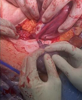
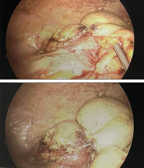
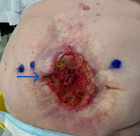
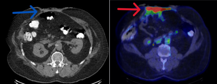
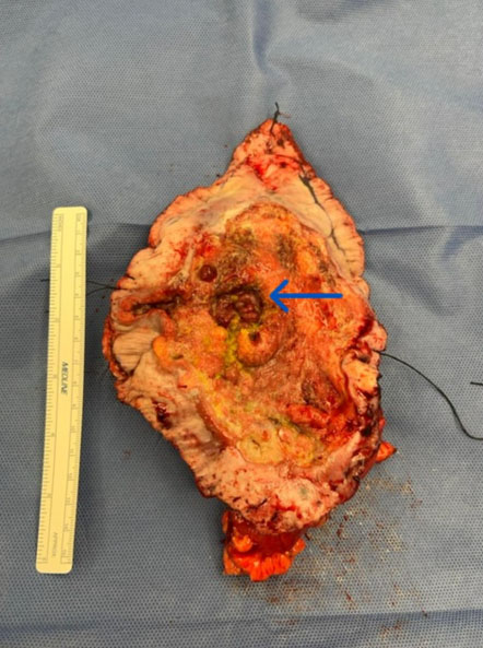
Over the subsequent years the patient continued to have a chronically open midline wound, managed with dressings in the community. Unfortunately, the ECF reopened, after 20 years. The skin edges of the wound also changed with the development of heaped up edges (Figure 1). A biopsy of the wound confirmed “invasive moderate to poorly differentiated SCC.” The ECF was moderate volume and was initially managed with total parenteral nutrition (TPN), reduced oral intake and stomal therapy. A staging computed tomography (CT) and 18F-fluorodeoxyglucose positron emission tomography (FDG-PET) scan was performed which showed diffuse involvement of the abdominal wall, but no distant nodal or visceral metastases (Figure 2). The patient proceeded to have a wide resection of the tumor and en bloc resection of the ECF. Due to loss of abdominal wall domain, an inlay dissolvable mesh and primary closure of the abdominal wall was performed with the aid of botulinum toxin preoperatively to help achieve closure. The resection margins were clear macroscopically and microscopically (Figure 3). Despite this, the patient postoperative stay was complicated by wound breakdown and formation of a new enteroatmospheric fistula, which was thought to be caused by a low volume anastomotic leak. The patient required multiple washouts of the wound as well partial explanting of the dissolvable mesh. This was further compounded by a high-output fistula with severe electrolyte derangement and renal impairment, which was managed non-operatively with TPN, electrolyte replacement, and use of anti-motility agents to slow down the output. The patient was discharged three months postoperatively, with no plans for adjuvant therapies due to clear resection margins and no regional or distant metastases.
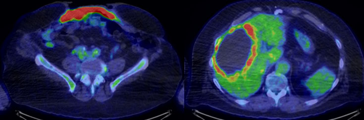
Eight months later, the patient developed a local recurrence of his MU, with a large centrally necrotic liver metastasis on PET scan (Figure 4). He was referred to medical oncology and initiated on immunotherapy. Shortly after, he was admitted with an adhesional small bowel obstruction that was non-operatively managed. Unfortunately, the patient deteriorated and died the following month.
Discussion
There is significant morbidity and mortality associated with MU, as they are more aggressive, with a higher metastatic potential, and greater rate of recurrence than de novo cutaneous malignancy [3]. Recurrence is also associated with higher mortality than other metastatic SCCs [4]. Marjolin’s ulcers most commonly arise from burns scars, and affect people in the fifth decade, with an average latency from initial wound to malignant degeneration of 29 years [5].
The pathogenesis of MU is multifactorial, and several pathophysiologic mechanisms have been proposed to explain the aggressive nature of these malignancies. Damage to the immune mechanisms of the skin impairs wound healing and cells damaged by chronic inflammation release toxins that promote mutations [1]. Mutations involving the HLA-DR4 and p53 have been identified to provide genetic predisposition to malignancy [5],[6]. Squamous cell carcinoma is identified on histopathologic examination in 80% to 90% of MU, although basal cell carcinoma (BCC) and melanoma are not rare [5]. Metastasis occurs commonly through lymphatics involving the regional lymph node drainage [5]. Hematogenous spread can still occur, albeit less commonly, most often involving the lungs [5]. The fibrous tissue surrounding the ulcer can serve as a mechanical or immunologic barrier to tumor spread, and lymphatic spread may be increased following excision [7].
A patient presenting with development of raised margins with ulceration, with or without surrounding induration within a chronic wound should raise suspicion of a MU, and should be biopsied via excision, incision, or punch [5],[6]. If incision or punch biopsy is performed, multiple areas should be sampled in a 4-quadrant distribution, with careful examination of local or regional lymph nodes by either ultrasound or lymphatic mapping [5]. Additional imaging modalities include CT, magnetic resonance imaging (MRI), and PET to clarify soft tissue involvement as well as evaluate for metastatic disease for staging [6],[8]. No specific tumor, nodes, and metastasis (TNM) staging criteria exist for MU, and the disease is usually staged according to criteria of the histopathology (SCC, BCC, or melanoma) [3]. There is no standardized treatment protocol for MU, with the most common treatment for non-metastatic disease is wide local excision with a 2 to 3 cm margin and sentinel lymph node biopsy, where possible [5],[6]. Definitive or neoadjuvant chemoradiation is not currently recommended. Adjuvant radiation may be utilized where an adequate margin cannot be achieved or in situations of unresectable involved lymph nodes or palliative cases [6]. Immunotherapies such as cemiplimab are monoclonal antibodies that bind to and inhibit programmed death receptor-1 (PD-1) on the surface of T cells, allowing T cells to recognize, target, and destroy cancer cells [9]. PD-1 inhibitors have demonstrated some efficacy in MU and are approved for use in Australia for metastatic or locally advanced MU not amenable to curative surgery or radiotherapy [10],[11]. Prevention and early detection remain the best practice for MU, which includes early excision and reconstruction or regular surveillance of chronic wounds [5].
The development of an MU within a laparotomy wound is exceedingly rare. A focused literature search was performed to identify published cases of MU arising within chronic non-healing laparotomy. PubMed/MEDLINE, Embase, and the Cochrane Library were searched for publications from 2016 to 2026. Search terms included combinations of “Marjolin ulcer,” “Marjolin’s ulcer,” “scar carcinoma,” “squamous cell carcinoma,” “chronic wound,” “non-healing wound,” “surgical wound,” “laparotomy,” “laparotomy scar,” “laparostomy,” “laparostomy scar,” and “abdominal wall.” Studies were included if they described histologically confirmed malignancy arising within a chronic non-healing laparotomy wound or scar. Reports involving burns, pressure ulcers, venous ulcers, or osteomyelitis without an abdominal surgical wound were excluded. Given the rarity of this condition, only one comparable case was identified in the literature over the past decade. Han et al. described a 67-year-old man who developed a SCC seven years after emergency open repair of a ruptured abdominal aortic aneurysm, followed by laparostomy for abdominal compartment syndrome [12]. Their patient presented with progressive wound dehiscence, necrotic skin, foul-smelling discharge, and hypercalcemia with CT demonstrating tumor invasion into the peritoneum that was deemed unresectable [12]. Their patient received best supportive care before dying one month later [12]. In contrast, while having a longer latency period, our case remained amenable to curative resection, despite recurrence and spread. Several similarities exist between their case and the case we present. Both patients developed enterocutaneous fistulation within a chronically inflamed wound environment and subsequently experienced rapid wound deterioration following a prolonged latency period, a pattern consistent with malignant transformation in chronic wounds. These shared features raise the possibility of identifiable risk factors for malignant degeneration in chronic laparotomy wounds, particularly in the presence of persistent inflammation and enterocutaneous fistulation. However, given the extreme rarity of this presentation, further case reports and larger case series are required before meaningful risk stratification can be established.
Conclusion
Marjolin’s ulcer arising within a chronic non-healing laparotomy wound is a rare phenomenon. This case highlights the potential for malignant transformation in longstanding abdominal wounds, particularly in the setting of chronic inflammation and enterocutaneous fistulation. Early recognition and definitive treatment of chronic wounds, together with vigilant surveillance of suspicious wound changes remain critical to improving outcomes. A multidisciplinary approach involving surgeons, dermatologists, oncologists, wound care specialists or stomal therapists, and dieticians offer an ideal model to achieve this. Further reports are required to better define risk factors for malignant degeneration within chronic laparotomy wounds and to guide strategies for prevention, surveillance, and management.
REFERENCES
1.
Bazaliński D, Przybek-Mita J, Barańska B, Więch P. Marjolin’s ulcer in chronic wounds – Review of available literature. Contemp Oncol (Pozn) 2017;21(3):197–202. [CrossRef]
[Pubmed]

2.
Junaid F. Marjolin ulcer. DermNet®. 2017. [Available at: https://dermnetnz.org/topics/marjolin-ulcer]
3.
Mitra S, Panda S, Sikka K, Mallick S, Thakar A. Multimodality management of locoregionally extensive Marjolin ulcer: A case report and review of the literature. Wounds 2024;36(5):166–9. [CrossRef]
[Pubmed]
4.
Elkins-Williams ST, Marston WA, Hultman CS. Management of the chronic burn wound. Clin Plast Surg 2017;44(3):679–87 [CrossRef]
[Pubmed]
5.
Marietta M, Crane JS. Marjolin ulcer. In: StatPearls. Treasure Island (FL): StatPearls Publishing; 2025.
[Pubmed]
6.
Khan K, Schafer C, Wood J. Marjolin ulcer: A comprehensive review. Adv Skin Wound Care 2020;33(12):629–34. [CrossRef]
[Pubmed]
7.
Mousa AK, Elshenawy AA, Maklad SM, Bebars SMM, Burezq HA, Sayed SE. Post-burn scar malignancy: 5-year management review and experience. Int Wound J 2022;19(4):895–909. [CrossRef]
[Pubmed]
8.
Dulet M, Taywade S, Verma V, Kumar R. FDG PET/CT in a case of Marjolin ulcer of the scalp over a longstanding burn scar. Clin Nucl Med 2025;50(6):e374–5. [CrossRef]
[Pubmed]
9.
Hughes BGM, Guminski A, Bowyer S, Migden MR, Schmults CD, Khushalani NI, et al. A phase 2 open-label study of cemiplimab in patients with advanced cutaneous squamous cell carcinoma (EMPOWER-CSCC-1): Final long-term analysis of groups 1, 2, and 3, and primary analysis of fixed-dose treatment group 6. J Am Acad Dermatol 2025;92(1):68–77. [CrossRef]
[Pubmed]
10.
Miodovnik M, Dolev Y, Buchen R, Brezis MR, Nikolaevski-Berlin A, Finkel I, et al. Squamous cell carcinoma arising in chronically damaged skin (Marjolin’s ulcer): Still an unmet need in the era of immunotherapy. Oncologist 2025;30(8):oyae326. [CrossRef]
[Pubmed]
11.
Pharmaceutical Benefits Scheme. Cemiplimab. Canberra: Australian Government Department of Health and Aged Care; 2026. [Available at: https://www.pbs.gov.au/medicine/search?term=13135H-13152F-13160P-13161Q-13162R-13169D-13153G-13159N]
12.
Han J, Gangalam V, Wilson P. Advanced squamous cell carcinoma arising in a laparostomy site. Ann R Coll Surg Engl 2022;104(3):e87–90. [CrossRef]
SUPPORTING INFORMATION
Author Contributions
Husein Al-Talib - Drafting the work, Final approval of the version to be published, Agree to be accountable for all aspects of the work in ensuring that questions related to the accuracy or integrity of any part of the work are appropriately investigated and resolved.
Sukhwant S Khanijaun - Conception of the work, Design of the work, Revising the work critically for important intellectual content, Final approval of the version to be published, Agree to be accountable for all aspects of the work in ensuring that questions related to the accuracy or integrity of any part of the work are appropriately investigated and resolved.
Corina Behrenbruch - Conception of the work, Design of the work, Revising the work critically for important intellectual content, Final approval of the version to be published, Agree to be accountable for all aspects of the work in ensuring that questions related to the accuracy or integrity of any part of the work are appropriately investigated and resolved.
Guarantor of SubmissionThe corresponding author is the guarantor of submission.
Source of SupportNone
Consent StatementWritten informed consent was obtained from the patient for publication of this article.
Data AvailabilityAll relevant data are within the paper and its Supporting Information files.
Conflict of InterestAuthors declare no conflict of interest.
Copyright© 2026 Husein Al-Talib et al. This article is distributed under the terms of Creative Commons Attribution License which permits unrestricted use, distribution and reproduction in any medium provided the original author(s) and original publisher are properly credited. Please see the copyright policy on the journal website for more information.