|
Case Report
Acute intragastric rupture of a giant splenic artery aneurysm requiring emergent multidisciplinary intervention: A case report
1 Attending General Surgeon, Department of General Surgery, Faculty of Medicine, Istanbul Medeniyet University, Goztepe Prof. Dr. Suleyman Yalcin City Hospital, Istanbul, Turkey
2 Resident General Surgeon, Department of General Surgery, Faculty of Medicine, Istanbul Medeniyet University, Goztepe Prof. Dr. Suleyman Yalcin City Hospital, Istanbul, Turkey
3 Associate Professor, Department of Radiology, Faculty of Medicine, Istanbul Medeniyet University, Goztepe Prof. Dr. Suleyman Yalcin City Hospital, Istanbul, Turkey
4 Associate Professor, Department of General Surgery, Faculty of Medicine, Istanbul Medeniyet University, Goztepe Prof. Dr. Suleyman Yalcin City Hospital, Istanbul, Turkey
5 Professor, Department of General Surgery, Faculty of Medicine, Istanbul Medeniyet University, Goztepe Prof. Dr. Suleyman Yalcin City Hospital, Istanbul, Turkey
Address correspondence to:
Leyla Zeynep Tigrel
MD, Attending General Surgeon, Department of General Surgery, Istanbul Medeniyet University Faculty of Medicine, Goztepe Prof. Dr. Suleyman Yalcin City Hospital, Goztepe 34722, Kadikoy, Istanbul,
Turkey
Message to Corresponding Author
Article ID: 101543Z01LT2026
Access full text article on other devices

Access PDF of article on other devices


How to cite this article
Tigrel LZ, Buyuker F, Ergun A, Asik M, Ekinci O, Alimoglu O. Acute intragastric rupture of a giant splenic artery aneurysm requiring emergent multidisciplinary intervention: A case report. Int J Case Rep Images 2026;17(2):17–21.ABSTRACT
Introduction: Splenic artery aneurysms (SAAs) are a rare pathology of the visceral vessels with a prevalence of less than 1%. While usually silent, spontaneous rupture represents a surgical emergency. This case report details the successful multidisciplinary management of an acute, massive intragastric rupture of a 9.5 cm giant SAA in a previously healthy 39-year-old male, emphasizing the diagnostic challenges and critical therapeutic transitions.
Case Report: A 39-year-old male experienced a sudden syncopal episode while on duty. He developed massive hematemesis, and an emergency upper gastrointestinal endoscopy revealed an ulcerated mass at the fundus of the stomach with active massive hemorrhage. Initial targeted embolization attempts failed to control the proximal inflow, and the patient rapidly deteriorated into severe hemorrhagic shock. Intraoperatively, a giant aneurysm was identified at the origin of the splenic artery, which had directly eroded through the posterior gastric wall into the gastric lumen. The splenic artery was rapidly dissected and ligated, the aneurysm was excised, the posterior gastric wall defect underwent primary repair, and a splenectomy was performed. Measuring 9.5 cm on pathological examination, the case presented herein represents the largest recorded splenic artery aneurysm presenting with an acute intragastric rupture in the medical literature.
Conclusion: Acute intragastric rupture of a giant splenic artery aneurysm is a life-threatening emergency that mimics routine upper gastrointestinal bleeding. When endovascular interventions fail or hemodynamic instability worsens, immediate open surgical control is definitive. This case highlights the critical importance of maintaining a high index of suspicion and utilizing rapid, multidisciplinary coordination between interventional radiology, surgery, and critical care.
Keywords: Aneurysm rupture, Case report, Gastrointestinal hemorrhage, Splenic artery aneurysm
Introduction
Splenic artery aneurysms (SAAs) are a rare pathology of the visceral vessels with a prevalence of less than 1%. Since SAAs are mostly asymptomatic, patients are rarely diagnosed. Retrospective data indicate a distinct female predominance (78%), with rupture carrying a catastrophic mortality rate ranging from 25% to 40% [1]. Despite their rarity, SAAs constitute approximately 60% to 70% of all visceral artery aneurysms (VAAs) [2]. While usually silent, spontaneous rupture represents a surgical emergency. This case report details the successful multidisciplinary management of an acute, massive intragastric rupture of a 9.5 cm giant SAA in a previously healthy 39-year-old male, emphasizing the diagnostic challenges and critical therapeutic transitions.
Case Report
A 39-year-old male, a hospital employee with no remarkable medical history, experienced a sudden syncopal episode while on duty. The patient was transferred to the department of radiology for a cranial computed tomography (CT) scan for a suspected head trauma, which showed no signs of acute intracranial pathology. He was admitted to the neurosurgery unit for observation, where he developed massive hematemesis, and an emergency upper gastrointestinal endoscopy was performed. The endoscopy revealed an ulcerated mass at the fundus of the stomach with active massive hemorrhage, which led to his transfer to the interventional radiology department for angiographic imaging and treatment.
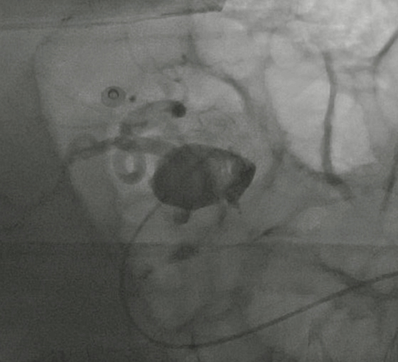
Imaging of the celiac trunk revealed a complex anatomy: a 7 × 8 mm aneurysmal dilatation at the origin of the splenic artery, adjacent to two irregularly lobulated, smooth-contoured aneurysms measuring 16 × 12 mm and 7 × 6 mm (Figure 1). Initial targeted embolization attempts failed to control the proximal inflow. During the endovascular procedure, the patient rapidly deteriorated into severe hemorrhagic shock (heart rate 128 bpm, blood pressure 80/40 mmHg). Laboratory evaluation confirmed severe anemia and coagulopathy [hemoglobin 6.3 g/dL, platelet count 134 × 103/µmL , prothrombin time international normalized ratio (INR) 1.83].
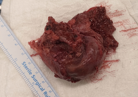
A joint decision by the general surgery and interventional radiology teams prompted an immediate transfer to the operating theatre for emergency laparotomy. Intraoperatively, a giant 10 × 10 cm aneurysm was identified at the origin of the splenic artery, which had directly eroded through the posterior gastric wall into the gastric lumen. The splenic artery was rapidly dissected and ligated at its origin. The aneurysm was excised, the posterior gastric wall defect underwent primary repair, and a splenectomy was performed (Figure 2). Despite aggressive resuscitation with packed red blood cells and inotropic support, the patient remained transiently hypotensive at the conclusion of the operation.
After the surgery, the patient was admitted to the intensive care unit (ICU) for close monitoring. Prophylactic low molecular weight heparin (LMWH) was safely initiated on the second postoperative day once stable hemostasis was confirmed and no active bleeding was recorded. Without any complications, he was transferred to the general surgery ward on the 5th postoperative day. During his stay, on the 11th postoperative day, he developed acute dyspnea. A CT pulmonary angiogram was performed, which revealed acute pulmonary emboli in the right pulmonary arteries; this was later managed conservatively.
Vaccinations against Streptococcus pneumoniae, Neisseria meningitidis, and Haemophilus influenzae type B were administered on postoperative day 14, and the patient was discharged without further complications on the 20th postoperative day.
Pathological examination of the removed specimens showed a 9.5 × 7 × 3.5 cm aneurysmal vessel with a hemorrhagic vascular wall structure, and a 12 × 7.8 × 4 cm spleen specimen containing dilated, congested blood vessels in focal areas. Given the patient’s young age and absence of risk factors, he was referred to the medical genetics department. Comprehensive genetic testing ruled out vascular Ehlers–Danlos syndrome and other major connective tissue disorders. He was discharged in stable condition on the 20th postoperative day.
Discussion
The etiology of SAAs involves a variety of predisposing factors. While pregnancy is strongly linked to SAA development in female patients due to increased intra-abdominal pressure and hormonal shifts, portal hypertension, liver cirrhosis, chronic pancreatitis, and congenital connective tissue disorders like Ehlers–Danlos syndrome are other factors that increase the risk [3],[4].
The incidence of SAA varies from 0.02% to 10.4% [5],[6]. Modern high-resolution imaging has improved incidental detection, yet presentation as an acute intraluminal gastric rupture remains an extraordinary clinical rarity. Rupture into adjacent viscera carries an extreme mortality rate of 25% to 70% [7],[8]. In this specific case, the patient’s presence in a hospital environment at the onset of syncope dramatically accelerated diagnostic and therapeutic timelines, directly altering an otherwise poor prognosis.
The management of a ruptured aneurysm depends on various factors, including the location and size of the aneurysm, the patient’s overall health, and the presence of complications. Various options are considered to address this critical condition. The selection of the appropriate management strategy is tailored to the individual circumstances of each case and involves a collaborative effort among a multidisciplinary medical team involving surgeons, interventional radiologists, and critical care specialists. The urgency of treatment cannot be overstated, as prompt intervention is critical to preventing further complications and reducing mortality associated with a ruptured aneurysm. Firstly, emergency resuscitation takes precedence, aiming to hemodynamically stabilize the patient. This involves the immediate administration of intravenous crystalloids, blood transfusions, and other supportive interventions to sustain adequate blood pressure and oxygenation [9].
A major step in the management of ruptured aneurysms is the use of interventional radiology techniques, such as angiography and embolization, to halt the bleeding. The reported success rate of embolization of ruptured SAA is 75% to 92% according to different studies [10],[11]. Early failure of embolization occurs at a rate of 20%, while late failure is observed at a rate of 40%. The so-called “Sandwich method”—selective embolization of the splenic artery proximal and distal to the aneurysm—is recommended [11],[12]. The common complications of the procedure include splenic infarction, abscess formation, and stent migration [10],[13]. The use of a covered stent-graft for aneurysm exclusion has been increasingly favored, demonstrating a success rate reaching 98%. However, challenging deployment arises due to the splenic artery’s tortuosity [13]. Surgical intervention may become necessary, especially if the patient is unstable or if embolization is not a viable option.
A comprehensive literature review spanning 1980 to 2023 identified only seven previously reported cases of an SAA rupturing directly into the stomach. Among these, five were managed successfully via open laparotomy and aneurysmal excision (with or without splenectomy), one was managed purely via embolization, and one patient with a mycotic SAA succumbed to exsanguination prior to intervention [14]. Measuring 9.5 cm on pathological examination, the case presented herein represents the largest recorded splenic artery aneurysm presenting with an acute intragastric rupture in the medical literature.
The acute pulmonary embolism on the 11th postoperative day emphasizes the profound hypercoagulable state caused by emergency laparotomy, hemorrhagic shock, and immobilization. Despite standard heparin prophylaxis, these factors significantly increase thromboembolic risk, highlighting the need for close clinical monitoring following major vascular interventions.
Conclusion
Acute intragastric rupture of a giant splenic artery aneurysm is a life-threatening emergency that mimics routine upper gastrointestinal bleeding. When endovascular interventions fail or hemodynamic instability worsens, immediate open surgical control is definitive. This case highlights the critical importance of maintaining a high index of suspicion and utilizing rapid, multidisciplinary coordination between interventional radiology, surgery, and critical care to rescue patients facing catastrophic visceral vascular events.
REFERENCES
1.
Kassem MM, Gonzalez L. Splenic artery aneurysm. In: StatPearls. Treasure Island (FL): StatPearls Publishing; 2026.
[Pubmed]

2.
Al-Habbal Y, Christophi C, Muralidharan V. Aneurysms of the splenic artery – A review. Surgeon 2010;8(4):223–31. [CrossRef]
[Pubmed]
3.
Lakin RO, Bena JF, Sarac TP, Shah S, Krajewski LP, Srivastava SD, et al. The contemporary management of splenic artery aneurysms. J Vasc Surg 2011;53(4):958–64; discussion 965. [CrossRef]
[Pubmed]
4.
Regus S, Lang W. Management of true visceral artery aneurysms in 31 cases. J Visc Surg 2016;153(5):347–52. [CrossRef]
[Pubmed]
5.
Akbulut S, Otan E. Management of giant splenic artery aneurysm: Comprehensive literature review. Medicine (Baltimore) 2015;94(27):e1016. [CrossRef]
[Pubmed]
6.
Misiakos EP, Gemenetzis G, Moris D, Sotiropoulos JR, Schizas D, Felekouras E, Papaconstantinou I. Ruptured splenic artery aneurysms: A series of 4 cases and a review of the literature. Ann Vasc Surg 2017;43:313.e1–e5. [CrossRef]
7.
Abbas MA, Stone WM, Fowl RJ, Gloviczki P, Oldenburg WA, Pairolero PC, et al. Splenic artery aneurysms: Two decades experience at Mayo clinic. Ann Vasc Surg 2002;16(4):442–9. [CrossRef]
[Pubmed]
8.
Perrot M, Bühler L, Deléaval J, Borisch B, Mentha G, Morel P. Management of true aneurysms of the splenic artery. Am J Surg 1998;175(6):466–8. [CrossRef]
[Pubmed]
9.
Chaer RA, Abularrage CJ, Coleman DM, Eslami MH, Kashyap VS, Rockman C, et al. The Society for Vascular Surgery clinical practice guidelines on the management of visceral aneurysms. J Vasc Surg 2020;72(1S):3S–39S. [CrossRef]
[Pubmed]
10.
Tulsyan N, Kashyap VS, Greenberg RK, Sarac TP, Clair DG, Pierce G, et al. The endovascular management of visceral artery aneurysms and pseudoaneurysms. J Vasc Surg 2007;45(2):276–83. [CrossRef]
[Pubmed]
11.
Hogendoorn W, Lavida A, Hunink MGM, Moll FL, Geroulakos G, Muhs BE, et al. Open repair, endovascular repair, and conservative management of true splenic artery aneurysms. J Vasc Surg 2014;60(6):1667–76.e1. [CrossRef]
[Pubmed]
12.
Guillaumon AT, Chaim EA. Splenic artery aneurysm associated with portal hypertension: Exclusion by endovascular sandwich technique. J Vasc Bras 2018;17(1):80–4.
13.
Etezadi V, Gandhi RT, Benenati JF, Rochon P, Gordon M, Benenati MJ, et al. Endovascular treatment of visceral and renal artery aneurysms. J Vasc Interv Radiol 2011;22(9):1246–53. [CrossRef]
[Pubmed]
14.
Dave SP, Reis ED, Hossain A, Taub PJ, Kerstein MD, Hollier LH. Splenic artery aneurysm rupture into the stomach: A case report and a 43-year literature review. Vasc Endovascular Surg 2004;38(4):379–83.
SUPPORTING INFORMATION
Acknowledgments
The authors would like to thank the clinical and administrative staff of Medeniyet University Göztepe Prof. Dr. Süleyman Yalçin City Hospital for their assistance and support in conducting this study.
Declaration of Generative AI and AI-assisted technologies in the manuscript preparation process
Generative AI technology was used during the preparation of this manuscript solely for the purpose of language editing, text structural formatting, and word-count optimization of the abstract and author contribution sections. No AI-generated scientific data, clinical findings, or medical literature interpretations were introduced into the text.
• Technology Name/Model: Gemini
• Version/Source: Gemini 1.5 Pro (Google)
• Specific Sections Utilizing Technology: Language refinement and structural separation of the Abstract (Introduction, Case Report, and Conclusion blocks) and alignment of the Author’s Contributions section to the journal’s template format.
Author ContributionsLeyla Zeynep Tigrel - Conception of the work, Design of the work, Acquisition of data, Analysis of data, Drafting the work, Revising the work critically for important intellectual content, Final approval of the version to be published, Agree to be accountable for all aspects of the work in ensuring that questions related to the accuracy or integrity of any part of the work are appropriately investigated and resolved.
Fatih Buyuker - Conception of the work, Design of the work, Acquisition of data, Analysis of data, Drafting the work, Revising the work critically for important intellectual content, Final approval of the version to be published, Agree to be accountable for all aspects of the work in ensuring that questions related to the accuracy or integrity of any part of the work are appropriately investigated and resolved.
Aysegul Ergun - Conception of the work, Design of the work, Acquisition of data, Analysis of data, Drafting the work, Revising the work critically for important intellectual content, Final approval of the version to be published, Agree to be accountable for all aspects of the work in ensuring that questions related to the accuracy or integrity of any part of the work are appropriately investigated and resolved.
Murat Asik - Conception of the work, Design of the work, Acquisition of data, Analysis of data, Drafting the work, Revising the work critically for important intellectual content, Final approval of the version to be published, Agree to be accountable for all aspects of the work in ensuring that questions related to the accuracy or integrity of any part of the work are appropriately investigated and resolved.
Ozgur Ekinci - Conception of the work, Design of the work, Acquisition of data, Analysis of data, Drafting the work, Revising the work critically for important intellectual content, Final approval of the version to be published, Agree to be accountable for all aspects of the work in ensuring that questions related to the accuracy or integrity of any part of the work are appropriately investigated and resolved.
Orhan Alimoglu - Conception of the work, Design of the work, Drafting the work, Revising the work critically for important intellectual content, Final approval of the version to be published, Agree to be accountable for all aspects of the work in ensuring that questions related to the accuracy or integrity of any part of the work are appropriately investigated and resolved.
Guarantor of SubmissionThe corresponding author is the guarantor of submission.
Source of SupportNone
Consent StatementWritten informed consent was obtained from the patient for publication of this article.
Data AvailabilityAll relevant data are within the paper and its Supporting Information files.
Conflict of InterestAuthors declare no conflict of interest.
Copyright© 2026 Leyla Zeynep Tigrel et al. This article is distributed under the terms of Creative Commons Attribution License which permits unrestricted use, distribution and reproduction in any medium provided the original author(s) and original publisher are properly credited. Please see the copyright policy on the journal website for more information.