|
Case Report
Small bowel ischemia, perforation, and enterocutaneous fistula as gastrointestinal manifestations of eosinophilic granulomatosis with polyangiitis: A case report
1 Higher Specialist General Surgery Trainee, Department of General and Vascular Surgery, Mater Dei Hospital, St. Paul’s Bay, Malta
2 Specialist Breast and General Surgeon, Department of General and Vascular Surgery, Mater Dei Hospital, St. Paul’s Bay, Malta
3 Consultant Breast and General Surgeon, Department of General and Vascular Surgery, Mater Dei Hospital, St. Paul’s Bay, Malta
Address correspondence to:
Marc Grech
9, Triq id-Duluri, St. Paul’s Bay,
Malta
Message to Corresponding Author
Article ID: 101542Z01MG2026
Access full text article on other devices

Access PDF of article on other devices


How to cite this article
Grech M, Ilgun AS, Agius J. Small bowel ischemia, perforation, and enterocutaneous fistula as gastrointestinal manifestations of eosinophilic granulomatosis with polyangiitis: A case report. Int J Case Rep Images 2026;17(2):12–16.ABSTRACT
Introduction: Eosinophilic granulomatosis with polyangiitis (EGPA), formerly known as Churg–Strauss syndrome, is a rare small- to medium-vessel vasculitis characterized by asthma, eosinophilia, sinonasal disease, and variable systemic involvement. Gastrointestinal involvement is less common but clinically important, as it may indicate severe disease and can lead to complications such as bowel ischemia, perforation and fistula formation.
Case Report: We report the case of a 46-year-old gentleman with a background of asthma and chronic sinusitis requiring functional endoscopic sinus surgery on two occasions, who initially presented with worsening shortness of breath, wheeze, productive cough, and lethargy. Initial investigations for pulmonary embolism were negative, and he was treated for an acute asthma exacerbation. During admission, he was found to have marked eosinophilia, raised inflammatory markers and positive antineutrophil cytoplasmic antibody (ANCA), supporting a diagnosis of EGPA. He improved following corticosteroid therapy and was discharged. One month later, after interruption of prednisolone therapy, he represented with severe abdominal pain and melaena. Computed tomography (CT) imaging demonstrated ischemic small bowel with portal and mesenteric venous gas. Laparotomy revealed diffuse dusky small bowel, most severe in the proximal jejunum, but no bowel resection was performed due to intraoperative improvement. He was managed in intensive care with high-dose corticosteroids, antibiotics, anticoagulation, nutritional support, and multidisciplinary input. Ongoing disease activity required cyclophosphamide, subsequently changed to rituximab. His course was complicated by bowel perforation, wound dehiscence, and enterocutaneous fistula, which were managed conservatively with bowel rest, total parenteral nutrition, antibiotics, intravenous immunoglobulin, and ongoing immunosuppression.
Conclusion: This case highlights the importance of considering EGPA in patients with difficult-to-control asthma, sinonasal disease, and eosinophilia. Gastrointestinal involvement may be life-threatening and should prompt urgent multidisciplinary assessment and escalation of immunosuppressive therapy.
Keywords: Churg–Strauss syndrome, Enterocutaneous fistula, Eosinophilic granulomatosis with polyangiitis, Mesenteric vasculitis, Small bowel ischemia
Introduction
Eosinophilic granulomatosis with polyangitis (EGPA), formerly known as Churg–Strauss syndrome, is a small to medium-vessel vasculitis which is characterized by allergic, eosinophilic, and vasculitic phases. It affects multiple systems, including lungs, kidneys, nervous system, heart, skin, and gastrointestinal tract and is associated with antineutrophil cytoplasmic antibody (ANCA) in 30–40% of the time [1].
Eosinophilic granulomatosis with polyangitis typically evolves through overlapping phases, beginning with an allergic or prodromal phase marked by adult-onset asthma, allergic rhinitis, chronic sinusitis, or nasal polyposis, followed by eosinophilic tissue infiltration and, in some patients, a systemic vasculitic phase [2]. Gastrointestinal involvement is less common but clinically important, as it may indicate severe systemic disease. Reported manifestations include abdominal pain, gastrointestinal bleeding, bowel wall inflammation, ischemia, perforation, and, rarely, fistula formation. These complications are thought to arise from eosinophilic infiltration and necrotizing vasculitis affecting the mesenteric circulation. Recognition of gastrointestinal vasculitis is essential because delayed diagnosis may lead to significant morbidity [1].
We present a case of a 46-year-old with EGPA presenting initially as an asthma exacerbation, followed shortly by severe mesenteric vasculitis causing small bowel ischemia, perforation and enterocutaneous fistula, managed without bowel resection and with multidisciplinary immunosuppressive therapy. This case highlights the need to consider EGPA in patients with difficult-to-control asthma and systemic features and underlines the potentially life-threatening complications of the condition.
Case Report
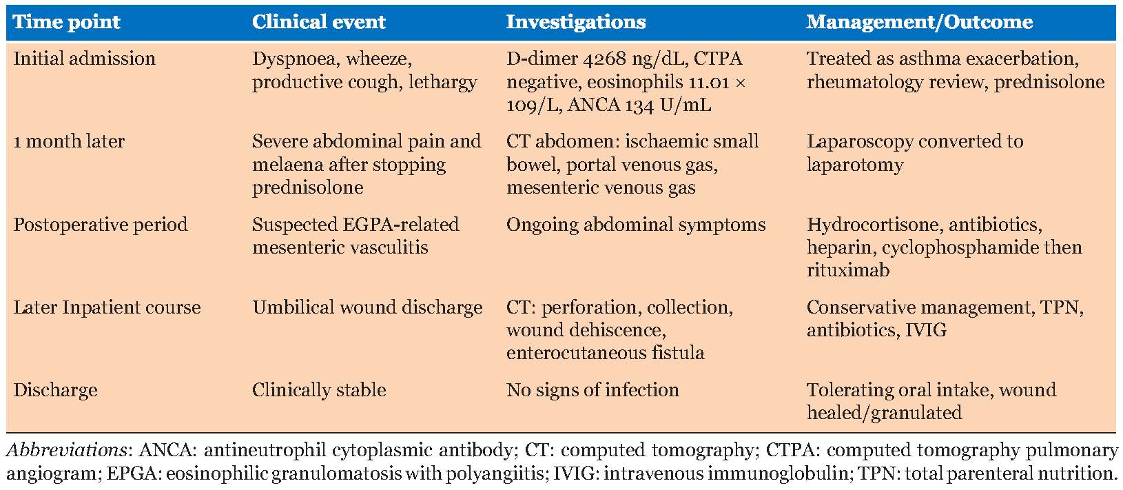
A 46-year-old gentleman with a history of asthma and chronic sinusitis requiring functional endoscopic sinus surgery twice, presented to the emergency department with worsening shortness of breath, wheeze, and productive cough, together with generalized lethargy. This presentation, together with a raised D-Dimer of 4268 ng/dL led to an investigation with electrocardiograms (ECGs), troponins, and CT pulmonary angiography, which yielded negative results, and he was admitted and treated for an acute exacerbation of asthma. An echocardiogram was carried out to exclude cardiac causes confidently, with a normal result. Renal function tests and urinalysis were also normal, thus no renal involvement was suspected.
During the admission, he was noted to have a high white cell count of 22.50 × 10⁹/L, eosinophil count of 11.01 × 10⁹/L, erythrocyte sedimentation rate of 45 mm/h and C-reactive protein of 321.6 mg/L. This, together with his systemic features, prompted investigation of ANCA levels, specifically the anti-myeloperoxidase antibody test, which was found to be raised at 134 U/mL. Applying the 2022 American College of Rheumatology (ACR)/European Alliance of Associations for Rheumatology (EULAR) classification criteria, the presence of obstructive airway disease and marked eosinophilia gave a score of 8, strongly supporting a diagnosis of EGPA. Other diagnosis such as bronchopulmonary aspergillosis, infection, and thromboembolic disease were considered less likely in view of negative findings on CT pulmonary angiography and CT abdomen. Furthermore, small bowel biopsies at gastroscopy showed no diagnostic abnormalities, which is common in EGPA in view of the superficial nature of the biopsies. A rheumatologist was involved in his care, and a course of Prednisolone 40 mg daily, tapering over four months, was prescribed, following which his symptoms improved and he was discharged home.
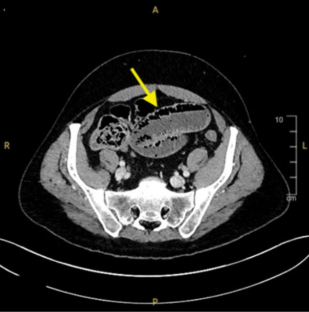
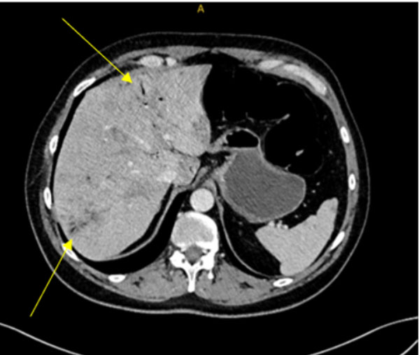
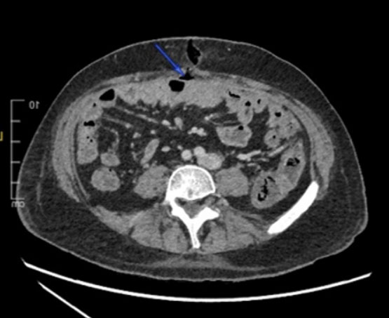
He presented one month later with severe abdominal pain with melaena. He reported that prednisolone had been discontinued because of presumed gastritis. The subsequent development of severe abdominal symptoms occurred in the context of interrupted corticosteroid therapy, although causality cannot be established. A CT of the abdomen and pelvis revealed ischemic small bowel with portal venous gas, mesenteric venous gas, and associated hepatic ischemic changes (Figure 1 and Figure 2). He underwent diagnostic laparoscopy converted to laparotomy under general anesthesia. Intraoperatively, diffuse dusky small bowel with patchy areas of normal bowel, most severe in the proximal jejunum, approximately 140 cm in length, starting around 10 cm from the ligament of Treitz, was reported. Initially, the mesenteric vessels were reported to have severely diminished pulses. The bowel was soaked in warm saline, and a decrease in duskiness was noted after a few minutes, with improvement of the mesenteric pulsation. The large bowel was reported to appear healthy. In view of the improvement in the bowel and no clear demarcations, no bowel resection was performed.
Postoperatively, he was managed in an intensive care setting. A multidisciplinary approach was adopted, with anesthetists, surgeons, rheumatologists, hematologists, pharmacists, nurses, and physiotherapists contributing to his care. He was treated with an initial 200 mg intravenous dose of hydrocortisone, followed by 100 mg three times daily. He was also given piperacillin with tazobactam 4.5 g three times daily, analgesia, nutritional support, and anticoagulation with intravenous heparin to improve bowel perfusion. In view of the global clinical picture, a diagnosis of small bowel ischemia secondary to mesenteric vasculitis was made. He received a dose of cyclophosphamide 15 mg/kg, which was, however, discontinued because of deranged liver function tests and persistent disease activity. Due to ongoing abdominal symptoms, he was prescribed rituximab 1 g, followed by a second dose two weeks later. The hydrocortisone dose was tailed down to 50 mg daily over the following three weeks, following which this was changed to prednisolone 12.5 mg daily, indefinitely.
In the interim, he developed copious greenish-brown discharge from the umbilical wound. Computed tomography of the trunk demonstrated bowel perforation with encapsulated fluid collection, wound dehiscence, and an enterocutaneous fistula in the periumbilical region (Figure 3). He was managed conservatively with bowel rest, total parenteral nutrition (TPN), broad-spectrum antibiotics, intravenous immunoglobulins (IVIG) for five days, and ongoing immunosuppression. The fistula output decreased over the following weeks, and the wound eventually granulated and healed through conservative measures. Following a total of two months of inpatient treatment, he was discharged once clinically stable, tolerating oral intake and free of signs of infection (Table 1).
Discussion
Eosinophilic granulomatosis with polyangiitis is a rare, systemic small- to medium-vessel vasculitis characterized by asthma, eosinophilia, and eosinophil-rich granulomatous inflammation. Although it is grouped within the antineutrophil cytoplasmic antibody-associated vasculitides, EGPA differs clinically from granulomatosis with polyangiitis and microscopic polyangiitis because asthma, chronic rhinosinusitis, nasal polyposis, and eosinophilic tissue infiltration are often dominant features. The diagnosis is frequently delayed since early manifestations may present as uncontrolled asthma, recurrent respiratory tract infection, allergic disease, or chronic sinus disease [3].
The combination of our patient’s presenting symptoms is clinically relevant, as asthma and upper airway involvement are among the most frequent and earliest manifestations of EGPA. The associated lethargy and chest symptoms suggest that the presentation was not simply an isolated asthma exacerbation, but part of a wider systemic inflammatory process. The differential diagnosis included infective asthma exacerbation, eosinophilic asthma, allergic bronchopulmonary aspergillosis, hypereosinophilic syndrome, pulmonary embolism and other ANCA-associated vasculitides. In suspected EGPA, investigations should assess both eosinophilic inflammation and end-organ involvement, including full blood count with eosinophil count, inflammatory markers, renal function, urinalysis, ANCA testing, chest imaging and cardiac assessment when chest pain or biomarkers suggest possible myocardial involvement [4].
This case is particularly notable because of the associated bowel presentation, with severe abdominal pain, small bowel ischemia and subsequent small bowel perforation and enterocutaneous fistula. Gastrointestinal involvement in EGPA may reflect eosinophilic infiltration of the bowel wall, vasculitic ischemia of mesenteric vessels, or a combination of both mechanisms [5].
The acute onset of severe abdominal pain and melaena, together with CT evidence of small bowel ischemia, portal venous gas and mesenteric venous gas, indicated significant intestinal hypoperfusion and threatened bowel viability. At laparotomy, dusky bowel, with intervening areas of relatively normal bowel and markedly diminished mesenteric pulsation, was considered more compatible with a diffuse mesenteric vascular process than with an isolated focal mechanical lesion. Improvement in bowel colour and mesenteric pulsation following warming further suggested a dynamic component of small-vessel hypoperfusion. Given this, together with the clinical context, EGPA was considered the leading clinical explanation.
The bowel presentation is clinically important because gastrointestinal involvement is considered a marker of more severe disease and poorer prognosis in EGPA. Reported gastrointestinal manifestations include abdominal pain, diarrhea, nausea, gastrointestinal bleeding, bowel ulceration, mesenteric ischemia and, in severe cases, bowel perforation. The presence of bowel symptoms in a patient with asthma, sinonasal disease, and eosinophilia should therefore prompt early consideration of EGPA rather than attributing the presentation solely to infection, inflammatory bowel disease or non-specific gastroenteritis [5].
The 2022 ACR/EULAR classification criteria support the diagnosis of EGPA when features such as obstructive airway disease, nasal polyps, peripheral eosinophilia, extravascular eosinophilic inflammation, neuropathy, and ANCA status are considered together. Gastrointestinal involvement is not a central weighted item in these criteria, but it remains clinically relevant because it influences prognosis and guides treatment intensity. Antineutrophil cytoplasmic antibody testing may assist phenotyping, but ANCA negativity does not exclude EGPA, particularly in patients with eosinophilic tissue-predominant disease involving the lungs, heart, or gastrointestinal tract [6].
Treatment should be guided by disease severity and the presence of organ-threatening manifestations. While systemic corticosteroids remain the cornerstone of initial therapy, gastrointestinal involvement may justify escalation to additional immunosuppression, particularly if there is evidence of bowel ischemia, ulceration, bleeding, perforation, severe systemic inflammation, or other major organ involvement. Cyclophosphamide or rituximab may be considered in severe disease, while mepolizumab has an established steroid-sparing role in relapsing or refractory EGPA with prominent eosinophilic disease. Glucocorticoid exposure should be minimized where possible, with dose reduction guided by clinical response and relapse risk [7].
Conclusion
This case highlights the importance of considering eosinophilic granulomatosis with polyangiitis in patients with difficult-to-control asthma, chronic sinonasal disease and marked eosinophilia, particularly when systemic or atypical features are present. Although EGPA most commonly presents with respiratory and upper airway manifestations, gastrointestinal involvement may be severe and life-threatening. In this patient, small bowel ischemia complicated by perforation and enterocutaneous fistula was consistent with severe mesenteric vasculitic involvement and required prolonged multidisciplinary management. Early recognition of gastrointestinal vasculitis in EGPA is essential, as prompt escalation of immunosuppressive therapy may prevent irreversible complications and improve outcomes.
REFERENCES
1.
Rout P, Maher L. Eosinophilic granulomatosis with polyangiitis (Churg-Strauss syndrome). In: StatPearls. Treasure Island (FL): StatPearls Publishing; 2026.
[Pubmed]

2.
Alam V, Nanzer AM. Eosinophilic granulomatosis with polyangiitis: Case report and literature review. Breathe (Sheff) 2022;18(4):220170. [CrossRef]
[Pubmed]
3.
Emmi G, Bettiol A, Gelain E, Bajema IM, Berti A, Burns S, et al. Evidence-based guideline for the diagnosis and management of eosinophilic granulomatosis with polyangiitis. Nat Rev Rheumatol 2023;19(6):378–93. [CrossRef]
[Pubmed]
4.
Comarmond C, Pagnoux C, Khellaf M, Cordier JF, Hamidou M, Viallard JF, et al. Eosinophilic granulomatosis with polyangiitis (Churg-Strauss): Clinical characteristics and long-term followup of the 383 patients enrolled in the French Vasculitis Study Group cohort. Arthritis Rheum 2013;65(1):270–81. [CrossRef]
[Pubmed]
5.
Assmann G, Molinger M, Pfreundschuh M, Bohle R, Zimmer V. Gastrointestinal perforation due to vasculitis at primary diagnosis of eosinophilic granulomatosis with polyangiitis (EGPA) despite a high dose glucocorticosteroids treatment. Springerplus 2014;3:404. [CrossRef]
[Pubmed]
6.
Grayson PC, Ponte C, Suppiah R, Robson JC, Craven A, Judge A, et al. 2022 American College of Rheumatology/European Alliance of Associations for Rheumatology Classification Criteria for Eosinophilic Granulomatosis with Polyangiitis. Ann Rheum Dis 2022;81(3):309–14. [CrossRef]
[Pubmed]
7.
Hellmich B, Sanchez-Alamo B, Schirmer JH, Berti A, Blockmans D, Cid MC, et al. EULAR recommendations for the management of ANCA-associated vasculitis: 2022 update. Ann Rheum Dis 2024;83(1):30–47. [CrossRef]
[Pubmed]
SUPPORTING INFORMATION
Acknowledgments
Artificial Intelligence (AI) Disclosure
Artificial Intelligence, Chat GPT version 5.5 was used in parts of the text solely to aid in grammar correction and in the improvement of sentence structure. All content was reviewed by the authors, and the authors take full responsibility for the accuracy and scientific integrity of the paper.
The corresponding author is the guarantor of submission.
Source of SupportNone
Consent StatementWritten informed consent for the write-up and publication of this case report, including the use of images, was obtained from the patient.
Data AvailabilityAll relevant data are within the paper and its Supporting Information files.
Conflict of InterestAuthors declare no conflict of interest.
Copyright© 2026 Marc Grech et al. This article is distributed under the terms of Creative Commons Attribution License which permits unrestricted use, distribution and reproduction in any medium provided the original author(s) and original publisher are properly credited. Please see the copyright policy on the journal website for more information.