|
Case Report
Primary Sjogren’s syndrome presenting as acute pancreatitis and interstitial nephritis: A diagnostic challenge
1 Postgraduate Resident, Department of Internal Medicine, Fortis Memorial Research Institute, Gurugram, Haryana, India
2 Senior Director and Unit Head, Department of Internal Medicine, Fortis Memorial Research Institute, Gurugram, Haryana, India
3 Senior Consultant, Department of Rheumatology, Fortis Memorial Research Institute, Gurugram, Haryana, India
4 Director, Renal Pathology, Agilus Diagnostics, New Delhi, India
5 Attending Consultant, Department of Radiology, Fortis Memorial Research Institute, Gurugram, Haryana, India
Address correspondence to:
Amitabh Parti
Senior Director and Unit Head, Department of Internal Medicine, Fortis Memorial Research Institute, Gurugram, Haryana,
India
Message to Corresponding Author
Article ID: 101541Z01CT2026
Access full text article on other devices

Access PDF of article on other devices


How to cite this article
Tatrari C, Parti A, Mendiratta N, Sabhikhi AK, Uberoi A. Primary Sjogren’s syndrome presenting as acute pancreatitis and interstitial nephritis: A diagnostic challenge. Int J Case Rep Images 2026;17(2):7–11.ABSTRACT
Primary Sjogren’s syndrome (pSS) is an autoimmune disorder primarily affecting the exocrine glands, with systemic involvement being increasingly recognized. Presentation with gastrointestinal involvement is a rare presentation of the disease. We report the case of a 72-year-old woman who presented with cholestatic jaundice, fever, and acute pancreatitis. During hospital stay, she also developed pulmonary involvement and an active urinary sediment, which significantly complicated the diagnosis. Serological testing revealed a strongly positive antinuclear antibody (ANA) titer along with anti-Smith antibodies, raising a strong suspicion for systemic lupus erythematosus (SLE). However, renal biopsy demonstrated a plasma cell predominant tubulointerstitial nephritis without immune complex deposition. This pattern effectively ruled out lupus nephritis and established the diagnosis of primary Sjogren’s syndrome. This case highlights the diagnostic challenge posed by overlapping autoimmune features and reinforces the importance of tissue biopsy in differentiating autoimmune mimics. Early recognition of atypical extra-glandular manifestations of pSS is essential for appropriate management and improved outcomes.
Keywords: Acute pancreatitis, Anti-Smith antibody, Primary Sjogren’s syndrome, Tubulointerstitial nephritis
Introduction
Primary Sjogren’s syndrome (pSS) is a chronic systemic autoimmune disease characterized by immune-mediated injury to the exocrine glands, leading predominantly to sicca symptoms [1],[2],[3]. Although extra-glandular involvement is well recognized, pancreatic disease in pSS is usually subclinical, and presentation with acute pancreatitis as the initial manifestation is very rare. Major diagnostic dilemma arises due to the clinical and serological overlap between pSS and systemic lupus erythematosus (SLE), particularly when antibodies such as anti-Smith, which are traditionally considered highly specific for SLE are detected. Accurate distinction between these two autoimmune entities is crucial, particularly when renal involvement develops, as both the treatment strategy and long-term renal prognosis differ substantially between lupus nephritis and Sjogren’s-associated tubulointerstitial nephritis [4].
Case Report
Initial Presentation and History
A 72-year-old Asian woman presented to the emergency department with a 15 day history of low to moderate grade fever, abdominal pain, generalized body aches and weakness. She also reported recurrent nausea and vomiting, persisting for four days.
Her past medical history included Hypothyroidism (on Thyroxine) and Osteoporosis (on Denosumab). She had undergone a laparoscopic cholecystectomy, ten years prior.
She had no history of alcohol consumption or any recent new drug exposure.
Clinical Examination
On admission, the patient was conscious, icteric, and clinically dehydrated. Vitals revealed tachycardia (heart rate: 110/min) and tachypnea (respiratory rate: 24/min). Her blood pressure (BP) was stable at 120/70 mmHg, and oxygen saturation was 99% on room air. Respiratory system examination revealed bilateral scattered rhonchi on chest auscultation.
Initial Laboratory Evaluation and Gastroenterology Workup
Laboratory evaluation showed leukocytosis, marked systemic inflammation with cholestatic pattern of liver injury:
- Hemoglobin: 13.3 g/dL (reference range 12–15 g/dL)
- Total leukocyte count (TLC): 14.39 × 10³/µL (reference range 4–10 × 103/µL)
- C-reactive protein (CRP) 340.4 mg/L (reference < 5 mg/L), Procalcitonin 2.31 ng/mL (reference < 0.5 ng/mL).
- Liver function tests: Total bilirubin 5.13 mg/dL (reference < 1.2 mg/dL), Direct bilirubin 3.86 mg/dL (reference = 0.3 mg/dL), alkaline phosphatase (ALP) 319 U/L (reference range 35–105 U/L), Gamma-glutamyl transferase (GGT) 370 U/L (reference range 5–36 U/L), serum glutamic-pyruvic transaminase (SGPT) 74 U/L (reference < 33 U/L).
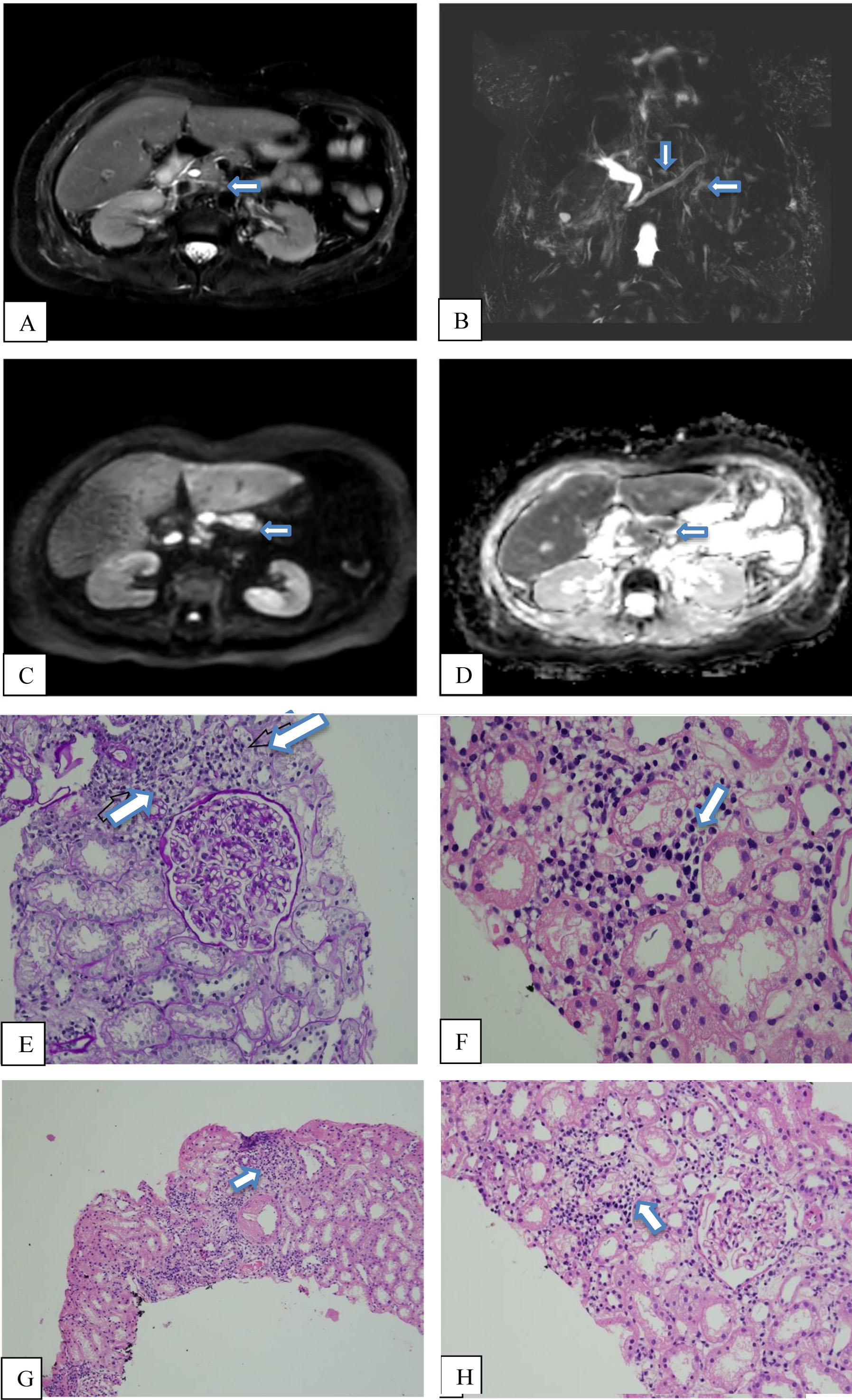
Viral serologies hepatitis B surface antigen (HBsAg), anti-hepatitis C virus (HCV) antibody, hepatitis A virus-immunoglobulin M (HAV-IgM), and hepatitis E virus-immunoglobulin M (HEV-IgM) were negative, and blood and urine cultures were sterile, thereby ruling out infectious causes. An abdominal ultrasound revealed hepatomegaly (16 cm) and a prominent Common Hepatic Duct. To investigate biliary anatomy and persistent pain abdomen, magnetic resonance cholangiopancreatography (MRCP) was performed, which showed (Figure 1):
- Subtle inflammatory changes in the pancreatic parenchyma with minimal peripancreatic inflammation.
- Focal areas of diffusion restriction.
- No intraductal calculi or side branch dilatation.
Gastroenterology consultation was obtained, and while serum amylase and serum lipase were within normal limits, a diagnosis of acute pancreatitis was established based on the clinical presentation and radiological findings.
Respiratory Involvement and Autoimmune Workup
During the course in hospital, the patient developed shortness of breath. High-resolution computed tomography (HRCT) revealed bilateral mild pleural effusions, patchy ground-glass opacities, and interlobular septal thickening. Given the multisystem involvement, an autoimmune profile workup was done:
- Antinuclear antibody (ANA): 3+ (1:320) Speckled
- Extractable nuclear antigen (ENA) profile: Positive for anti-Smith, anti-U1 ribonucleoprotein (RNP), anti-Jo-1, anti-proliferating cell nuclear antigen (PCNA), borderline anti-Ro-52
- Anti-dsDNA Antibody: Negative
- Complement: C4 < 2.0 (reference range 10–40 mg/dL); C3 91 (reference range 90–180 mg/dL)
- Immunoglobulins: Total immunoglobulin G (IgG) 8.96 g/L (reference range 7–16 g/L) and IgG4 0.202 g/L (reference range 0.03–2.01 g/L) were normal
Nephrology Consultation and the Diagnostic Dilemma
Urinalysis demonstrated evidence of renal involvement despite an initial normal serum creatinine level (0.87 mg/dL). The findings were as follows:
- Protein: 2+
- Ketones: Trace
- Microscopy: 15–20 pus cells and 10–15 red blood cells (RBCs) per High-Power Field (HPF)
Over the course of hospitalization, serum creatinine increased to 2.73 mg/dL. The presence of active urinary sediment in association with reduced C4 levels and a positive anti-Smith antibody raised a high index of suspicion for lupus nephritis [1]. In view of this diagnostic uncertainty, a renal biopsy was undertaken.
Histopathology and Final Diagnosis
Renal biopsy findings were pivotal in establishing the final diagnosis:
- Primary finding: Marked plasma cell predominant interstitial nephritis.
- Key negative findings: Absence of immune complex deposition, no evidence of IgG4-positive plasma cells, and no morphological evidence of storiform fibrosis.
- Interpretation: The lack of immune complex deposition, including the absence of a “full-house” immunofluorescence pattern, effectively excluded lupus nephritis. Instead, the histological appearance, together with the absence of storiform fibrosis and other characteristic morphological features of IgG4-related disease, was characteristic of Sjogren’s-syndrome-associated tubulointerstitial nephritis (TIN) [5].
- Final Diagnosis: Primary Sjogren’s syndrome presenting with acute pancreatitis and Tubulo-interstitial nephritis.
Management and Outcome
A comprehensive, multidisciplinary treatment approach was initiated:
- Empiric Antimicrobial Coverage: Broad-spectrum antibiotics (meropenem and teicoplanin) were administered initially to cover possible infective etiology but there was no clinical response to antibiotics. Subsequently, given the negative cultures and positive autoimmune serology, corticosteroid therapy was initiated.
- Immunosuppression: High dose intravenous methylprednisolone (125 mg twice daily) was started to control systemic autoimmune inflammation. This was subsequently transitioned to oral prednisolone for maintenance therapy.
- Fluid Optimization and Respiratory Support: In the setting of dyspnea and pleural effusion documented on HRCT, loop diuretics were administered to manage volume overload. As the patient’s fluid balance normalized and respiratory parameters improved, diuretics were progressively tapered and discontinued.
- Disease-Modifying Therapy: Hydroxychloroquine at a dose of 200 mg twice daily was commenced for long-term immunomodulation and disease control along with Mycophenolate 360 mg twice a day.
The patient showed a favorable clinical response, with resolution of fever, jaundice, abdominal pain, and respiratory distress. She was discharged in stable condition on a tapering regimen of corticosteroids, along with advice for regular follow up with Rheumatology and Nephrology department.
Discussion
Sjogren’s vs lupus nephritis
This case illustrates a classic example of autoimmune diagnostic overlap. At presentation, the patient fulfilled various clinical and serological features which are commonly attributed to SLE, including:
- Serositis: Pleural effusion
- Renal involvement: Proteinuria with an active urinary sediment
- Serology: High-titer ANA (1:320), reduced C4 levels, and the presence of anti-Smith antibodies
Anti-Smith antibodies are regarded as highly specific for SLE [1]. However, anti-dsDNA antibody testing was negative in our patient, creating a diagnostic discordance between serological findings and the eventual histopathological diagnosis. Serological markers alone are insufficient to establish the diagnosis when clinical features overlap. In this patient, renal histopathology became the decisive investigation that clarified the underlying disease process.
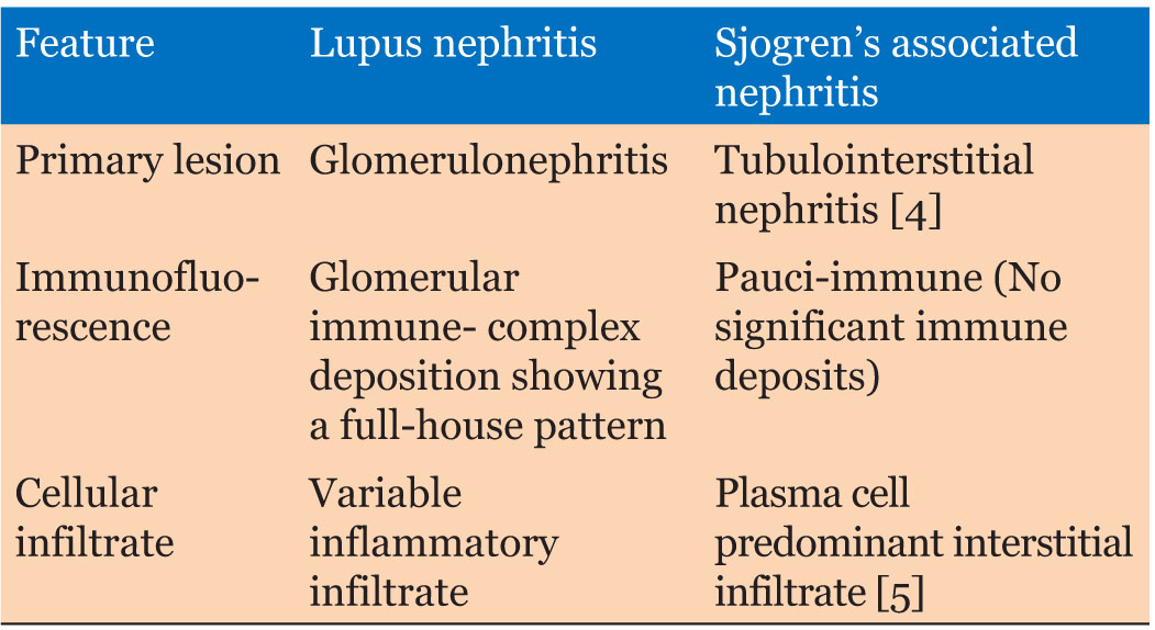
The key differentiating features between lupus nephritis and Sjogren’s-associated nephritis are summarized in Table 1.
Renal involvement in pSS is most often mediated through tubulointerstitial inflammation rather than immune-complex glomerular disease. Plasma-cell predominant interstitial nephritis without immune deposits represents the pathological hallmark of Sjogren’s-related renal disease and is reported in approximately 5% of patients [5]. In addition, the absence of IgG4-positive plasma cells, absence of storiform fibrosis, and normal serum IgG4 levels in this case effectively excluded IgG4-related disease, another important differential that may present with pancreatitis and interstitial nephritis.
The diagnosis was based on the combined assessment of clinical presentation, serological findings, and renal histopathology, with the latter proving decisive in excluding lupus nephritis and IgG4-related disease.
Although rare, pancreatic involvement in pSS has been described and is believed to result from immune-mediated inflammation of the pancreatic parenchyma.
Conclusion
Acute pancreatitis may represent an uncommon initial manifestation of primary Sjogren’s syndrome. This case highlights that even in the presence of so-called “lupus-specific” antibodies such as anti-Smith, histopathological confirmation remains indispensable. The absence of immune-complex deposition on renal biopsy correctly identified the renal pathology as Sjogren’s associated tubulointerstitial nephritis rather than lupus nephritis, thus directing appropriate immunosuppressive therapy and prognostication.
REFERENCES
1.
Satoh M, Chan EK, Sobel ES, Kimpel DL, Yamasaki Y, Narain S, et al. Clinical implication of autoantibodies in patients with systemic rheumatic diseases. Expert Rev Clin Immunol 2007;3(5):721–38. [CrossRef]
[Pubmed]

2.
Ramos-Casals M, Brito-Zerón P, Sisó-Almirall A, Bosch X. Primary Sjogren syndrome. BMJ 2012;344:e3821. [CrossRef]
[Pubmed]
3.
Moutsopoulos HM. Sjögren's syndrome: Autoimmune epithelitis. Clin Immunol Immunopathol 1994;72(2):162–5. [CrossRef]
[Pubmed]
4.
Evans RDR, Laing CM, Ciurtin C, Walsh SB. Tubulointerstitial nephritis in primary Sjögren syndrome: Clinical manifestations and response to treatment. BMC Musculoskelet Disord 2016;17:2. [CrossRef]
[Pubmed]
5.
Bossini N, Savoldi S, Franceschini F, Mombelloni S, Baronio M, Cavazzana I, et al. Clinical and morphological features of kidney involvement in primary Sjögren's syndrome. Nephrol Dial Transplant 2001;16(12):2328–36. [CrossRef]
[Pubmed]
SUPPORTING INFORMATION
Author Contributions
Chetan Tatrari - Conception of the work, Design of the work, Acquisition of data, Analysis of data, Drafting the work, Revising the work critically for important intellectual content, Final approval of the version to be published, Agree to be accountable for all aspects of the work in ensuring that questions related to the accuracy or integrity of any part of the work are appropriately investigated and resolved.
Amitabh Parti - Conception of the work, Design of the work, Acquisition of data, Analysis of data, Drafting the work, Revising the work critically for important intellectual content, Final approval of the version to be published, Agree to be accountable for all aspects of the work in ensuring that questions related to the accuracy or integrity of any part of the work are appropriately investigated and resolved.
Naval Mendiratta - Conception of the work, Design of the work, Acquisition of data, Analysis of data, Drafting the work, Revising the work critically for important intellectual content, Final approval of the version to be published, Agree to be accountable for all aspects of the work in ensuring that questions related to the accuracy or integrity of any part of the work are appropriately investigated and resolved.
Abha K Sabhikhi - Conception of the work, Design of the work, Acquisition of data, Analysis of data, Drafting the work, Revising the work critically for important intellectual content, Final approval of the version to be published, Agree to be accountable for all aspects of the work in ensuring that questions related to the accuracy or integrity of any part of the work are appropriately investigated and resolved.
Anmol Uberoi - Conception of the work, Design of the work, Acquisition of data, Analysis of data, Drafting the work, Revising the work critically for important intellectual content, Final approval of the version to be published, Agree to be accountable for all aspects of the work in ensuring that questions related to the accuracy or integrity of any part of the work are appropriately investigated and resolved.
Guarantor of SubmissionThe corresponding author is the guarantor of submission.
Source of SupportNone
Consent StatementWritten informed consent was obtained from the patient for publication of this article.
Data AvailabilityAll relevant data are within the paper and its Supporting Information files.
Conflict of InterestAuthors declare no conflict of interest.
Copyright© 2026 Chetan Tatrari et al. This article is distributed under the terms of Creative Commons Attribution License which permits unrestricted use, distribution and reproduction in any medium provided the original author(s) and original publisher are properly credited. Please see the copyright policy on the journal website for more information.