 |
Case Report
Could tractography help in the diagnosis of motor neuron diseases\amyotrophic lateral sclerosis? A case report
1 Medical student at Iguaçu University - UNIG/RJ, Nova Iguaçu - RJ, Brazil
2 Physician, Neurologist, Adjunct Professor of Medicine at Universidade Iguaçu - UNIG/Nova Iguaçu, RJ, Brazil
3 Neuroradiologist, Federal University of Rio de Janeiro - UFRJ, Rio de Janeiro - RJ, Brazil
4 Department of Neurology of Hospital Geral de Nova Iguaçu, PhD student in Neurology at the Federal University of the State of Rio de Janeiro - UNIRIO, Adjunct Professor of Medicine at Iguaçu University - UNIG/Nova Iguaçu, RJ, Brazil
Address correspondence to:
Antonio Marcos da Silva Catharino
Rua Gavião Peixoto 70, Room 811, CEP 24.2230-100, Icaraí, Niterói-RJ,
Brazil
Message to Corresponding Author
Article ID: 101418Z01FF2023
Access full text article on other devices

Access PDF of article on other devices


How to cite this article
Felicio FC, Campos MB, Neves MAO, Pereira DA, Toomassini LAB, da Silva Catharino AM. Could tractography help in the diagnosis of motor neuron diseasesamyotrophic lateral sclerosis? A case report. Int J Case Rep Images 2023;14(2):97–101.ABSTRACT
Introduction: Amyotrophic lateral sclerosis (ALS) is a disease that affects motor neurons, progressively degenerating them. This degeneration process has a complex and multifactorial etiology, culminating in the motor disability of the carriers.
Case Report: The case report discusses a clinical suspicion of motor neuron disease (ALS) in the patient NVF, male, 72 years old, with a report of falls and paresis in the left distal crural third for two years. His condition evolved with the four limbs and trunk involvement, but it does not show bulbar involvement. In addition, atrophy, fasciculations, and paresis have been identified from lower motor neuron injury in all four limbs. Regarding the pyramidal pathway, only lively but symmetrical reflexes.
Conclusion: In the last two decades, there have been significant advances in non-invasive imaging techniques, which allow the evaluation of brain structure, as is the case with tractography. Although this technique does not diagnose ALS, it can help early detection.
Keywords: ALS amyotrophic lateral sclerosis, Motor neuron diseases, Tractography
Introduction
The term motor neuron disease refers to a category of diseases that result in progressive motor neuron degeneration. This term is often synonymously with amyotrophic lateral sclerosis (ALS), the most common disease in this category. Amyotrophic lateral sclerosis is a progressive neurodegenerative disease characterized by losing motor neurons in the spinal cord, brainstem, and motor cortex, dramatically reducing life expectancy [1],[2].
The degenerative process of this disease comprises a complex and multifactorial etiology and may be of hereditary or acquired origin. Current hypotheses about this entity's underlying pathological mechanisms suggest a complex interplay between various mechanisms, including genetic factors, oxidative damage, accumulation of intracellular aggregates, mitochondrial dysfunction, axonal transport defects, glial cell pathology, and excitotoxicity [3].
In amyotrophic lateral sclerosis, both the lower and upper motor neurons can be affected, but in most cases, only the lower motor neuron is involved. Injury to this lower neuron is responsible for the characteristic clinical hallmark of motor neuron disease, related to signs of atrophy, weakness, and fasciculations of affected motor units [1].
The diagnosis of the disease is based on its clinical signs, observing its involvement in upper, lower, or brainstem motor neurons. It must also be differentiated into its forms: Primary lateral sclerosis, progressive bulbar palsy, and Frank ALS. Clinical symptoms, distinguished by their location and origin, associated with diagnostic tests, usually magnetic resonance imaging and electroneuromyography, are tabulated to arrive at confirmation of the disease [4].
Over the past two decades, neuroscientists and clinicians have witnessed enormous growth in non-invasive imaging techniques, which allow the assessment of brain structure. Among the most recent advances in imaging is tractography or diffusion tensor imaging (DTI) [5].
Tractography is a technique derived from magnetic resonance imaging that allows visualization of the orientation and integrity of cerebral white matter tracts in vivo by measuring the diffusion of water molecules in neural fibers. The technique allows the study of pre-defined areas of interest, such as specific tracts or the brain. Any microstructural alteration in the white matter, such as those found in ALS, appears on the tractography, indicating the lesion’s site [5].
Diffusion of water occurs preferentially in the direction parallel to the axons, being restricted in the plane perpendicular to them in the absence of pathologies. An ellipse or a tensor can represent this phenomenon graphically or mathematically. Based on these principles, the technique provides quantitative information on the degree and direction of water diffusion in the analyzed region, allowing visualization and characterization of white matter tracts in vivo [6],[7].
With the improvement of this technique over the years, it became possible to characterize tissue and locate lesions, in addition to mapping tracts [5], showing a fundamental role in monitoring the evolution, prognosis, and treatment of several neurodegenerative diseases.
Currently, tractography is useful in planning surgery or biopsies, early diagnosis of asymptomatic diseases of the nervous system, evaluation of disease extension, monitoring of its evolution, response to treatment and prognosis [7].
The tractography is mainly based on two measures of DTI to assess white matter: anisotropy fractional (AF) and mean diffusivity (DM). Fractional anisotropy is a measure of the directional diffusivity of water along bundles of nerve fibers, while average diffusivity is related to the overall movement of molecules on a microscopic scale. Low values of fractional anisotropy reflect axonal loss and/or demyelination, and high values may be related to inflammatory responses, such as axonal swelling or cytotoxic edema [8],[9].
Diffusion tensor imaging may prove useful in understanding the pathophysiology of this disease by providing quantitative biomarkers that can potentially aid diagnosis, identify at-risk populations, and assess therapeutic interventions [10].
Case Report
NVF, 72 years old, retired, without co-morbidities. He reports that he began to experience unexplained falls and paresis in the left distal crural third approximately two years ago. The picture evolved and compromised the four limbs and trunk; it does not present, at the moment, bulbar involvement.
Atrophy, fasciculations, and paresis were identified from lower motor neuron lesion in the four limbs. The patient no longer walks with progressive loss of strength in the lower and upper left limbs.
Only live but symmetrical reflexes were observed concerning the involvement of the pyramidal pathway.
Lab: normal.
Electroneuromyography (ENM): Observed abolition of left tibial nerve responses and reduced amplitudes and conduction velocity in median, ulnar, peroneal, and right tibial nerves. Observed a marked decrease in the number of motor units and an increase in the size of motor units:
MUNIX 16 right APB; MUSIX 82
APB left MUNIX 6; MUSIX 51
Demonstrating chronic motor peripheral neurogenic involvement.
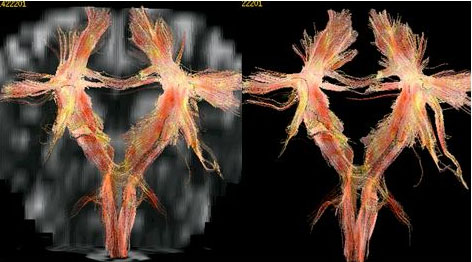
Cranial MRI with tractography: Pyramidal pathway and spinal cortical tract with good bilateral thickness (Figure 1).
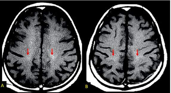
A tenuous hyper signal was noted on the T1 sequence with the magnetization transfer technique (T1MP) in the topography of the pyramidal pathway bilaterally, suggesting its impairment (Figure 2).
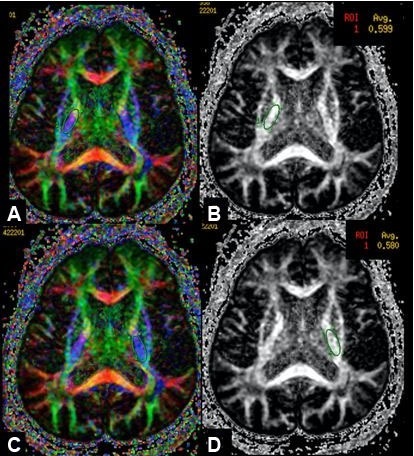
Bilaterally, fractional anisotropy within the limits of normality was observed (Figure 3).
Discussion
Amyotrophic lateral sclerosis (ALS) rarely appears before the age of 30, reaching its peak around the age of sixty and seventy, being more prevalent in male adults [8].
It is defined as a progressive neurological disease and, with about 80% of cases of unknown etiology, however, it most commonly affects the motor neuron. It manifests with cramps, fasciculations, weakness, and progressive muscle atrophy, which are the most common complaints of ALS. It progresses with unilateral and distal muscle weakness in a single segment, however it does not show sensory alterations and associated bladder dysfunction [8],[9]. As demonstrated in the reported case.
In terms of age and gender, our patient is in accordance with the indicator parameters, being male and aged 60 or over.
According to Moura et al. (2016), the estimated incidence of ALS in Brazil is 0.4 cases per 100,000 inhabitants, but the authors corroborate that there is a lack of epidemiological studies at the national level [9].
Worldwide, more precisely in the United States of America (USA) and Europe, the incidence of ALS is on average 1–3 cases per 100,000 inhabitants, with a prevalence in these same places of 3–5 cases per 100,000 inhabitants [10].
The diagnosis of ALS is usually based on a combination of clinical symptoms, neurological examinations, nerve conduction tests, electroneuromyography, and exclusion of other conditions that may cause similar symptoms. Magnetic resonance imaging can be used to rule out other conditions that may present with symptoms similar to ALS, but tractography is not considered a standard diagnostic tool for this specific disease [11].
The onset of clinical symptoms differs according to their location and origin of manifestations, associated with diagnostic tests such as magnetic resonance imaging, electroneuromyography, and nerve conduction studies. These tests are useful to differentiate suspicion from the definitive diagnosis [12]. Following this context, tactography can be used in research to better understand brain connectivity in patients with ALS.
Tractography is a magnetic resonance technique that allows detailed mapping of the nerve fibers of the white matter of the brain, obtaining qualitative and also quantitative images by means of the brain image diffusion tensor (DTI) [13].
The most common applications of tractography are: the study of white matter tracts and their topographic relationship with tumors and infarcts; the study of the pathophysiology of white matter lesions, such as multiple sclerosis and amyotrophic lateral sclerosis, cerebral palsy, epilepsy [13],[14].
However, tractography, when performed alone, is insufficient for the diagnosis of amyotrophic lateral sclerosis, since this exam is restricted in the evaluation of white matter fiber mapping, failing in a more specific evaluation for the condition in question [15].
Therefore, it can be said that tractography is not an efficient test to aid in the diagnosis of amyotrophic lateral sclerosis, as it demonstrates insufficient findings when associated with the clinical examination presented by the patient. However, this test can be used to monitor the evolution of the disease. With the progression of ALS, the integrity of the pyramidal pathway decreases, evidenced by the decrease in fractional anisotropy, showing the integrity of the pyramidal pathway. However, for the diagnosis of ALS, magnetic resonance imaging is the main neuroimaging study of the brain and spinal cord, using the T1 sequence with the magnetization transfer technique (T1MT) is the most recommended, in which brightness and degeneration are evident of the pyramidal pathway in 80% of the cases.
There is no known cure for ALS, but there are treatment approaches that can help manage symptoms and improve patients’ quality of life. This may include medication to relieve symptoms, physical and occupational therapy to maintain muscle function, and improve mobility, assistive devices for communication and breathing, among others [16],[17].
Among them we have nutritional and respiratory support, which are important for survival and based on the control of the main symptoms, such as sialorrhea, bulbar effects on emotional lability, sleep disorders, respiratory failure, pain, fatigue, muscle spasticity, laryngospasm, and chronic constipation [17].
Conclusion
Amyotrophic lateral sclerosis is a complex disease, responsible for a severe progressive condition, which may result in the patient’s absolute motor incapacity. For this reason, despite its low prevalence, it deserves special attention with regard to its early detection.
In this sense, tractography is an important auxiliary tool in the diagnosis of ALS, especially in cases in which the patient’s condition is suggestive of the disease, but there is little evidence in the physical examination that corroborates this finding. However, only the tractography is not enough for its diagnosis, since this is complex and requires a rigorous evaluation, based on the combination of clinical information, physical exams, laboratory exams, and imaging tests.
REFERENCES
1.
Tiryaki E, Horak HA. ALS and other motor neuron diseases. Continuum (Minneap Minn) 2014;20(5 Peripheral Nervous System Disorders):1185–207. [CrossRef]
[Pubmed]

2.
Batra G, Jain M, Singh RS, et al. Novel therapeutic targets for amyotrophic lateral sclerosis. Indian J Pharmacol 2019;51(6):418–25. [CrossRef]
[Pubmed]
3.
Linden-Junior E, Becker J, Schestatsky P, Rotta FT, Marrone CD, Gomes I. Prevalence of amyotrophic lateral sclerosis in the city of Porto Alegre, in Southern Brazil. Arq Neuropsiquiatr 2013;71(12):959–62. [CrossRef]
[Pubmed]
4.
Bertazzi RN, Martins FR, Saade SZZ, Guedes VR. Esclerose lateral amiotrófica. Revista de Patologia do Tocantins 2017;4(3):54–65. [CrossRef]
5.
Gierek T, Paluch J, Pencak P, Kaźmierczak B, Klimczak-Golab L. Magnetic resonance tractography in neuroradiological diagnostic aspects. [Article in Polish]. Otolaryngol Pol 2009;63(5):403–6. [CrossRef]
[Pubmed]
6.
Prakash KM, Tan EK. Development of Parkinson's disease biomarkers. Expert Rev Neurother 2010;10(12):1811–25. [CrossRef]
[Pubmed]
7.
Lipton ML, Gellella E, Lo C, et al. Multifocal white matter ultrastructural abnormalities in mild traumatic brain injury with cognitive disability: A voxel-wise analysis of diffusion tensor imaging. J Neurotrauma 2008;25(11):1335–42. [CrossRef]
[Pubmed]
8.
Marin B, Fontana A, Arcuti S, et al. Age-specific ALS incidence: A dose-response meta-analysis. Eur J Epidemiol 2018;33(7):621–34. [CrossRef]
[Pubmed]
9.
Moura MC, Casulari LA, Carvalho Garbi Novaes MR. Ethnic and demographic incidence of amyotrophic lateral sclerosis (ALS) in Brazil: A population based study. Amyotroph Lateral Scler Frontotemporal Degener 2016;17(3–4):275–81. [CrossRef]
[Pubmed]
10.
Brown RH, Al-Chalabi A. Amyotrophic lateral sclerosis. N Engl J Med 2017;377(2):162–72. [CrossRef]
[Pubmed]
11.
Borghetti VS, Cintra VP, Ramos JO, et al. Misdiagnoses in a Brazilian population with amyotrophic lateral sclerosis. Arq Neuropsiquiatr 2022;80(7):676–80. [CrossRef]
[Pubmed]
12.
Nagae LM, Pinho MDC, Funari MBDG. Tractography. Einstein (Sao Paulo) 2010;8(2):252–3. [CrossRef]
[Pubmed]
13.
Garcia LN, da Silva AV, Carrete H Jr, et al. Correlation between corticospinal tract degeneration through magnetic resonance imaging, and functional scale (ALSFRS) in patients with amyotrophic lateral sclerosis. [Article in Portuguese]. Arq Neuropsiquiatr 2007;65(3B):869–74. [CrossRef]
[Pubmed]
14.
Foerster BR, Dwamena BA, Petrou M, et al. Diagnostic accuracy of diffusion tensor imaging in amyotrophic lateral sclerosis: A systematic review and individual patient data meta-analysis. Acad Radiol 2013;20(9):1099–106. [CrossRef]
[Pubmed]
15.
Li J, Pan P, Song W, Huang R, Chen K, Shang H. A meta-analysis of diffusion tensor imaging studies in amyotrophic lateral sclerosis. Neurobiol Aging 2012;33(8):1833–8. [CrossRef]
[Pubmed]
16.
Filippi M, Rocca MA. Magnetization transfer magnetic resonance imaging of the brain, spinal cord, and optic nerve. Neurotherapeutics 2007;4(3):401–13. [CrossRef]
[Pubmed]
17.
Linden EJ, Linden D, Bareta GM, et al. Esclerose lateral amiotrófica: Artigo de atualização. FAAE 2016;47–62.
SUPPORTING INFORMATION
Author Contributions
Felipe de Castro Felicio - Conception of the work, Design of the work, Drafting the work, Final approval of the version to be published, Agree to be accountable for all aspects of the work in ensuring that questions related to the accuracy or integrity of any part of the work are appropriately investigated and resolved.
Marcello de Brito Campos - Conception of the work, Design of the work, Drafting the work, Final approval of the version to be published, Agree to be accountable for all aspects of the work in ensuring that questions related to the accuracy or integrity of any part of the work are appropriately investigated and resolved.
Marco Antônio Orsini Neves - Conception of the work, Design of the work, Acquisition of data, Analysis of data, Drafting the work, Revising the work critically for important intellectual content, Final approval of the version to be published, Agree to be accountable for all aspects of the work in ensuring that questions related to the accuracy or integrity of any part of the work are appropriately investigated and resolved.
Daniel Antunes Pereira - Conception of the work, Design of the work, Drafting the work, Final approval of the version to be published, Agree to be accountable for all aspects of the work in ensuring that questions related to the accuracy or integrity of any part of the work are appropriately investigated and resolved.
Lara Alexandre Brandão Toomassini - Conception of the work, Design of the work, Acquisition of data, Analysis of data, Drafting the work, Revising the work critically for important intellectual content, Final approval of the version to be published, Agree to be accountable for all aspects of the work in ensuring that questions related to the accuracy or integrity of any part of the work are appropriately investigated and resolved.
Antonio Marcos da Silva Catharino - Conception of the work, Design of the work, Analysis of data, Drafting the work, Revising the work critically for important intellectual content, Final approval of the version to be published, Agree to be accountable for all aspects of the work in ensuring that questions related to the accuracy or integrity of any part of the work are appropriately investigated and resolved.
Guarantor of SubmissionThe corresponding author is the guarantor of submission.
Source of SupportNone
Consent StatementWritten informed consent was obtained from the patient for publication of this article.
Data AvailabilityAll relevant data are within the paper and its Supporting Information files.
Conflict of InterestAuthors declare no conflict of interest.
Copyright© 2023 Felipe de Castro Felicio et al. This article is distributed under the terms of Creative Commons Attribution License which permits unrestricted use, distribution and reproduction in any medium provided the original author(s) and original publisher are properly credited. Please see the copyright policy on the journal website for more information.


