 |
Case Report
Candida infection of zygomatic implants/fillers in an HIV-positive patient: A case report
1 Northern Ontario School of Medicine, Sudbury, ON P3E 2C6, Canada
2 Department of Radiology, Health Sciences North, Northern Ontario School of Medicine, Sudbury, ON P3E 5J1, Canada
Address correspondence to:
Yasmine Sallam
41, Ramsey Lake Road, Sudbury, ON,
Canada
Message to Corresponding Author
Article ID: 101488Z01ZG2025
Access full text article on other devices

Access PDF of article on other devices


How to cite this article
Gagné Z, Mensour E, Mejía MEM, Elnayal A, Sallam Y. Candida infection of zygomatic implants/fillers in an HIV-positive patient: A case report. Int J Case Rep Images 2025;16(1):1–4.ABSTRACT
Introduction: Facial fillers are a common procedure that has gradually been increasing in popularity. While most cases of facial fillers have good outcomes, a small percentage have associated complications, with infection being one of the most common.
Case Report: We present a case of a 63-year-old human immunodeficiency virus (HIV)-positive male who presented to the emergency department following a two-week history of a swollen and tender right cheek. The patient had a history of facial cosmetic procedures, including facial fillers and cheek implants. Infection was suspected and the patient was sent for a computed tomography (CT) scan that revealed the presence of a peripherally enhancing organized fluid collection overlying the right zygomatic bone. Analysis of the fluid revealed the presence of Candida albicans. The abscess was drained and treated with a two-week course of fluconazole.
Conclusion: For oral and maxillofacial surgeons as well as radiologists, this case report highlights the importance of being familiar with the normal anatomical location and radiologic appearance of facial fillers and their associated complications.
Keywords: Candida, Facial fillers, Head and neck radiology, HIV
Introduction
Facial fillers are common cosmetic procedures that have increased in popularity over the past few decades [1]. A large variety of facial fillers are currently available on the market each with their associated indications and longevity, including calcium hydroxylapatite, hyaluronic acid, polyalkylimide, polylactic acid, and polymethyl-methacrylate microspheres (PMMA). While most of these procedures are performed cosmetically for facial rejuvenation, other indications exist, such as post-traumatic facial disfiguration and facial lipoatrophy associated with certain HIV medications [2]. Complications for all types of facial fillers can arise a few days to weeks following the procedure (short-term), or several months to years afterward (long-term). Short-term complications include hypersensitivity reactions, infection, skin necrosis, and discoloration, while long-term complications include delayed hypersensitivity reactions, filler migration, infection, foreign-body granulomas, and scarring [3]. We report a case of an HIV-positive 63-year-old male who presented to the emergency department following a two-week history of a swollen right cheek.
Facial lipodystrophy in HIV-positive patients is a common side effect of antiviral treatments with multiple studies reporting prevalence rates between 11% and 83% [4]. Facial fillers are an accepted treatment for these side effects, and while increased infection rates among HIV-positive patients have not been reported [5], HIV-positive patients have been shown more susceptible to certain opportunistic infections [6]. While the overall rate of infections associated with facial filler procedures remains fairly low, with estimates between 0.04% and 0.2% [7], their increased use over the past few decades ensures that these radiological findings will become more common in practice.
Case Report
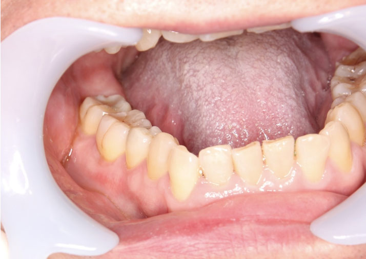
A 63-year-old male presented to the emergency department following a two-week history of a swollen and tender right cheek. The patient had a history of diabetes, with the most recent test performed six weeks prior to the infection showing an A1C of 10.5. The patient also had a history of cirrhosis, and HIV, for which he was being treated with Cabenuva. The patient reported having multiple cosmetic procedures performed in the past, including zygomatic implants, injectable facial fillers, with his last cosmetic procedure being implant replacements following an infection of an unknown organism in 2012. Upon examination, infection was suspected, and the patient was sent for a CT scan as well as a needle aspiration of the right cheek for analysis. The CT scan indicated the presence of a peripherally enhancing organized fluid collection overlying the right zygomatic bone, surrounding the right zygomatic implant measuring about 5 × 1.3 cm axially and 2.3 cm craniocaudally (Figure 1). The fluid collection was surrounded by mild fat stranding as well as mild asymmetric thickening of the overlying skin. The CT scan also showed multiple other hypodense structures located on the left side over the left zygomatic bone (Figure 2), along the inferior margin of the left zygomatic facial implant and lateral to the left maxilla (Figure 3), all favored to represent cosmetic facial fillers. The abscess was surgically drained three days later, and the implant as well as the surrounding fillers were removed. Only the implants and fillers on the affected side were removed as the physician was concerned about the possible risk of spreading the infection. The patient was seen one week later, and no signs of any residual infection were seen, but the patient was still experiencing swelling and discomfort. At this time, the patient was informed that the results from the needle aspiration came back positive for Candida albicans, for which the patient was treated with a two-week course of fluconazole. The patient was seen again in clinic three weeks later and showed no signs of swelling or discomfort following the completion of the 2-week course of fluconazole.


Discussion
We bring you a rare case of a Candida albicans-infected facial filler that resulted in a zygomatic abscess in an HIV-positive patient. Complications associated with facial fillers and implants remain rare [7] and while HIV-positive patients have been shown to be at an increased risk of surgical site infections, sepsis, and opportunistic infections [8], there is still debate as to whether patients on antiviral therapy have a higher risk of complications following cosmetic facial procedures [5],[9]. Many factors have been shown to influence the infection risk in HIV-positive patients, with CD4+ count being shown as the strongest predictor of surgical and procedural complications [10],[11]. The current research indicates that surgical complications are highest in patients with a CD4+ count of less than 200 cells/mm3, and therefore, elective procedures should be avoided in these patients [11].
With cosmetic procedures increasing significantly in the past few decades, the number of complications associated with these procedures will continue to rise proportionately. Abscesses can be difficult to differentiate from normal filler materials on CT and magnetic resonance imaging (MRI) as they can have a similar density on these modalities [3]. Familiarity with the specific imaging features of facial fillers as well as their typical anatomical locations can help make these distinctions. This case also highlights the importance of correlating clinical information with radiological findings. While rare in immunologically healthy patients, Candida albicans infections are one of the most common opportunistic infections seen in HIV-positive patients [12]. Human immunodeficiency virus-positive patients make up a significant proportion of individuals undergoing these cosmetic procedures performed. Given that these patients are more susceptible to developing opportunistic infections, special attention should be given when presented with a possible case of infected facial fillers in this patient population.
Conclusion
Given the increasing number of cosmetic facial procedures being performed, this case report highlights the importance for oral and maxillofacial surgeons and radiologists to be familiar with the specific imaging features and normal anatomical locations of facial fillers to avoid misinterpretations. This case report also highlights a patient population undergoing these procedures that might be more susceptible to developing infectious complications.
REFERENCES
1.
Gutowski KS, Chwa ES, Weissman JP, et al. Practice profile of practicing plastic surgeons: A 20-year review of plastic surgery statistics. Plast Reconstr Surg Glob Open 2023;11(12):e5486. [CrossRef]
[Pubmed]

2.
Sánchez-Carpintero I, Candelas D, Ruiz-Rodríguez R. Dermal fillers: Types, indications, and complications. [Article in Spanish]. Actas Dermosifiliogr 2010;101(5):381–93. [CrossRef]
[Pubmed]
3.
Ginat DT, Schatz CJ. Imaging features of midface injectable fillers and associated complications. AJNR Am J Neuroradiol 2013;34(8):1488–95. [CrossRef]
[Pubmed]
4.
Lana LGC, Junqueira DRG, Perini E, Menezes de Pádua C. Lipodystrophy among patients with HIV infection on antiretroviral therapy: A systematic review protocol. BMJ Open 2014;4(3):e004088. [CrossRef]
[Pubmed]
5.
Chirico F, Rauso GM, Fragola R, et al. Complications following non-surgical aesthetic treatments in HIV+ patients receiving antiretroviral therapy: A 12-years experience. Appl Sci 2021;11(9):4059. [CrossRef]
6.
Morales DR, Moreno-Martos D, Matin N, McGettigan P. Health conditions in adults with HIV compared with the general population: A population-based cross-sectional analysis. EClinicalMedicine 2022;47:101392. [CrossRef]
[Pubmed]
7.
Ferneini EM, Beauvais D, Aronin SI. An overview of infections associated with soft tissue facial fillers: Identification, prevention, and treatment. J Oral Maxillofac Surg 2017;75(1):160–6. [CrossRef]
[Pubmed]
8.
Zhao R, Ding R, Zhang Q. What are the risk factors for surgical site infection in HIV-positive patients receiving open reduction and internal fixation of traumatic limb fractures? A retrospective cohort study. AIDS Res Hum Retroviruses 2021;37(7):551–6. [CrossRef]
[Pubmed]
9.
Rauso R, Freda N, Parlato V, Gherardini G, Amore R, Tartaro G. Polyacrylamide gel injection for treatment of human immunodeficiency virus-associated facial lipoatrophy: 18 months follow-up. Dermatol Surg 2011;37(11):1584–9. [CrossRef]
[Pubmed]
10.
Ma R, Zhang Q, Zhao CS, et al. The consensus guideline of perioperative antiviral therapy for AIDS patients in China based on clinical practice. Front Med (Lausanne) 2023;10:1267236. [CrossRef]
[Pubmed]
11.
Shin SJ, Vail RM, Shah SS, et al. Perioperative Care in Adults With HIV [Internet]. Baltimore (MD): Johns Hopkins University; 2024.
[Pubmed]
12.
Anwar KP, Malik A, Subhan KH. Profile of candidiasis in HIV infected patients. Iran J Microbiol 2012;4(4):204–9.
[Pubmed]
SUPPORTING INFORMATION
Author Contributions
Zacharie Gagné - Conception of the work, Design of the work, Acquisition of data, Analysis of data, Drafting the work, Final approval of the version to be published, Agree to be accountable for all aspects of the work in ensuring that questions related to the accuracy or integrity of any part of the work are appropriately investigated and resolved.
Emma Mensour - Analysis of data, Drafting the work, Final approval of the version to be published, Agree to be accountable for all aspects of the work in ensuring that questions related to the accuracy or integrity of any part of the work are appropriately investigated and resolved.
Mauricio Enrique Moreno Mejía - Conception of the work, Design of the work, Revising the work critically for important intellectual content, Final approval of the version to be published, Agree to be accountable for all aspects of the work in ensuring that questions related to the accuracy or integrity of any part of the work are appropriately investigated and resolved.
Amr Elnayal - Conception of the work, Design of the work, Revising the work critically for important intellectual content, Final approval of the version to be published, Agree to be accountable for all aspects of the work in ensuring that questions related to the accuracy or integrity of any part of the work are appropriately investigated and resolved.
Yasmine Sallam - Conception of the work, Design of the work, Revising the work critically for important intellectual content, Final approval of the version to be published, Agree to be accountable for all aspects of the work in ensuring that questions related to the accuracy or integrity of any part of the work are appropriately investigated and resolved.
Guarantor of SubmissionThe corresponding author is the guarantor of submission.
Source of SupportNone
Consent StatementAs the patient's identity was not revealed, consent was not sought.
Data AvailabilityAll relevant data are within the paper and its Supporting Information files.
Conflict of InterestAuthors declare no conflict of interest.
Copyright© 2025 Zacharie Gagné et al. This article is distributed under the terms of Creative Commons Attribution License which permits unrestricted use, distribution and reproduction in any medium provided the original author(s) and original publisher are properly credited. Please see the copyright policy on the journal website for more information.
