 |
Case Report
Embryotomy for head last retention: A case report from Thiès, Senegal
1 Gynecology and Obstetrics Department, Thiès Regional Hospital Center (CHRT), Thiès, Senegal
2 Health Sciences Training and Research Unit of Thiès, Iba Der THIAM University of Thiès, Senegal
3 Gynecology and Obstetrics Department, Saint-Louis Regional Hospital Center, Health Sciences Training and Research Unit of Gaston Berger University of Saint-Louis, Senegal
4 Gynecology and Obstetrics Department, Nabil Choucair Hospital in Dakar, Faculty of Medicine, Cheikh Anta Diop University of Dakar, Senegal
Address correspondence to:
Lamine Gueye
Gynecology and Obstetrics Department, Thiès Regional Hospital Center,1 Avenue Malick SY Extended, Thiès,
Senegal
Message to Corresponding Author
Article ID: 101477Z01LG2024
Access full text article on other devices

Access PDF of article on other devices


How to cite this article
Gueye L, Thiam O, Thiam M, Gassama O, Ba PA, Cissé ML. Embryotomy for head last retention: A case report from Thiès, Senegal. Int J Case Rep Images 2024;15(2):85–88.ABSTRACT
Introduction: Hydrocephalus is a pathology of heterogeneous nature and complex pathogenesis. It corresponds to an active distension of the ventricular system. Faced with hydrocephalus with retained head in a dead child, embryotomy by craniotomy is indicated.
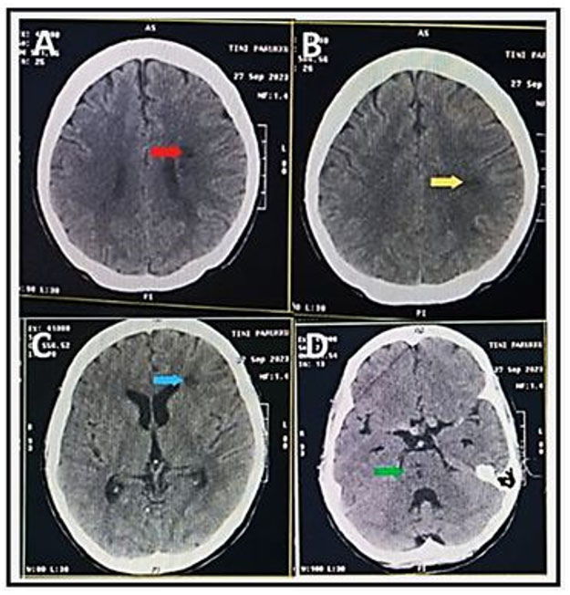
Case Report: We report a case of intrapartum fetal death with head-last retention due to hydrocephalus treated with craniotomy performed at the regional hospital of Thiès (CHRT). This was a 21-year-old primigravida patient seen for premature delivery in a pregnancy of 33 weeks 5 days. The ultrasound concluded that the pregnancy had stopped at 33 weeks 5 days with major hydrocephalus. A craniotomy followed by gradual evacuation of the cerebrospinal fluid allowed easy delivery of a fresh stillborn weighing 1950 grams. The afterbirth was simple.
Conclusion: Embryotomy by craniotomy constitutes a good option to complete delivery by natural means in the face of head retention due to hydrocephalus with intrapartum death. It is therefore necessary to master this simple and less morbid technique than a cesarean section in such a situation.
Keywords: Craniotomy, Hydrocephalus, Intrapartum fetal death
Introduction
Hydrocephalus is a pathology of heterogeneous nature and complex pathogenesis. It corresponds to an active distension of the ventricular system [1]. A late diagnosis during the expulsive phase of labor can lead to intrapartum fetal death due to retained head after breech presentation. In this scenario, embryotomy remains a good indication. It is an operation to mutilate the dead fetus, aimed at reducing its volume in order to facilitate vaginal delivery if it does not take place normally due to a mechanical obstacle. Various types of embryotomies were performed including cephalic embryotomies (craniotomy, cranioclasia, and basiotripsy) and spinal embryotomies used in neglected shoulder presentations. At present, there remains only one indication for embryotomy: craniotomy with possibly cranioclasis of hydrocephalus in dead children [2]. We report a case of craniotomy performed at the CHRT due to intrapartum fetal death with head-last retention resulting from hydrocephalus.
Case Report
Patient information: This is a 21-year-old patient with no prior pathological history, primigravida evacuated from the Pout/Thiès Health Center for a threat of premature delivery in a 33-week pregnancy of amenorrhea (AS) according to an early ultrasound. She had carried out 04 prenatal consultations of poor quality, a prenatal assessment not carried out, an obstetric ultrasound in the first trimester of pregnancy without particularity.
Results of the clinical examination: The clinical examination found blood pressure (BP) at 110/80 mmHg, good general condition, colored mucous membranes, a flexible, ovoid uterus with a long longitudinal axis with a uterine height of 33 cm. Fetal heart sounds were heard and regular. Vaginal examination revealed a cervix dilated to 8 cm and an engaged breech presentation. Faced with this picture of inevitable premature delivery with an engaged breech presentation, the patient was admitted to the delivery room. During the expulsive phase we discovered head-last retention.
Diagnostic approach: The ultrasound carried out revealed a pregnancy stopped at 33 weeks 5 days with major hydrocephalus. The biological assessment carried out (blood count, rhesus blood grouping, prothrombin level, and activated partial thromboplastin time) was normal.
Therapeutic intervention and follow-up: The patient was transferred to the operating room. Under general anesthesia, we performed a craniotomy by opening the spinal canal with scissors then gradually separating it (Figure 1), which allowed us to evacuate the cerebrospinal fluid, obtain a collapse of the cranial vault and easy expulsion of the last head (Figure 2 and Figure 3). This procedure allowed us to extract a fresh stillborn female weighing 1950 grams. We carried out a directed delivery followed by a uterine revision. Subsequently, we performed a valve examination followed by an injection of 10 IU of syntocinon by slow intravenous injection and 10 IU by infusion and antibiotic prophylaxis with 1 g of ceftriaxone. No incident or accident was noted. The uterus was empty and intact, no vaginal, cervical, or perineal tears. The postpartum period was simple and the patient was discharged on the third day postpartum after psychosocial assistance and long-acting contraception such as nexplanon.


Discussion
In utero fetal death refers by definition to the spontaneous cessation of cardiac activity of the fetus, at a gestational stage greater than 14 weeks of amenorrhea. This termination of pregnancy can take place before labor (antepartum fetal death) or during labor (intrapartum fetal death) [3]. In our developing countries, this intrapartum fetal death is most often encountered in evacuated patients, presenting an unnoticed fetopelvic disproportion. When intrapartum fetal death occurs in the face of fetal hydrocephalus, the performance of embryotomy, an operation which has become exceptional, should be favored. Two scenarios must be considered depending on whether the hydrocephalus presents through the head or whether it is in a breech position. In case of cephalic presentation, simple perforation using a large trocar or a branch of long scissors. In case of breech presentation, it is preferable to evacuate the cerebrospinal fluid (CSF) according to the Van Huevel-Tarnier maneuver [2]. Indeed, Van Huevel-Tarnier recommended the introduction of a rigid metal probe into the spinal canal then the evacuation of the CSF to complete the delivery by natural means [2]. For our case, we performed a simple scissor craniotomy in the operating room under general anesthesia. The delivery was easy and no maternal complications were observed in our patient. Ewacs J.O. in his historical series of 21 cases of embryotomies did not find any complications [4]. The use of embryotomy scissors by an experienced person can therefore be recommended for this regrettable procedure, but sometimes essential in certain situations that have become exceptional. In our developing countries where prenatal surveillance is still not up to par and where women find themselves in active labor with intrauterine fetal death or with undiagnosed fatal fetal malformations, every obstetrician should be trained in all operations destructive. However, given the high rate of complications, procedural failure, and declining skills in modern obstetrics, embryotomy should be attempted at least under the supervision of an experienced obstetrician and in a well-equipped location only [5].
Conclusion
Cephalic embryotomy by craniotomy is an obstetric practice that has become exceptional even. It constitutes a good option to complete delivery by natural means in the face of head-end retention due to hydrocephalus with intrapartum death. It is therefore necessary to master this simple and less morbid technique than a cesarean section in such a situation.
REFERENCES
1.
Tully HM, Dobyns WB. Infantile hydrocephalus: A review of epidemiology, classification and causes. Eur J Med Genet 2014;57(8):359–68. [CrossRef]
[Pubmed]

2.
Merger R, Lévy J, Melchior J. Obstetrics Notes. 6ed. Paris: Masson; 2001.
3.
Quibel T, Bultez T, Nizard J, Subtil D, Huchon C, Rozenberg P. In utero fetal death. J Gynecol Obstet Biol Reprod (Paris) 2014;43(10):883–907. [CrossRef]
[Pubmed]
4.
Obed JY. Fetal decapitation: The application and safety of the stout embryotomy scissors. Trop Doct 1994;24(3):139–40. [CrossRef]
[Pubmed]
5.
Rohilla M, Aggarwal N, Singh P, Jain V. Evisceration as fetal destructive operation: An art revisited. Arch Gynecol Obstet 2015;291(3):701–3. [CrossRef]
[Pubmed]
SUPPORTING INFORMATION
Author Contributions
Lamine Gueye - Conception of the work, Design of the work, Acquisition of data, Drafting the work, Revising the work critically for important intellectual content, Final approval of the version to be published, Agree to be accountable for all aspects of the work in ensuring that questions related to the accuracy or integrity of any part of the work are appropriately investigated and resolved.
Ousmane Thiam - Conception of the work, Design of the work, Drafting the work, Revising the work critically for important intellectual content, Final approval of the version to be published, Agree to be accountable for all aspects of the work in ensuring that questions related to the accuracy or integrity of any part of the work are appropriately investigated and resolved.
Mariétou Thiam - Conception of the work, Design of the work, Drafting the work, Revising the work critically for important intellectual content, Final approval of the version to be published, Agree to be accountable for all aspects of the work in ensuring that questions related to the accuracy or integrity of any part of the work are appropriately investigated and resolved.
Omar Gassama - Conception of the work, Design of the work, Drafting the work, Revising the work critically for important intellectual content, Final approval of the version to be published, Agree to be accountable for all aspects of the work in ensuring that questions related to the accuracy or integrity of any part of the work are appropriately investigated and resolved.
Papa Abdoulaye Ba - Conception of the work, Design of the work, Drafting the work, Revising the work critically for important intellectual content, Final approval of the version to be published, Agree to be accountable for all aspects of the work in ensuring that questions related to the accuracy or integrity of any part of the work are appropriately investigated and resolved.
Mamadou Lamine Cissé - Conception of the work, Design of the work, Drafting the work, Revising the work critically for important intellectual content, Final approval of the version to be published, Agree to be accountable for all aspects of the work in ensuring that questions related to the accuracy or integrity of any part of the work are appropriately investigated and resolved.
Guarantor of SubmissionThe corresponding author is the guarantor of submission.
Source of SupportNone
Consent StatementWritten informed consent was obtained from the patient for publication of this article.
Data AvailabilityAll relevant data are within the paper and its Supporting Information files.
Conflict of InterestAuthors declare no conflict of interest.
Copyright© 2024 Lamine Gueye et al. This article is distributed under the terms of Creative Commons Attribution License which permits unrestricted use, distribution and reproduction in any medium provided the original author(s) and original publisher are properly credited. Please see the copyright policy on the journal website for more information.


