 |
Case Report
Presence of multiple supernumerary premolars, distomolars, and anterior maxillary teeth in a non-syndromic dizygotic twin: A case report
1 BSc (Hons), BDS (Hons), MFDS RCS (Eng), Dental Core Trainee in Oral and Maxillofacial Surgery, Oral and Maxillofacial Surgery Department, Broomfield Hospital, Mid and South Essex NHS Trust, Essex, England
2 Statutory Exams (UK), BDS, MFDS RCSEd, MSc IOE UCL, MAcadMED, Registered Hypnotherapist (GHR), DCH, GQHP, CNHC Registered, Associate Specialist in Oral Surgery, Oral and Maxillofacial Surgery Department, Broomfield Hospital, Mid and South Essex NHS Trust, Essex, England
Address correspondence to:
Amandeep Singh Sarkaria
Broomfield Hospital, Mid and South Essex NHS Trust, Essex,
England
Message to Corresponding Author
Article ID: 101437Z01AS2024
Access full text article on other devices

Access PDF of article on other devices


How to cite this article
Sarkaria AS, Ogadako R. Presence of multiple supernumerary premolars, distomolars, and anterior maxillary teeth in a non-syndromic dizygotic twin: A case report. Int J Case Rep Images 2024;15(1):17–21.ABSTRACT
Introduction: While supernumerary teeth are occasionally encountered by the dentist and multidisciplinary team, it is relatively rare for multiple supernumeraries to be found in a non-syndromic patient.
Case Report: This report describes a case in which a dizygotic twin with a history of anterior maxillary supernumeraries presents with asymptomatic late developing bilateral mandibular premolar supernumeraries and distomolars. The premolar supernumeraries were surgically removed, while the distomolars managed conservatively.
Conclusion: The importance of periodic follow-up for patients with a history of supernumeraries is highlighted alongside the significance of high quality referrals.
Keywords: Dizygotic twin, Multiple supernumeraries, Non-syndromic
Introduction
A supernumerary tooth is one that has developed in addition to the normal complement of teeth within the dentition [1]. Supernumerary teeth may be singular or multiple in number and can occur in both the primary and permanent dentition, but are more common in the latter where their prevalence varies from 1% to 3% [2],[3]. Additionally they can be found anywhere within the dental arch; however, the majority (>90%) occur in the anterior maxilla [4].
Supernumeraries can be classified according to their location, which includes mesiodens, paramolar, or distomolar. Conversely they can be grouped based on their morphology, such as conical, tuberculate, supplemental, and odontomes, which may be complex or compound. Conical supernumeraries are the most commonly [5] observed and tend to be found in the anterior maxilla, whereas supplemental supernumeraries are more likely in the premolar region [6].
While supernumeraries are largely asymptomatic and tend to be detected as a chance finding during radiographic examination, they can also have a series of effects on the dentition. These include, preventing eruption, causing crowding, spacing, displacement, rotation and resorption of surrounding teeth, and occasionally they may even undergo pathological change, such as contributing to the formation of cysts [7]. In addition, they have also been found to prevent or delay space closure during orthodontic treatment [8].
The etiology of supernumeraries is unclear; however, hyperactivity of the dental lamina has been suggested as the most likely mechanism [9]. This theory proposes that lingual extension of an accessory tooth bud leads to development of a supplemental supernumerary. However, other types of supernumeraries develop due to proliferation of epithelial remnants of the dental lamina [3].
Nevertheless, there certainly appears to be a genetic element, supported by children of parents with supernumerary teeth having a six times increased risk of their development [10]. There also appears to be a sex link, as supernumeraries are twice as common in males when compared to females [4]. But interestingly this pattern is not seen in the primary dentition. Further evidence for genetic involvement is supported by supernumeraries being associated with syndromes such as Cleidocranial Dysplasia, Gardeners syndrome, Cleft Lip, and Palate and Ehlers Danlos with identification of causative genes like RUNX2 [11]. In such syndromes, multiple supernumeraries can be found, and it is relatively rare to find multiple supernumeraries in non-syndromic patients.
Case Report
A fit and well Caucasian male was referred to the Oral and Maxillofacial Surgery team via Orthodontic Department of the hospital at age 9 years and 11 months for removal of three impacted supernumeraries in the anterior maxilla that had impeded the eruption of both the upper central incisors (Figure 1A and Figure 1B).
Having extracted the supernumeraries in the anterior maxilla, the incisors were exposed and space made with an upper removable appliance. At age 13 the central incisors had spontaneously erupted, and the patient was discharged by the hospital back to his own orthodontist for alignment.
At age 14 years and 8 months, the patient was once again referred to the Oral and Maxillofacial Department, but this time by his orthodontist via his general dentist, for removal of all first premolars and interestingly asymptomatic developing lower supernumerary premolars (Figure 2).
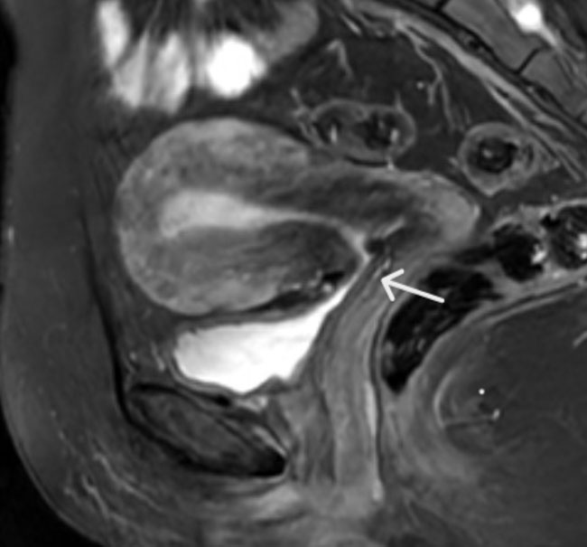
As the patient previously had supernumeraries, a detailed assessment excluded any family history of additional teeth and on examination there were no obvious signs of a syndrome. The only atypical finding was heterochromia, which was not immediately present at birth (Figure 3). The patient also had a twin sister who had undergone orthodontic treatment but had no history of supernumeraries and brown eyes.
Intra-orally, the lower supernumerary premolars were not visible or palpable and therefore to aid visualization and planning for surgical removal, a cone beam computed tomography (CBCT) scan was performed. This highlighted the lingually positioned supernumeraries that were in close proximity to the mental nerve, but also developing distomolars bilaterally, LL9 and LR9. In addition, a small radio-opacity was detected in the anterior maxilla (Figure 4 and Figure 5).
As the distomolars were visible on the CBCT and not the Orthopantogram (OPG) sent via the referral, the original was requested from the orthodontist. This higher quality image clearly demonstrated the developing mandibular distomolars (Figure 6).
On discussion with the patient and parents of all the options, a plan was made for surgical removal of all the first premolars and lower supernumerary premolars, and to manage the developing distomolars and anterior maxillary radio-opacity conservatively for now.
Three months after removal of the mandibular supernumeraries, an OPG and upper standard occlusal (USO) (Figure 7A and Figure 7B) were taken which showed no change in the maxillary radio-opacity. Therefore it is likely that this radio-opacity was a remnant after surgical removal of the anterior maxillary supernumeraries.


Discussion
Calcification of a mandibular premolar occurs between 18 and 24 months; however, it may not be visible radiographically until age 4 [5]. But in this patient, the mandibular premolar supernumeraries were detected at nearly age 15. While it is known that development of supernumeraries is delayed, as indicated by their incomplete root development compared to adjacent teeth, it is uncommon for them to form as late as demonstrated.
Nevertheless, it has been found that 24% of patients with an anterior supernumerary later develop mandibular premolar supernumeraries [12], and this pattern has been documented with similar case reports [13],[14]. In addition, solely late developing premolar supernumeraries have also been reported [8],[15].
Although what distinguishes this case is the further development of the fourth molars or distomolars. Incidence of fourth molars has been reported as 0.32% [16] and they tend to develop unilaterally and in the maxilla [17]. However, this patient had bilateral mandibular distomolars, similar to the late developing premolar supernumeraries. It is worth noting that the distomolars were not outlined at the time of the referral and were not clearly visible on the attached OPG due to its low quality. They only became apparent to the surgical team once a CBCT was performed and thus reinforce the importance of high quality radiographic images and referrals from the referring practitioner.
The presence of heterochromia can sometimes be associated with syndromes. For instance, Nance-Horan syndrome can be associated with both heterochromia and supernumerary teeth [18]. However, this patient did not exhibit any other features of a syndrome and therefore it is unlikely that the heterochromia was indicative of a syndrome.
The patient also had a twin sister who did not develop any supernumeraries. Indeed it has been reported that even among monozygotic twins, supernumeraries may not always develop in both twins and when they do, there can be discordance in their expression [19]. Therefore, this suggests that factors other than genetics may contribute to the development of supernumeraries. In addition, when four families with a history of non-syndromic supernumerary teeth underwent whole-exome sequencing, no common gene variant could be identified among all the families [20]. However, there were common gene variants between some of the families, but the authors reported that currently the identified gene variants are not known to be involved in tooth development.
For the ongoing care of such a patient, a periodic OPG would be beneficial to assess the status of current supernumeraries and also the development of further ones. This is especially important considering that supernumeraries may be associated with pathology, such as cyst formation [21]. For instance, it has been found that when assessed with a CBCT, over 20% of supernumeraries had caused resorption of adjacent teeth [22]. Interestingly, the study reported that resorption was most likely with supernumerary premolars and least likely for supernumerary canines, paramolars, and distomolars.
Conclusion
Although rare, multiple supernumeraries can present in non-syndromic patients. Clinicians should therefore ensure appropriate clinical and radiographic follow-up for patients with a history of supernumeraries. If detected, risks of both conservative and surgical management must be discussed, highlighting possible sequelae. Finally, if further advice or management is required, high quality referrals to the multi-disciplinary team are desired.
REFERENCES
1.
Cobourne MT. Supernumerary teeth. In: Orthodontic Management of the Developing Dentition: An Evidence-based Guide. Cham: Springer; 2017. p. 53–65.

2.
Mahabob MN, Anbuselvan GJ, Kumar BS, Raja S, Kothari S. Prevalence rate of supernumerary teeth among non-syndromic South Indian population: An analysis. J Pharm Bioallied Sci 2012;4(Suppl 2):S373–5. [CrossRef]
[Pubmed]
3.
Meade MJ. Supernumerary teeth: An overview for the general dental practitioner. Dent Update 2020;47(9):729–38. [CrossRef]
4.
Rajab LD, Hamdan MAM. Supernumerary teeth: Review of the literature and a survey of 152 cases. Int J Paediatr Dent 2002;12(4):244–54. [CrossRef]
[Pubmed]
5.
Cochrane SM, Clark JR, Hunt NP. Late developing supernumerary teeth in the mandible. Br J Orthod 1997;24(4):293–6. [CrossRef]
[Pubmed]
6.
Khalaf K, Al Shehadat S, Murray CA. A review of supernumerary teeth in the premolar region. Int J Dent 2018;2018:6289047. [CrossRef]
[Pubmed]
7.
Demiriz L, Durmuşlar MC, Mısır AF. Prevalence and characteristics of supernumerary teeth: A survey on 7348 people. J Int Soc Prev Community Dent 2015;5(Suppl 1):S39–43. [CrossRef]
[Pubmed]
8.
Shah A, Hirani S. A late-forming mandibular supernumerary: A complication of space closure. J Orthod 2007;34(3):168–72. [CrossRef]
[Pubmed]
9.
Lu X, Yu F, Liu J, et al. The epidemiology of supernumerary teeth and the associated molecular mechanism. Organogenesis 2017;13(3):71–82. [CrossRef]
[Pubmed]
10.
Kawashima A, Nomura Y, Aoyagi Y, Asada Y. Heredity may be one of the etiologies of supernumerary teeth. Pediatr Dent J 2006;16(1):115–7. [CrossRef]
11.
Subasioglu A, Savas S, Kucukyilmaz E, Kesim S, Yagci A, Dundar M. Genetic background of supernumerary teeth. Eur J Dent 2015;9(1):153–8. [CrossRef]
[Pubmed]
12.
Solares R, Romero MI. Supernumerary premolars: A literature review. Pediatr Dent 2004;26(5):450–8.
[Pubmed]
13.
Breckon JJ, Jones SP. Late forming supernumeraries in the mandibular premolar region. Br J Orthod 1991;18(4):329–31. [CrossRef]
[Pubmed]
14.
Hall A, Onn A. The development of supernumerary teeth in the mandible in cases with a history of supernumeraries in the pre-maxillary region. J Orthod 2006;33(4):250–5. [CrossRef]
[Pubmed]
15.
Gibson N. A late developing mandibular premolar supernumerary tooth. Aust Dent J 2000;45(4):277–8. [CrossRef]
[Pubmed]
16.
Bamgbose BO, Okada S, Hisatomi M, et al. Fourth molar: A retrospective study and literature review of a rare clinical entity. Imaging Sci Dent 2019;49(1):27–34. [CrossRef]
[Pubmed]
17.
Shahzad KM, Roth LE. Prevalence and management of fourth molars: A retrospective study and literature review. J Oral Maxillofac Surg 2012;70(2):272–5. [CrossRef]
[Pubmed]
18.
Guven Y, Saracoglu HP, Aksakal SD, et al. Nance-Horan syndrome: Characterization of dental, clinical and molecular features in three new families. BMC Oral Health 2023;23(1):314. [CrossRef]
[Pubmed]
19.
Townsend GC, Richards L, Hughes T, Pinkerton S, Schwerdt W. Epigenetic influences may explain dental differences in monozygotic twin pairs. Aust Dent J 2005;50(2):95–100. [CrossRef]
[Pubmed]
20.
Takahashi M, Hosomichi K, Yamaguchi T, et al. Whole-exome sequencing analysis of supernumerary teeth occurrence in Japanese individuals. Hum Genome Var 2017;4:16046. [CrossRef]
[Pubmed]
21.
Shah KM, Karagir A, Adaki S, Pattanshetti C. Dentigerous cyst associated with an impacted anterior maxillary supernumerary tooth. BMJ Case Rep 2013;2013:bcr2012008329. [CrossRef]
[Pubmed]
22.
Mossaz J, Kloukos D, Pandis N, Suter VGA, Katsaros C, Bornstein MM. Morphologic characteristics, location, and associated complications of maxillary and mandibular supernumerary teeth as evaluated using cone beam computed tomography. Eur J Orthod 2014;36(6):708–18. [CrossRef]
[Pubmed]
SUPPORTING INFORMATION
Author Contributions
Amandeep Singh Sarkaria - Conception of the work, Design of the work, Acquisition of data, Drafting the work, Revising the work critically for important intellectual content, Final approval of the version to be published, Agree to be accountable for all aspects of the work in ensuring that questions related to the accuracy or integrity of any part of the work are appropriately investigated and resolved.
Rhiyoma Ogadako - Conception of the work, Design of the work, Acquisition of data, Revising the work critically for important intellectual content, Final approval of the version to be published, Agree to be accountable for all aspects of the work in ensuring that questions related to the accuracy or integrity of any part of the work are appropriately investigated and resolved.
Guarantor of SubmissionThe corresponding author is the guarantor of submission.
Source of SupportNone
Consent StatementWritten informed consent was obtained from the patient for publication of this article.
Data AvailabilityAll relevant data are within the paper and its Supporting Information files.
Conflict of InterestAuthors declare no conflict of interest.
Copyright© 2024 Amandeep Singh Sarkaria et al. This article is distributed under the terms of Creative Commons Attribution License which permits unrestricted use, distribution and reproduction in any medium provided the original author(s) and original publisher are properly credited. Please see the copyright policy on the journal website for more information.


