 |
Case Report
Unilateral gemination: A case report
1 General Dentist, Dentistry, Ministry of Health, Taif, Mecca, Saudi Arabia
Address correspondence to:
Majed Mansour Alsuwaida
Taif, Mecca,
Saudi Arabia
Message to Corresponding Author
Article ID: 101420Z01MA2023
Access full text article on other devices

Access PDF of article on other devices


How to cite this article
Alsuwaida MM. Unilateral gemination: A case report. Int J Case Rep Images 2023;14(2):106–109.ABSTRACT
Introduction: Dental gemination is an uncommon developmental anomaly characterized by an enlarged bifid tooth crown.
Case Report: This case report presents clinical and radiographic findings of gemination affecting a mandibular left lateral incisor in a 20-year-old female patient. The patient presented for a routine dental examination, which revealed an abnormal crown morphology of the mandibular left lateral incisor. There was no associated symptomology. Radiographic analysis confirmed the presence of a single root and root canal beneath the bifid tooth crown. Based on the clinical and radiographic features, a diagnosis of gemination was made. The treatment plan options were discussed with the patient, who opted not to restore the anomalous tooth. Instead, the geminated tooth will be kept under observation with regular monitoring and preventive care.
Conclusion: This case provides an example of the importance of proper diagnosis of developmental dental anomalies. It also underscores the role of patient preferences in determining appropriate management of incidental findings like asymptomatic geminated teeth.
Keywords: Bifid crown, Gemination, Twin tooth, Unilateral gemination
Introduction
Dental gemination is a rare developmental anomaly that results in a tooth exhibiting a bifid crown with a single root and root canal [1]. During odontogenesis, the incomplete splitting of the tooth bud leads to this enlarged tooth that resembles two partially fused teeth [2]. The anomalous crown is characterized by a larger mesiodistal diameter, while the root morphology remains relatively normal [3].
This condition can affect both primary and permanent dentitions, though the maxillary incisors and canines are most frequently impacted [4],[5]. Unilateral presentation is more common than bilateral, with gemination more often occurring in the maxilla compared to the mandible [6]. Though the abnormal morphology can result in aesthetic concerns and occlusion disturbances, geminated teeth also have an increased risk of dental caries, pulpitis, and periodontal complications that require careful monitoring and management [7].
Radiographic examination is critical for confirming the number of roots present, as most geminated teeth have a single root and root canal despite the bifid anatomy [1]. Treatment ranges from restoration and endodontic therapy to extraction depending on pulp vitality and symptomology. As a dental anomaly, understanding the embryological and morphological characteristics of gemination allows for early detection and appropriate intervention.
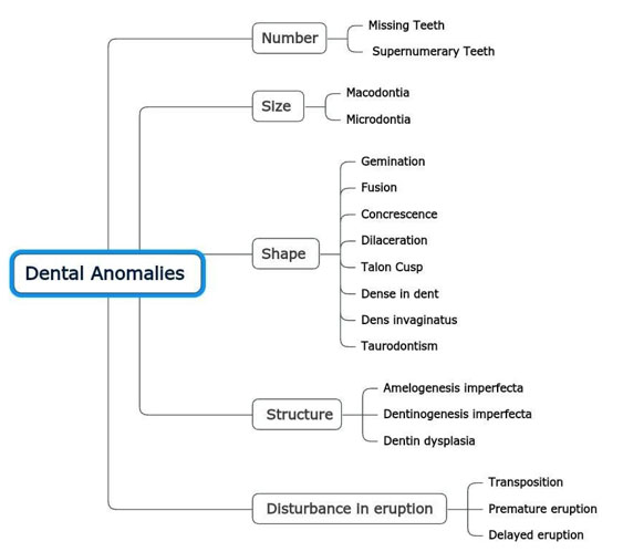
A diagram showing different types of dental anomalies. Also tooth gemination can increase the risk of pulp exposure due to caries due to the abnormal tooth morphology and proximity of the pulp to the tooth surface. Here are some key points on this issue:
- The incomplete cleavage of the tooth bud and extra crown anatomy in geminated teeth results in deep developmental grooves or fissures that are prone to plaque accumulation and caries [8].
- Geminated teeth often have a large pulp chamber and pulp horns that extend close to the surface, especially in the fissures, putting the pulp at high risk for exposure with even small amounts of decay [9].
- Studies have shown much higher rates of pulp exposure in geminated primary molars with caries compared to normal primary molars with caries, indicating the pulp is far more vulnerable in these anomalous teeth [8],[10].
- Once caries reaches the dentin in a geminated tooth, pulp exposure can occur rapidly due to the thin partition between the pulp and heavily fissured occlusal surface [11].
- Early and thorough fissure sealant placement is imperative in geminated teeth to prevent caries and subsequent pulp involvement [8].
In summary, the unique morphology of geminated teeth with deep fissures and close pulp proximity significantly increases susceptibility to caries and risk of pulp exposure compared to normal teeth [8],[10],[11]. Preventive sealants and prompt restorative treatment help reduce this risk.
Case Report
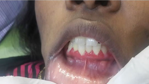
A 20-year-old female patient came to a routine checkup. Clinical examination revealed multiple carious lesions on anterior and posterior teeth, normal soft tissues, multiple restorations on maxillary and mandibular molars, mild anterior crowding and gemination of mandibular lateral incisor (Figure 1). Radiographic examination with periapical radiography (PA) showed the presence of geminated tooth with a single root and root canal (Figure 2). Treatment options were explained to the patient and the treatment consisted of observation with regular monitoring and preventive care according to patient preferences.


Discussion
This case illustrates a classic presentation of dental gemination affecting the mandibular left lateral incisor. The enlarged bifid crown and single root confirmed radiographically are characteristics of this developmental dental anomaly [1],[12]. Gemination arises during odontogenesis when the tooth bud attempts to split into two teeth but the division is incomplete, resulting in a larger anomalous structure [2].
Maxillary incisors are more commonly impacted, making the mandibular presentation in this case less typical [13]. The unilateral manifestation follows expected patterns, with bilateral gemination being very rare [6]. Though the patient was asymptomatic, the abnormal crown shape and size can lead to aesthetic concerns, occlusal interferences, and increased susceptibility to caries and periodontal defects over time [7],[14]. Therefore, preventive measures and monitoring are recommended.
Critical radiographic analysis was vital for confirming the presence of a single root canal in this geminated tooth [1],[15]. This guided the appropriate endodontic therapy prior to placement of a full-coverage restoration. Though extraction can be considered, conservation is preferred for geminated teeth with no pulpal involvement to maintain function and esthetics [16],[17]. Protecting the tooth structure through caries prevention and monitoring the occlusion are important long-term preferences [18].
Differentiating gemination from fusion is also essential, as the latter exhibits two separate roots radiographically with joined crowns [19]. Making this distinction allows proper diagnosis and management of abnormalities in tooth number and morphology [4]. Early detection of anomalies through comprehensive examination facilitates optimal treatment planning and follow-up.
While dental gemination may primarily affect esthetics if asymptomatic, this case demonstrates the need to address associated complications. With a thorough diagnostic workup, evidence-based treatment, and preventive care, developmental anomalies like gemination can be successfully maintained for excellent prognosis. This underscores the importance of careful monitoring and individualized management when such dental irregularities are encountered clinically.
Conclusion
This case highlights the importance of accurate diagnosis of dental anomalies through comprehensive clinical and radiographic assessment. The bilateral crown enlargement and single root confirmed gemination of the mandibular lateral incisor. After discussing treatment options, the asymptomatic tooth was left untreated per patient preference and will be monitored preventively long term. Though restorative intervention was possible, this case demonstrates the role of patient goals in managing incidental findings like gemination. Regular ongoing care can maintain function and esthetics despite dental irregularities.
REFERENCES
1.
Brook AH, Winter GB. Double teeth. A retrospective study of ‘geminated’ and ‘fused’ teeth in children. Br Dent J 1970;129(3):123–30. [CrossRef]
[Pubmed]

2.
Vojdani M, Khoshandam R. Gemination in primary teeth: A review article. Int J Clin Pediatr Dent 2022;15(3):357–62.
3.
Karayilmaz H, Kirzioglu Z, Ozcan I. Radiographic diagnosis of gemination and fusion in nonsyndromic primary anterior teeth. Imaging Sci Dent 2021;51(1):75–1.
4.
Afify AR, Zawawi KH. Prevalence of dental anomalies in the western region of Saudi Arabia. Saudi Dent J 2020;32(8):437–43.
5.
Tomizawa M, Shimizu A, Hayashi S, Noda T. Bilateral maxillary fused primary incisors accompanied by succedaneous supernumerary teeth: Report of a case. Int J Paediatr Dent 2002;12(3):223–7. [CrossRef]
[Pubmed]
6.
Saygili G, Durmuslar MC. Unilateral molar-premolar transposition and bilateral canine fusion-gemination: A very rare combination of dental anomalies. Am J Orthod Dentofacial Orthop 2020;157(1):132–9.
7.
Neville BW, Damm DD, Allen CM, Bouquot JE. Oral and Maxillofacial Pathology. 3ed. St. Louis, MO: Saunders Elsevier; 2009.
8.
Fernandes A, Lambor R, Maheshwari S. Unilateral primary tooth gemination: A rare case report. Cureus 2020;12(6):e8473.
9.
Pawar R, Pawar M, Kokate S. Endodontic management of a fused and germinated tooth. J Conserv Dent 2019;22(5):544–7.
10.
da Silva Neto JJ, Medina AC, da Silva FW, Machado AW, Pereira KM. Gemination or fusion: A case report of a supernumerary primary tooth. Gen Dent 2010;58(4):e127–9.
11.
Dankner E, Harari D, Rotstein I. Conservative treatment of dens invaginatus type 2 associated with a talon cusp in a permanent maxillary lateral incisor: A case report. Quintessence Int 1996;27(6):373–6.
12.
Mader CL. Fused teeth: A correlation of the histology and radiology. J Am Dent Assoc 1979;98(3):420–2.
13.
Nik-Hussein NN, Abdul Majid Z. Dental anomalies in the primary dentition: Distribution and correlation with the permanent dentition. J Clin Pediatr Dent 1996;21(1):15–9.
[Pubmed]
14.
Hattab FN, Yassin OM, al-Nimri KS. Talon cusp—Clinical significance and management: Case reports. Quintessence Int 1995;26(2):115–20.
[Pubmed]
15.
Gursel O, Oztas B, Gundogdu M. Unusual radicular patterns in geminated teeth: Two case reports. J Dent Child (Chic) 2018;85(3):176–80.
16.
Paduano S, Márquez SC, Rivera Silvia M. Conservative esthetic and functional treatment of a geminated/taloned maxillary central incisor. Quintessence Int 2019;50(10):800–5.
17.
Rana T, Sandhu SV, Sharma R, Padwal A. Endodontic management of a geminated tooth: A case report and review. Int J Clin Pediatr Dent 2022;15(4):518–21.
18.
Salman M, Tümen EC, Yıldırım E, Tosun G. Endodontic treatment of a geminated maxillary central incisor: A case report. Eur Endod J 2022;7(2):200–4.
19.
Levitas TC. Gemination, fusion, twinning and concrescence. J Dent Child (Chic) 1965;32:93–100.
[Pubmed]
SUPPORTING INFORMATION
Author Contributions
Majed Mansour Alsuwaida - Conception of the work, Design of the work, Acquisition of data, Analysis of data, Drafting the work, Revising the work critically for important intellectual content, Final approval of the version to be published, Agree to be accountable for all aspects of the work in ensuring that questions related to the accuracy or integrity of any part of the work are appropriately investigated and resolved.
Guarantor of SubmissionThe corresponding author is the guarantor of submission.
Source of SupportNone
Consent StatementWritten informed consent was obtained from the patient for publication of this article.
Data AvailabilityAll relevant data are within the paper and its Supporting Information files.
Conflict of InterestAuthor declares no conflict of interest.
Copyright© 2023 Majed Mansour Alsuwaida. This article is distributed under the terms of Creative Commons Attribution License which permits unrestricted use, distribution and reproduction in any medium provided the original author(s) and original publisher are properly credited. Please see the copyright policy on the journal website for more information.

