 |
Case Report
Chondroid chordoma within dorsal spine
1 Radiology Department, Ibn Sina Hospital, Rabat, Morocco
Address correspondence to:
El Houssni Jihane
Radiology Department, Ibn Sina Hospital, Rabat,
Morocco
Message to Corresponding Author
Article ID: 101290Z01EJ2022
Access full text article on other devices

Access PDF of article on other devices


How to cite this article
Jihane EH, Nabil MB, Ittimade N. Chondroid chordoma within dorsal spine. Int J Case Rep Images 2022;13:101290Z01EJ2022.ABSTRACT
Introduction: Chordoma is an extremely rare malignant bone tumor that mainly affects the sacrococcygeal and spheno-occipital regions. Localization in the spine is exceptional and occurs mainly in young individuals.
Case Report: We report the case of a 32-year-old male patient with a chondroid chordoma in the dorsal spine which is a very rare histological subtype of chordoma.
Conclusion: The chondroid chordoma is an uncommon malignant bone tumor. An intrathoracic chordoma manifesting as a posterior mediastinal tumor is unusual, it appears especially in young individuals. The imaging helps to allow a better characterization of the tumor.
Keywords: Cartilaginous matrix, Chondroid chordoma, Thoracic spine
Introduction
Chordoma is a very rare, insidious, locally invasive, malignant primary bone tumor that develops in exceptional cases from the embryonic remnants of the notochord of the axial skeleton [1],[2],[3]. Chondroid chordoma is a rare histological variant of chordoma, it is characterized by the presence of a cartilaginous matrix, and it has a better prognosis compared to the classical chordoma [2].
Case Report
The patient is a 32 years old man, with a history of asthma under bronchodilators and corticosteroids. He was admitted for chest pain with respiratory distress, the clinical examination was without abnormal findings.
The thoracic computed tomography (CT) scan showed a posterior mediastinal tissue process, occupying the right and left costovertebral gutters, extending over the three upper, middle and lower mediastinal levels, well limited, with regular contours, heterogeneous, containing arciform and popcorn calcifications, suggestive of a cartilaginous matrix, with erosion of the bony cortex of the dorsal vertebrae D5, D6, and D7, with no clear enhancement after injection of product contrast. This process measured 187×127×180 mm (Craniocaudal × Anteroposterior × Transverse diameters), displacing adjacent anatomical structures, compressing the aerodigestive tract (esophagus, trachea, and stem bronchi), left atrium and inferior pulmonary veins with no clear interface, it encompassed the aorta over 180°, with spinal cord extension via the right D4–D5 and D5–D6 foramen (Figure 1).
On the abdominal level, suspicious hepatic lesions were noted in segments VI and VIII.
The patient underwent an additional spinal cord magnetic resonance imaging (MRI) showing a posterior mediastinal mass, multi-lobulated, moderate T2 hypersignal, T1 isosignal, heterogeneously enhanced after injection of Gadolinium, hypersignal diffusion, high apparent diffusion coefficient (ADC), containing cartilaginous calcifications, cystic areas and fine septa, T2 hyposignal, regular enhancement after injection of Gadolinium, with spinal cord extension, pushing back the medullary canal at D6 and arriving at its contact at D5 via the right neural foramina without any medullary signal abnormality opposite (Figure 2).
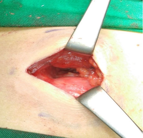
A scan-guided biopsy was performed, which showed tumoral micro-fragments whose morphological appearance and imunohistochemical results were in favor of a chondroid chordoma.


Discussion
Chordoma represents 1–4% of primary malignant bone tumors [1]. It is predominantly male (sex ratio M/F: 2.7/1–1.6/1) [3]. Chordoma is mainly found in the sacrococcygeal and spheno-occipital region, and is rare in the axial skeleton (0.8 to 1/100,000 persons/year) [4],[5]. Vertebral chordoma mainly affects young individuals [1]. The cervical spine is the most frequent site of involvement [4]. Intrathoracic chordoma is extremely rare (2–5%) [1], it usually appears as a posterior mediastinal mass [6], this is the case of our patient.
Chordoma is metastatic in 20–60% of cases [3], mostly in lymph nodes, lungs, liver, and bone [7].
The clinical manifestations of chordoma are location-dependent, with symptoms secondary to compression and invasion of the surrounding organs. In the dorsal spine, patients may present chest pain, dyspnea, and spinal pain, especially if the tumor is large [3],[7].
Imaging plays a very important role in the management of patients with chordoma, it has a triple interest: Diagnostic, allowing a better characterization of the tumor, prognostic and in the postoperative monitoring.
Chordoma most commonly presents itself as a large tumor with a predominantly extraosseous component. Due to its slow growth, it tends to compress adjacent structures rather than to invade them. The paraspinal component may extend to adjacent vertebral bodies, but usually spares the intervertebral discs. A spinal cord extension via the neural foramina is very rare [8].
The CT scan is used to assess the location of the tumor, its connection to adjacent anatomical structures, the involvement of the vertebral bodies, and the assessment of eventual extension [5].
The CT scan shows a non-infiltrative tissue lesion centered on both sides of the spine, a “Dumbbell shape” or “mushroom appearance,” multi-lobulated, osteolytic, exophytic, and expansive, with erosion of the bone cortex of the dorsal vertebrae [2][8],[9]. The chordoma may extend along the vertebral bodies [8],[10]. The enhancement after contrast injection is poor due to the presence of myxoid-like tissue [9]. The intratumoral calcifications are amorphous, peripheral, in case of chondroid chordoma the calcifications are more numerous and cartilaginous [7],[8]. The cartilaginous matrix is better highlighted on the CT scan [10]. There may also be hypodense areas related to cystic degeneration of the chordoma [1].
In our case, the thoracic CT scan showed the absence of clear enhancement of the chondroid chordoma after injection of iodinated contrast, with the presence of cartilaginous calcifications within this process, associated with erosion of the bone cortex of the dorsal vertebrae. The lobulated character is not visible in our scan.
Magnetic resonance imaging is more sensitive than CT to assess extension into the epidural and intradural space, especially to look for spinal cord compression, epidural metastases, or epiduritis [3].
Magnetic resonance imaging consistently shows the characteristics lobulated appearance of the chordoma, with intense T2 hypersignal due to the high concentration of water in the myxoid matrix, intermediate or hyposignal T1, heterogeneous contrast of variable intensity, hypo-T2 septa, enhanced after injection of Gadolinium [5],[7],[11], a diffusion hypersignal with a high ADC. Diffusion is of prognostic interest, low ADC is an element in favor of tumor progression, in which case chondrosarcoma must also be evoked [7],[8]. There may also be a pseudocapsule in hyposignal on all sequences, some areas in T1 hypersignal in relation to bleeding or mucinous material [7],[9].
The chondroid chordoma has a short T1 and T2 relaxation time, compared to classical chordoma because the gelatinous matrix is replaced by cartilaginous tissue, which explains the moderate T2 hypersignal of this type of chordoma [12].
The above signs are highly suggestive of chordoma but none of these signs are pathognomonic [8].
The MRI of our patient showed the lobulated character of the chordoma, with moderate T2 hypersignal, which are characteristics of chondroid type chordoma, a T1 isosignal, heterogeneous enhancement after injection of Gadolinium, cartilaginous calcifications, some areas of cystic remodeling, thin regular septa in T2 hyposignal enhanced after injection of Gadolinium, and a spinal cord extension.
In front of a posterior mediastinal mass, the differential diagnoses are very numerous, the main diagnoses that pose a real problem of differential diagnosis are: Chondrosarcoma which often develops from the posterior arch of the vertebrae, it is often extra medial in location, it shows the same MRI signal of chordoma, cartilaginous calcifications which are also characteristic of the chondroid form of chordoma [7],[13].
Neurogenic tumors, especially the schwannoma, particularly in the face of chordomas extending into the intervertebral foramina [8].
Solitary plasmacytoma, which presents itself as an infiltrating lesion of the vertebral body, with a compression fracture. The MRI appearance is hyposignal T1, hypersignal T2, with an intense and homogeneous contrast [7],[9].
Giant cell tumors: occur mostly in young individuals, without T2 hypersignal since they are rich in hemosiderin and collagen [7],[9].
Other diagnoses are: Adenocarcinoma metastases, myxoid liposarcoma, lymphangioma, hemangioma... [2],[6].
The NCCN guidelines 2020 recommend extensive resection of operable tumors; adjuvant radiotherapy should be given if resection margins are positive or if the mass is larger than 7 cm [5]. However, conventional chemotherapy is not recommended in the therapeutic arsenal of chordoma [14].
The 5-year survival rate of chondroid chordoma is generally between 10% and 70% [3]. Only 10–20% of chordoma are curable by surgery with a local recurrence rate between 26% and 68% [3]. Adjuvant radiotherapy is often used to improve local control [5]. The study by Park et al. revealed high survival rates (93% at five years and 91% at ten years) for patients treated with local radiotherapy after complete surgical resection [5]. For non-operable tumors, high-dose radiotherapy may be an alternative treatment; Chen et al. reported a survival rate of 78% at five years for patients with non-operable chordoma treated with high-dose radiotherapy [5].
Conclusion
The chondroid chordoma is a very rare primary malignant bone tumor, the intrathoracic localization is unusual, and it is mostly seen in young individuals. Imaging plays an important role in the management of chordoma. The presence of a cartilaginous matrix within the tumor is a very characteristic radiological sign of chordoma but not pathognomonic, the moderate T2 hypersignal points to the chondroid type variant. Therefore, imaging can provide several very useful elements for the diagnostic and prognostic approach.
REFERENCES
1.
Topsakal C, Bulut S, Erol FS, Ozercan I, Yildirim H. Chordoma of the thoracic spine – Case report. Neurol Med Chir (Tokyo) 2002;42(4):175–80. [CrossRef]
[Pubmed]

2.
Demireli P, Yilmaz Ovah G, Yegen G, Temiz C, Tarhan S. Condroid chordoma of the thoracic spine: Case report. Pathology 2007;39(2):280–2. [CrossRef]
[Pubmed]
3.
Crespo IR, Rivas de Andrés JJ, Flor RE, Cortés Franco S. Chondroid chordoma in an atypical location. Arch Bronconeumol 2013;49(11):419–93. [CrossRef]
[Pubmed]
4.
Tanaka K, Sakakima H, Hida K, Hatanaka K, Ijiri K. A case of C5 vertebral chordoma in a 73-year-old patient with more than 8 years of follow-up after total piecemeal spondylectomy. Case Rep Orthop 2017;2017:3284131. [CrossRef]
[Pubmed]
5.
Bai R, Zhao ZQ, Wang YX, et al. Sacral and thoracic chordoma with pulmonary metastases: A case report and review of the literature. Mol Clin Oncol 2021;14(1):17. [CrossRef]
[Pubmed]
6.
Taki S, Kakuda K, Kakuma K, Yamashita R, Kosugi M, Annen Y. Posterior mediastinal chordoma: MR imaging findings. AJR Am J Roentgenol 1996;166(1):26–7. [CrossRef]
[Pubmed]
7.
Dodin G, Dupres R, Beuret F, et al. Imagerie des chordomes du typique à l'atypique. Service de Neuroradiologie – CHRU Nancy. Poster JFR 2017.
8.
Benson JC, Vizcaino MA, Kim DK, et al. Exophytic lumbar vertebral body mass in an adult with back pain. AJNR Am J Neuroradiol 2020;41(10):1786–90. [CrossRef]
[Pubmed]
9.
Rodallec MH, Feydy A, Larousserie F, et al. Diagnostic imaging of solitary tumors of the spine: What to do and say. Radiographics 2008;28(4):1019–41. [CrossRef]
[Pubmed]
10.
Motamedi K, Ilaslan H, Seeger LL. Imaging of the lumbar spine neoplasms. Semin Ultrasound CT MRI 2004;25(6):474–89. [CrossRef]
[Pubmed]
11.
Shah R, Pope T. Chordoma. Appl Radiol 2007.
12.
Sebag G, Dubois J, Beniaminovitz A, Lelouch-Tubiana A, Brunelle F. Extraosseous spinal chordoma: Radiographic appearance. AJNR Am J Neuroradiol 1993;14(1):205–7.
[Pubmed]
13.
Santegoeds RGC, Temel Y, Beckervordersandforth JC, Van Overbeeke JJ, Hoeberigs CM. State-of-the-art imaging in human chordoma of the skull base. Curr Radiol Rep 2018;6(5):16. [CrossRef]
[Pubmed]
14.
Wang TJ, Shu SH, Lin CW, et al. Thoracic chordoma: An unusual presentation of the spinal tumor. Am J Med Sci 2008;335(3):239–41. [CrossRef]
[Pubmed]
SUPPORTING INFORMATION
Author Contributions
El Houssni Jihane - Conception of the work, Design of the work, Acquisition of data, Analysis of data, Drafting the work, Revising the work critically for important intellectual content, Final approval of the version to be published, Agree to be accountable for all aspects of the work in ensuring that questions related to the accuracy or integrity of any part of the work are appropriately investigated and resolved.
Moatassim Billah Nabil - Conception of the work, Design of the work, Acquisition of data, Analysis of data, Drafting the work, Revising the work critically for important intellectual content, Final approval of the version to be published, Agree to be accountable for all aspects of the work in ensuring that questions related to the accuracy or integrity of any part of the work are appropriately investigated and resolved.
Nassar Ittimade - Conception of the work, Design of the work, Acquisition of data, Drafting the work, Revising the work critically for important intellectual content, Final approval of the version to be published, Agree to be accountable for all aspects of the work in ensuring that questions related to the accuracy or integrity of any part of the work are appropriately investigated and resolved.
Guarantor of SubmissionThe corresponding author is the guarantor of submission.
Source of SupportNone
Consent StatementWritten informed consent was obtained from the patient for publication of this article.
Data AvailabilityAll relevant data are within the paper and its Supporting Information files.
Conflict of InterestAuthors declare no conflict of interest.
Copyright© 2022 El Houssni Jihane et al. This article is distributed under the terms of Creative Commons Attribution License which permits unrestricted use, distribution and reproduction in any medium provided the original author(s) and original publisher are properly credited. Please see the copyright policy on the journal website for more information.


