 |
Clinical Image
Kaposi sarcoma
1 Internal Medicine Resident, Mountain Vista Medical Center, Midwestern University-OPTI, Mesa, Arizona, USA
2 Hospitalist, Mountain Vista Medical Center, Internal Medicine, Mesa, Arizona, USA
Address correspondence to:
Lyndie Wilkins Parker
1301 S. Crismon Road, Mesa, Arizona 85209,
USA
Message to Corresponding Author
Article ID: 101278Z01LP2022
Access full text article on other devices

Access PDF of article on other devices


How to cite this article
Parker LW, Mele S. Kaposi sarcoma. Int J Case Rep Images 2022;13:101278Z01LP2022.ABSTRACT
No Abstract
Keywords: Antiretroviral therapy, HHV-8, HIV, Kaposi sarcoma
Case Report
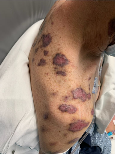
A 47-year-old heterosexual man with history of intravenous (IV) methamphetamine use presented to the Emergency Department with dyspnea and raised lesions throughout his arms, legs, and chest as depicted in Figure 1. Biopsy of a right leg lesion demonstrated atypical vascular proliferation within the dermis producing slit-like/irregular architecture consistent with Kaposi sarcoma. Human herpesvirus 8 (HHV-8) staining was consistent with Kaposi sarcoma. HIV-1 antibody testing was positive and the patient had an absolute CD4 count of 19. Kaposi sarcoma is relatively rare in the United States as antiretroviral therapy has become more accessible. The appearance of a vascular tumor with inflammation and mucosal involvement should raise suspicion for HHV-8.
Discussion
Kaposi sarcoma (KS) is a rare type of cancer associated with vascular tumors secondary to infection of human herpesvirus 8 (HHV-8). There are four epidemiologic forms of KS, all of which are attributed to infection with HHV-8 [1],[2]. In HIV patients, KS is the most common tumor and is considered to be an AIDS-defining illness. Since the initiation of widespread antiretroviral therapy (ART), incidence of KS has rapidly declined [3]. Typically, AIDS-related KS is more common in homosexual or bisexual men, while seen less frequently in heterosexual injection drug users. The most important predisposing factor related to AIDS-KS is the CD4 count [4].
AIDS-KS can present in a multitude of clinical conditions—ranging from asymptomatic to rapidly progressive. Manifestations can be seen in the oral cavity, lung, lymph nodes, and gastrointestinal tract in the disseminated form [2].
When evaluating a patient for KS, the physical exam is paramount. Identifying lesions in the most typical locations—lower extremities, face, and mucosa—is essential to staging and targeting treatment. Some societies recommend measuring the largest lesion if possible, but due to confluence of KS lesions this is not always practical [2]. Further testing should be guided by both symptoms and lab abnormalities. Fecal occult blood testing is the best initial indicator for gastroenterological involvement, with positive screening requiring endoscopy for further investigation. Chest X-ray is the primary modality used to screen for pulmonary lesions, with bronchoscopy reserved for those found to have abnormalities. CD4 count and HIV viral load are important in both staging and prognosis [2],[3],[5],[6].
Staging for KS is based on the AIDS Clinical Trial Group (ACTG) of the National Institute of Health. It is divided into three considerations: extent of tumor, immune status, and severity of systemic symptoms [2]. While societies disagree on the significance of the immune status of the patient, most societies are in agreement that it does have an important role, with the general consensus appreciating that improvement in CD4 count can lead to regression and better response of KS to therapies.
Prevention of disease progression, symptom palliation, and shrinkage of tumor are the main goals of treatment [2]. All patients should be started on combination of ART. In some patient populations, the initiation of ART can be the only treatment required, whereas others will require more systemic modalities. With the widespread use of ART therapies, the incidence of KS has rapidly declined [2]. The effectiveness of ART leading to immune reconstitution is thought to be the underlying mechanism. With initiation of ART therapy, there is the risk of immune reconstitution inflammatory syndrome. As such, the initiation of ART therapy often requires concurrent local treatment of KS as well. However, even in patients chronically on ART with appropriate CD4 counts, KS has been identified. Thus, some patients require further treatment beyond systemic immune support [2].
Local therapy is targeted at decreasing the tumor burden and cosmetic improvement. The three major forms of local therapy are intralesional chemotherapy, radiation therapy, and topical alitretinoin. Intralesional chemotherapy with vinblastine is used in small lesions with regression noted on average four months after the initial injection. Radiation treatment is often reserved for patients with extensive KS that would not benefit from localized chemotherapy injection, but not widespread enough to necessitate systemic therapy. Patients with widespread KS do not benefit from radiation therapy. Topical alitretinoin is effective for cutaneous KS lesions, but is now rarely used due to pigmentation changes and inflammation [2].
Systemic therapy is reserved for patients with rapidly progressive or advanced KS. The most common indications for systemic therapy are the presence of more than 25 lesions, cutaneous KS that is unresponsive to localized treatment, immune reconstitution inflammatory syndrome, and progression of KS on ART. The risks of further immunosuppression and insult must be weighed carefully in each patient. While pegylated liposomal doxorubicin is preferred, other chemotherapy agents such as paclitaxel, bleomycin, and vinblastine have also been used with success and the literature demonstrates results are equivocal when all therapies are compared in a head-to-head analysis [2]. The selection of the most appropriate agent is outside the scope of this discussion.
Conclusion
Cases of Kaposi sarcoma have greatly decreased with introduction of HIV therapies. Identification, staging, and treatment are paramount to preventing progression of both HHV-8 and HIV.
REFERENCES
1.
Dezube BJ. Clinical presentation and natural history of AIDS-related Kaposi's sarcoma. Hematol Oncol Clin North Am 1996;10(5):1023–9. [CrossRef]
[Pubmed]

2.
Groopman JE. AIDS-related Kaposi sarcoma: Staging and treatment. UpToDate 2021. [Available at: https://www.uptodate.com/contents/aids-related-kaposi-sarcoma-staging-and-treatment?search=kaposi%20sarcoma]
3.
Mocroft A, Kirk O, Clumeck N, et al. The changing pattern of Kaposi sarcoma in patients with HIV, 1994–2003: The EuroSIDA Study. Cancer 2004;100(12):2644–54. [CrossRef]
[Pubmed]
4.
Gbabe OF, Okwundu CI, Dedicoat M, Freeman EE. Treatment of severe or progressive Kaposi's sarcoma in HIV-infected adults. Cochrane Database Syst Rev 2014;(9):CD003256. [CrossRef]
[Pubmed]
5.
Krown SE, Metroka C, Wernz JC. Kaposi's sarcoma in the acquired immune deficiency syndrome: A proposal for uniform evaluation, response, and staging criteria. AIDS Clinical Trials Group Oncology Committee. J Clin Oncol 1989;7(9):1201–7. [CrossRef]
[Pubmed]
6.
El Amari EB, Toutous-Trellu L, Gayet-Ageron A, et al. Predicting the evolution of Kaposi sarcoma, in the highly active antiretroviral therapy era. AIDS 2008;22(9):1019–28. [CrossRef]
[Pubmed]
SUPPORTING INFORMATION
Author Contributions
Lyndie Wilkins Parker - Conception of the work, Design of the work, Acquisition of data, Analysis of data, Drafting the work, Revising the work critically for important intellectual content, Final approval of the version to be published, Agree to be accountable for all aspects of the work in ensuring that questions related to the accuracy or integrity of any part of the work are appropriately investigated and resolved.
Sandra Mele - Acquisition of data, Revising the work critically for important intellectual content, Final approval of the version to be published, Agree to be accountable for all aspects of the work in ensuring that questions related to the accuracy or integrity of any part of the work are appropriately investigated and resolved.
Guarantor of SubmissionThe corresponding author is the guarantor of submission.
Source of SupportNone
Consent StatementWritten informed consent was obtained from the patient for publication of this article.
Data AvailabilityAll relevant data are within the paper and its Supporting Information files.
Conflict of InterestAuthors declare no conflict of interest.
Copyright© 2022 Lyndie Wilkins Parker et al. This article is distributed under the terms of Creative Commons Attribution License which permits unrestricted use, distribution and reproduction in any medium provided the original author(s) and original publisher are properly credited. Please see the copyright policy on the journal website for more information.


