 |
Case Report
Biliary cystadenoma: A typical radiological presentation
1 Institute National d’Oncologie, Université Mohammed V, Rabat, Morocco
Address correspondence to:
Kaoutar Imrani
Radiology Department, Oncology National Institute, Mohammed V University, Rabat,
Morocco
Message to Corresponding Author
Article ID: 101272Z01KI2021
Access full text article on other devices

Access PDF of article on other devices


How to cite this article
Imrani K, Messaoud O, Oubaddi T, Jerguigue H, Latib R, Omor Y. Biliary cystadenoma: A typical radiological presentation. Int J Case Rep Images 2021;12:101272Z01KI2021.ABSTRACT
Biliary cystadenoma is a rare benign hepatobiliary cystic tumor, more common in middle-aged women. The clinical manifestation is variable. It can be asymptomatic or symptomatic presenting with abdominal pain, nausea, vomiting, or a palpable mass. We report a case of a 36-year-old female patient, with a biliary cystadenoma mimicking a hydatid cyst (HC) complicated by local recurrence. The radiological diagnosis can be difficult and may present a problem of differential diagnosis with the HC and complicated liver cyst. Surgical management is based on complete resection given the high risk of recurrence and malignant degeneration.
Keywords: Cystadenoma, Liver, MRI
Introduction
Biliary cystadenoma is a rare benign cystic tumor with high potential for recurrence and malignant transformation, common in middle-aged women. It is often asymptomatic and has no specific clinical or biological presentation, hence the difficulty of a preoperative diagnosis. Imaging using ultrasound, computed tomography (CT) and magnetic resonance imaging (MRI) of the liver plays an essential role in the diagnostic orientation, the preoperative workup, and the follow-up. However, in some cases it can be hard to differentiate it from a hydatid cyst (HC), hepatic cyst, liver abscess, liquefied hematoma, biloma, and cystic metastasis [1].
Case Report
We report a case of a 36-year-old woman, with no medical history, who underwent surgery two years ago for a cystic liver mass initially diagnosed as HC. A resection of the protruding dome was performed, consisting in evacuation of the HC and closure of the biliary communications in one simple surgical procedure. The anatomical-pathological study was in favor of a biliary serous cystadenoma. The patient has consulted for a second time due to right hypochondrium pain evolving for two months. The clinical examination revealed a patient in good general condition, apyretic, with an epigastric mass slightly sensitive to palpation. Hydatid serology was negative.
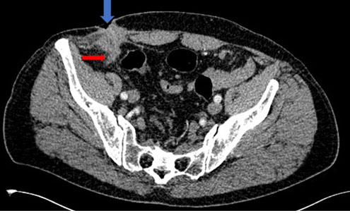
Abdominal ultrasound showed a thin-walled multiloculated cystic mass without vegetation or tissue buds. Magnetic resonance imaging was performed showing a thin-walled multiloculated cystic mass of the left liver, enhanced after injection of contrast medium, containing thin partitions with poor contrast uptake and no solid component (Figure 1A, Figure 1B, Figure 1C). The lesion was measuring 112×87 mm. It was realizing a mass effect on the adjacent intra-hepatic bile ducts without vascular invasion.
The patient underwent a left hepatectomy with no postoperative complications. Histological examination was in favor of a recurrence of serous biliary cystadenoma. The surgical margin was 0.6 mm from the lesion.

Discussion
Biliary cystadenoma is a rare benign cystic tumor (less than 5% of cystic lesions of the liver). It is marked by a risk of local recurrence and malignant degeneration to cystadenocarcinoma. It is most frequently observed in middle-aged women (40 years) with a variability in size ranging from 1.5 to 30 cm.
The clinical presentation is nonspecific, usually asymptomatic. However, in the case of a large palpable mass, abdominal pain, nausea, and vomiting can be manifested [1],[2].
On abdominal ultrasound, biliary cystadenoma presents as a multilocular mass with thin septa. It may be an echogenic or echogenic depending on the content of the cyst (serous, mucinous, biliary, and hemorrhagic). Computed tomography scan often shows a large uniloculated or multiloculated cystic mass with homogeneous CT attenuation of liquid density, or sometimes spontaneously hyperdense, with thin wall and partitions, without vegetation or tissue buds. It also allows to delineate to adjacent organs and vascular structures. The MRI appearance is a mass that may have a liquid signal (T1 hyposignal, T2 hypersignal), or a hypersignal in T1- and T2-weighted sequences in case of a cyst with mucinous or hemorrhagic content. The wall and septa are thin and regular, hyposignal in T2, with poor or no contrast uptake [2],[3],[4].
Complications that can be encountered in biliary cystadenoma are hemorrhage, infection, and biliary compression, however, malignant degeneration to cystadenocarcinoma remains the most feared complication [4],[5].
Cystadenocarcinoma usually has nodular septa with nodules larger than 10 mm and a thick irregular wall with early intense contrast uptake. The presence of papillary projections and calcifications at the septum may also suggest malignant transformation [3],[5].
Histological study of the mass provides the diagnosis of certainty. Biliary cystadenoma is a bulky, multilocular, encapsulated tumor lined by cuboidal epithelium. There are two histological varieties, the most frequent is the mucinous type and the serous type that is less common.
The differential diagnoses of cystadenoma are simple hepatic cyst, HC type III, abscess (infectious syndrome), liquefied hematoma (post-traumatic context, anti-coagulant treatment), biloma (post-traumatic or iatrogenic context), and cystic metastasis [2],[3][5].
The simple hepatic cyst is easily diagnosed on ultrasound, it is a cystic lesion with no visible wall, hypodense on CT and a liquid signal on MRI without contrast uptake [1],[2]. When complicated by hemorrhage, the hemorrhagic hepatic cyst presents on CT and ultrasound and an irregular wall with endo-cystic vegetations with a T1-weighted sequence hypersignal appearance on MRI [1].
Type III HC can also be confused with biliary cystadenoma, as in our reported case, knowing that hydatic serology can be negative in 10% of cases. However, certain radiological criteria can be used to orient the diagnosis. On ultrasound, the HC presents a thick wall, with peripheral calcifications in 50% of cases. On MRI, it is surrounded by a hyposignal crown in T1-weighted sequence, the daughter vesicles have a variable signal but have no contrast uptake [2],[5].
The treatment of biliary cystadenoma is surgical. Total resection of the tumor is the first line treatment due to the risk of recurrence and malignant transformation to cystadenocarcinoma [3],[4],[5].
Conclusion
Biliary cystadenoma is a rare benign tumor. In this article, we underline the problem of differential diagnosis of biliary cystadenoma with the hydatid cyst, in particular in endemic countries, as well as the risk of recurrence which remains the most dreaded complication. The diagnosis of certainty is histological, however, a good radiological analysis associated with clinical and biological data can orientate and suspect the diagnosis.
REFERENCES
1.
Poggio PD, Buonocore M. Cystic tumors of the liver: A practical approach. World J Gastroenterol 2008;14(23):3616–20. [CrossRef]
[Pubmed]

2.
Mavilia M, Pakala T, Molina M, Wu GY. Differentiating cystic liver lesions: A review of imaging modalities, diagnosis and management. J Clin Transl Hepatol 2018;6(2):208–16.
[Pubmed]
3.
Kim JY, Kim SY, Eun HW, et al. Differentiation between biliary cystic neoplasms and simple cysts of the liver: Accuracy of CT. AJR Am J Roentgenol 2010;195(5):1142–8. [CrossRef]
[Pubmed]
4.
Soares KC, Arnaoutakis DJ, Kamel I, et al. Cystic neoplasms of the liver: Biliary cystadenoma and cystadenocarcinoma. J Am Coll Surg 2014;218(1):119–28. [CrossRef]
[Pubmed]
5.
Gómez-Martín C, Rodríguez A, Malón D, Cortés-Funes H. Biliary cystadenocarcinoma with mesenchymal stroma. Clin Transl Oncol 2010;12(3):234–7. [CrossRef]
[Pubmed]
SUPPORTING INFORMATION
Author Contributions
Kaoutar Imrani - Conception of the work, Design of the work, Acquisition of data, Analysis of data, Drafting the work, Revising the work critically for important intellectual content, Final approval of the version to be published, Agree to be accountable for all aspects of the work in ensuring that questions related to the accuracy or integrity of any part of the work are appropriately investigated and resolved.
Ola Messaoud - Conception of the work, Design of the work, Acquisition of data, Analysis of data, Drafting the work, Revising the work critically for important intellectual content, Final approval of the version to be published, Agree to be accountable for all aspects of the work in ensuring that questions related to the accuracy or integrity of any part of the work are appropriately investigated and resolved.
Tlaite Oubaddi - Conception of the work, Design of the work, Acquisition of data, Analysis of data, Drafting the work, Revising the work critically for important intellectual content, Final approval of the version to be published, Agree to be accountable for all aspects of the work in ensuring that questions related to the accuracy or integrity of any part of the work are appropriately investigated and resolved.
Hounayda Jerguigue - Conception of the work, Design of the work, Acquisition of data, Analysis of data, Drafting the work, Revising the work critically for important intellectual content, Final approval of the version to be published, Agree to be accountable for all aspects of the work in ensuring that questions related to the accuracy or integrity of any part of the work are appropriately investigated and resolved.
Rachida Latib - Conception of the work, Design of the work, Acquisition of data, Analysis of data, Drafting the work, Revising the work critically for important intellectual content, Final approval of the version to be published, Agree to be accountable for all aspects of the work in ensuring that questions related to the accuracy or integrity of any part of the work are appropriately investigated and resolved.
Youssef Omor - Conception of the work, Design of the work, Acquisition of data, Analysis of data, Drafting the work, Revising the work critically for important intellectual content, Final approval of the version to be published, Agree to be accountable for all aspects of the work in ensuring that questions related to the accuracy or integrity of any part of the work are appropriately investigated and resolved.
Guarantor of SubmissionThe corresponding author is the guarantor of submission.
Source of SupportNone
Consent StatementWritten informed consent was obtained from the patient for publication of this article.
Data AvailabilityAll relevant data are within the paper and its Supporting Information files.
Conflict of InterestAuthors declare no conflict of interest.
Copyright© 2021 Kaoutar Imrani et al. This article is distributed under the terms of Creative Commons Attribution License which permits unrestricted use, distribution and reproduction in any medium provided the original author(s) and original publisher are properly credited. Please see the copyright policy on the journal website for more information.


