
 |
Case Report
Vaginal ureterocele: Rare finding in complicated pyelonephritis
1 Department of Urology, Clinical Hospital of the Federal University of the Triangulo Mineiro, Uberaba, Brazil
2 Institute of Health Sciences, Federal University of Triangulo Mineiro, Uberaba, Brazil
Address correspondence to:
Paulo Silveira Campos Soares
Department of Urology, Clinical Hospital of the Federal University of the Triangulo Mineiro, Uberaba,
Brazil
Message to Corresponding Author
Article ID: 101219Z01PS2021
Access full text article on other devices

Access PDF of article on other devices


How to cite this article
Soares PSC, Guimarães VHA, Freitas-Silva AL, Rodrigues AR, Cobo VAA, Lara RC, Monti PR. Vaginal ureterocele: Rare finding in complicated pyelonephritis. Int J Case Rep Images 2021;12:101219Z01PS2021.ABSTRACT
Introduction: Pyelonephritis is a urinary tract infection that can frequently be complicated by anatomical abnormalities such as in ectopic ureteroceles, pathologies which are rare in the literature.
Case Report: A 23-year-old woman with a clinical history, physical and laboratory examinations compatible with pyelonephritis was admitted. Ultrasonography revealed a ureterocele and hydronephrosis of the upper collecting system of the left kidney. We evoked a complicated pyelonephritis and the patient underwent a computed tomography (CT) scan that confirmed bilateral pyelocalyceal and ureteral duplicity, pyonephrosis, and implantation of the upper ureter of the left kidney into the vaginal dome, forming a left ureterocele. The patient was placed on antibiotics and the ureterocele was drained through the vaginal canal. After twelve days of hospitalization, she underwent an open partial nephrectomy of the upper portion of the left kidney. On the third postoperative day, she was asymptomatic and was discharged.
Conclusion: Despite the typical clinical picture of complicated pyelonephritis, this case stands out for the unusual and challenging anatomical findings, such as the pyelocalyceal bilateral duplicity and a rare infected ectopic ureterocele with vagina implantation. These challenging findings required transvaginal drainage and heminephrectomy, what shows that anatomical rare abnormalities can be hidden in a typical clinical presentation of pyelonephritis and demand an intense radiologic investigation and unusual treatments.
Keywords: Nephrectomy, Pyelonephritis, Ureterocele
Introduction
Pyelonephritis is a frequent urinary tract infection, with global estimates ranging from 10.5 million to 25.9 million cases annually. Each year, pyelonephritis causes 4000 deaths in the United States and its treatment costs USD 2 billion [1].
However, despite being common in the population and well known by the medical community, urinary tract infections, especially pyelonephritis, can be challenging in some cases with an underlying anomalous anatomy because these rare anomalies can make the diagnosis and treatment substantially more difficult [2].
Among these rare anomalies, we highlight ureterocele, a congenital condition formed by the dilatation of the distal segment of the ureter. It occurs in 1 in every 5000 to 12,000 births and is frequently diagnosed in childhood. Thus, in adults, this condition usually presents as an incidental radiological finding and an orthotopic insertion. When uncommonly symptomatic, ureterocele is related to lithiasis or its own prolapse [3],[4].
However, we describe a rare case of an adult woman diagnosed with complicated pyelonephritis who was found to have several rare anatomical abnormalities, which were challenging to treat.
Case Report
A 23-year-old married woman was admitted to the hospital for a 5-day progressive, severe colicky pain in the suprapubic region, irradiating to the left labia majora. This pain was associated with dysuria, fever, and hyporexia. Two days earlier, the pain began to irradiate to the left flank with a progressive increase in intensity and concentrated diuresis with a fetid odor.
The patient’s general condition was stable, with intense pain on deep palpation of the left hemiabdomen and Giordano's sign present on the left. She was tachycardic (HR = 110 bpm), eupneic, dehydrated (+1/+4), pale (+1/+4), febrile (T = 37.9°C), and hypotensive (BP = 90/50 mmHg). Laboratory tests confirmed an acute infectious process with marked neutrophilia (32,000 leukocytes with 29% neutrophils) and a positive urine culture for multi-sensitive Escherichia coli.
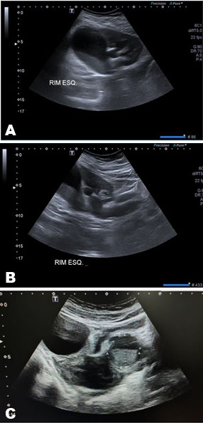
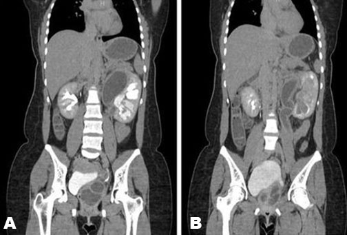
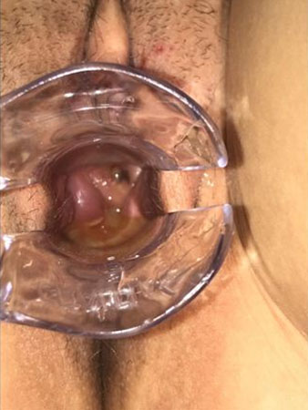
Ultrasonography of the kidneys (Figure 1), urinary tract, and pelvis showed a duplicated left kidney collecting system, hydronephrosis of the upper collecting system with probable pyogenic content, and a left ureterocele with thickened content. To better evaluate the case, the patient underwent contrast-enhanced computed tomography (CT) of the whole abdomen and pelvis, which demonstrated bilateral pyelocalyceal and ureteral duplicity, left pyonephrosis, and anomalous implantation of the upper left ureter into the vagina, forming a ureterocele with a probable pyogenic content (Figure 2). Tapering of the renal parenchyma of the left kidney's upper unit was also associated with hydronephrosis, without contrast uptake. The magnetic resonance and radioisotope renography weren’t available in the moment. Speculum examination confirmed the presence of a ureteral meatus inserted into the vagina (Figure 3). Thus, we could schematically represent the main anatomical alterations of the patient (Figure 4).
She was started on ceftriaxone 1 g every 12 h, and ureterocele drainage was performed through an intravaginal scalpel incision in the wall facing the ureterocele, with drainage of approximately 100 mL of purulent content. After 12 days of hospitalization, the clinical picture had significantly improved and an open upper left polar nephrectomy with closure of the ureteral stump was performed. The patient evolved satisfactorily and was discharged on the third postoperative day.
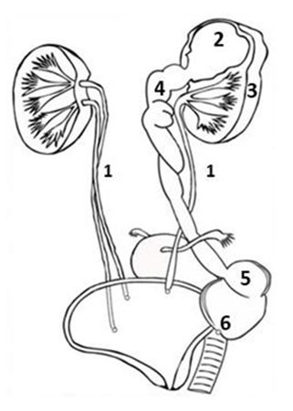
1. Bilateral pyelocalyceal and ureteral duplication. 2. Pyonephrosis. 3. Tapering of the renal parenchyma.
4. Ureteral dilatation. 5. Ureterocele. 6. Ectopic insertion of the ureter of the upper unit of the left kidney.
Discussion
The most common pyelonephritis symptoms are systemic inflammation (fever, chills, headache, sweating, and malaise), bladder inflammation (dysuria, urgency, altered urination, and polyuria), gastrointestinal symptoms (anorexia, hyporexia, nausea, vomiting, and abdominal pain), and renal pain symptoms (pain in the lumbar region and flank ipsilateral to the affected kidney) [1],[2]. In this case, we immediately suspected pyelonephritis based on clinical symptoms and laboratory tests [1],[5].
However, this case is peculiar due to the infrequent findings on imaging exams. Bilateral duplication of the collecting system occurs in 0.07–0.11% of the population [6] and ureterocele occurs in 1 among 5000 to 12,000 autopsies [7]. However, the rarest finding is the insertion of the left ureter into the vagina forming a ureterocele, which is described in very few case reports. This rare insertion may be diagnosed due to urinary incontinence. Still, as the patient did not report any history of incontinence, but she answered about similar attacks of pain and fever, previous finding in occasional gynecology vaginal examination or recurrent urinary tract infection (UTI) during childhood, even when directly questioned. It is believed that the ureteral meatus in the vagina was not adequately formed, preventing urine outflow [8].
The clinical management and the drainage of the infectious content of the ureterocele were fundamental for the patient's recovery. Even with the scarce literature on the approach of vaginal ectopic ureterocele with infected content in adult patients, drainage was necessary due to the purulent collection in the ectopic ureterocele. Drainage of the ureterocele with bladder insertion ureter is usually done by the transurethral endoscopic approach [9], but vaginal drainage was chosen for the only possibility of access.
A partial open nephrectomy was indicated for pyonephrosis with destruction of the left kidney's upper unit. Complete duplicity of the ureter may become a challenge for urologists in pyelonephritis management due to anatomical variations. If the renal function in one segment is severely impaired, heminephrectomy becomes necessary [10]. The ureteric stump was closed with suture and kept in the patient's body.
Conclusion
Rare urinary tract malformations such as ureterocele with ectopic vaginal insertion greatly complicate the management of pyelonephritis. In this rare situation, intravaginal drainage is a possible intervention. In complete duplications of the pyeloureteral system, we must consider ureteral implantation in the vagina, even in the absence of urinary incontinence.
REFERENCES
1.
Johnson JR, Russo TA. Acute pyelonephritis in adults. N Eng J Med 2018;378(1):48–59. [CrossRef]
[Pubmed]

2.
Kasat P, Bhosale Y, Muthiyan G. A cadaveric study of variations in the urological system. Int J Anat Res 2018;6(3.3):5686–94.
3.
Ghosh B, Shridhar K, Pal DK, Banerjee M. Ectopic ureter draining into the uterus. Urol Ann 2016;8(1):105–7. [CrossRef]
[Pubmed]
4.
Campobasso D, Lanzi A, Pozzoli GL, Frattini A. Prolapsed ureterocele in an adult woman after endoscopic incision. Urol Ann 2018;10(1):100–2. [CrossRef]
[Pubmed]
5.
Gupta K, Grigoryan L, Trautner B. Urinary tract infection. Ann Intern Med 2017;167(7):ITC49–64. [CrossRef]
[Pubmed]
6.
Gonzalez F, Canning DA, Hyun G, Casale P. Lower pole pelvi-ureteric junction obstruction in duplicated collecting systems. BJU Int 2006;97(1):161–5. [CrossRef]
[Pubmed]
7.
Wu CY, Lee CY, Yang IJ, Shen H, Torng PL. Double collecting system with ectopic ureterocele masquerading as an ovarian torsion. Taiwan J Obstet Gynecol 2018;57(6):867–70. [CrossRef]
[Pubmed]
8.
Narula H, Mittal S, Vohra A, et al. Ectopic vaginal ureterocele presenting as vaginal Gartner's cyst. Journal of OBGYN 2016;3(1):61–5.
9.
Timberlake MD, Corbett ST. Minimally invasive techniques for management of the ureterocele and ectopic ureter: Upper tract versus lower tract approach. Urol Clin North Am 2015;42(1):61–76. [CrossRef]
[Pubmed]
10.
Choi H, Oh SJ. The management of children with complete ureteric duplication: Selective use of uretero-ureterostomy as a primary and salvage procedure. BJU Int 2020;86(4):508–12. [CrossRef]
[Pubmed]
SUPPORTING INFORMATION
Author Contributions
Paulo Silveira Campos Soares - Conception of the work, Design of the work, Acquisition of data, Analysis of data, Drafting the work, Revising the work critically for important intellectual content, Final approval of the version to be published, Agree to be accountable for all aspects of the work in ensuring that questions related to the accuracy or integrity of any part of the work are appropriately investigated and resolved.
Vinícius Henrique Almeida Guimarães - Conception of the work, Design of the work, Acquisition of data, Analysis of data, Drafting the work, Revising the work critically for important intellectual content, Final approval of the version to be published, Agree to be accountable for all aspects of the work in ensuring that questions related to the accuracy or integrity of any part of the work are appropriately investigated and resolved.
Ana Letícia Freitas-Silva - Conception of the work, Design of the work, Acquisition of data, Analysis of data, Drafting the work, Revising the work critically for important intellectual content, Final approval of the version to be published, Agree to be accountable for all aspects of the work in ensuring that questions related to the accuracy or integrity of any part of the work are appropriately investigated and resolved.
André Rezek Rodrigues - Acquisition of data, Revising the work critically for important intellectual content, Final approval of the version to be published, Agree to be accountable for all aspects of the work in ensuring that questions related to the accuracy or integrity of any part of the work are appropriately investigated and resolved.
Vitor Augusto Alves Cobo - Acquisition of data, Revising the work critically for important intellectual content, Final approval of the version to be published, Agree to be accountable for all aspects of the work in ensuring that questions related to the accuracy or integrity of any part of the work are appropriately investigated and resolved.
Rolf Carvalho Lara - Analysis of data, Revising the work critically for important intellectual content, Final approval of the version to be published, Agree to be accountable for all aspects of the work in ensuring that questions related to the accuracy or integrity of any part of the work are appropriately investigated and resolved.
Paulo Ricardo Monti - Analysis of data, Revising the work critically for important intellectual content, Final approval of the version to be published, Agree to be accountable for all aspects of the work in ensuring that questions related to the accuracy or integrity of any part of the work are appropriately investigated and resolved.
Guarantor of SubmissionThe corresponding author is the guarantor of submission.
Source of SupportNone
Consent StatementWritten informed consent was obtained from the patient for publication of this article.
Data AvailabilityAll relevant data are within the paper and its Supporting Information files.
Conflict of InterestAuthors declare no conflict of interest.
Copyright© 2021 Paulo Silveira Campos Soares et al. This article is distributed under the terms of Creative Commons Attribution License which permits unrestricted use, distribution and reproduction in any medium provided the original author(s) and original publisher are properly credited. Please see the copyright policy on the journal website for more information.


