 |
Case Report
Case report on Best vitelliform macular dystrophy: A cause of fixation disparity
1 MBBS, MS, Ophthalmology, Professor and Head, Department of Ophthalmology, G.S.V.M. Medical College, Kanpur, Uttar Pradesh, India
2 DOMS, DNB Ophthalmology, Assistant Professor, Department of Ophthalmology, G.S.V.M. Medical College, Kanpur, Uttar Pradesh, India
3 MBBS, Junior Resident, Department of Ophthalmology, G.S.V.M. Medical College, Kanpur, Uttar Pradesh, India
Address correspondence to:
Shweta Singh
14, PG Girls Hostel, G.S.V.M. Medical College, Kanpur, Uttar Pradesh,
India
Message to Corresponding Author
Article ID: 101198Z01PK2021
Access full text article on other devices

Access PDF of article on other devices


How to cite this article
Khan P, Singh P, Singh S, Srivastava A. Case report on Best vitelliform macular dystrophy: A cause of fixation disparity. Int J Case Rep Images 2021;12:101198Z01PK2021.ABSTRACT
Introduction: To evaluate a case of bilateral diminution of vision with metamorphopsia and hyperdeviation. This is a retrospective and descriptive study and data was gathered from a series of questionnaire and investigative analysis.
Case Report: A 27-year-old female presented with complains of diminution of vision in both eyes which was associated with metamorphopsia. Visual acuity on Snellen chart was 20/60 and 20/200 in right and left eye, respectively. The patient was evaluated with detailed history taking and ocular examination assisted by a battery of investigations. Anterior segment was unremarkable but the patient was detected to have fixation disparity in left eye. Characteristic fundal findings of egg yolk-like lesion were presents in both the eyes. Macular optical coherence tomography (OCT) scan revealed homogenous deposits beneath the photoreceptor layer. Multifocal electroretinogram was normal but electro-oculogram showed decreased Arden index which confirmed our diagnosis of Best disease. No relevant family history was present. The patient was counseled about the progression of disease and advised six monthly follow-up.
Conclusion: Best disease is a vitelliform macular dystrophy that occurs due to mutation in a gene encoding for bestrophin protein which leads to lipofuscin accumulation in the central macula, causing progressive central vision loss due to retinal pigment epithelial (RPE) degeneration. Patients might present with fixation disparity. No cure available as such and management strategies target only choroidal neovascularization (CNV) resulting due to advanced disease.
Keywords: Best disease, Fixation disparity, Heterotropia, Metamorphopsia
Introduction
Best disease, also termed vitelliform macular dystrophy, is typically an autosomal dominant disorder, which classically presents in childhood with the striking appearance of a yellow or orange yolk-like lesion in the macula. It is a rare entity and occurs in about 1 in 10,000 individuals. Dr. Franz Best, a German ophthalmologist, described the first pedigree in 1905 [1]. A hallmark of the disease is a markedly abnormal electro-oculogram in all stages of progression and in phenotypically normal carriers [2].
Lesions in Best disease are restricted to the eye. The mutations responsible for Best disease are found in a gene called VMD2. It encodes a transmembrane protein named bestrophin-1 (hBest1) [3]. The protein is located in the basolateral plasma membrane of RPE cells. A dysfunction of the protein bestrophin results in abnormal fluid and ion transport by the RPE [4]. Lipofuscin (periodic acid-Schiff [PAS] positive) accumulates within the RPE cells and in the sub-RPE space, particularly in the foveal area. The RPE appears to have degenerative changes in some cases, and secondary loss of photoreceptor cells has been noted [5]. Breakdown of RPE/Bruch’s membrane can allow CNV to develop as a late complication.
Visual acuity is good in the previtelliform stage. Even with the egg-yolk appearance, visual acuity is maintained in the range of 20/20 to 20/50 for many years. It is the final stages of geographic RPE atrophy with possible development of choroidal neovascular membrane that is associated with further deterioration in acuity [6],[7]. Usual onset of Best disease is from 3 to 15 years. The condition often is not detected until much later in the disease because visual acuity may remain good for many years. The atrophic stage usually occurs after age 40 years. Some individuals will never have progression beyond the earliest stages of the disease and will maintain better than 20/40 vision in both eyes. In general, most people will maintain reading vision in at least one eye throughout life [8].
Case Report
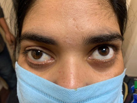
This is a clinical case of a 27-year-old female, teacher by occupation, who presented to our department with the complaint of reduced visual acuity along with metamorphopsia of central vision in both eyes for several months. The visual impairment though bilateral was asymmetric with best corrected visual acuity measured via Snellen chart was 20/60 in right eye and 20/200 in left eye. The patient, however, had 15° of hypertropia in left eye, as measured by Hirschberg corneal reflex test, but there was no complain of diplopia (Figure 1). The anterior segment on slit lamp examination was normal. The anterial chamber depth (ACD) was of van Herick grade 4. Pupillary reaction to light was normal and ocular movements were full in all gazes. Intraocular pressure taken was 15 and 16 mmHg in the right and left eyes respectively. Color vision was intact. Her medical and family history was unremarkable.
Cycloplegic refraction revealed myopia of −6.0 diopters with −1.0 cylinder in right eye and −6.0 diopters in left eye. The fixation was paramacular as detected by the fixation star of the direct ophthalmoscope, hence causing fixation disparity. Posterior segment examination via indirect ophthalmoscopy revealed a well circumscribed small yellowish lesion at the macula in the right eye (Figure 2). In the left eye a bigger roundish lesion roughly delineated with a small center of yellow material was seen at the posterior pole (Figure 3). No other abnormality was detected in the peripheral retina. On fundus auto fluorescence, the yellow material was intensely hyperautofluorescent in right eye and the same was surrounded by hypoautofluorescent area in left eye.
The macular OCT scan revealed thin band of homogenous material deposit beneath the photoreceptor layer in right and thinning at macula in the left eye (Figure 4 and Figure 5). Electro-oculogram revealed Arden index of 0.81 in right eye and 0.76 in left eye. Multifocal electroretinogram was normal. Taking into consideration all the above findings, the diagnosis of Best vitelliform macular dystrophy with left hypertropia was made. The right eye lesion was identified as early vitelliform and left eye as late vitelliruptive with accompanying atrophic areas. The patient was counseled and advised for use of base-up Fresnel glasses for correction of hypertropia and also explained the progression of Best disease. Her parents were examined thereafter and none revealed any similar fundal findings. The patient was advised six monthly reviews for early detection of development of CNV.


Discussion
The incidence of macular dystrophies in North India remains unknown as very limited sporadic reporting is done. Snow-ball sampling is the only way to recruit subjects for any further study. Majority of the patients remain unknown of their diagnosis and often present in their later life when their symptoms exceed a certain degree. Symptoms are identified at younger age only by keen parents when their child complains of deterioration of vision or might point the child of squinting. Early diagnosis is possible only if every child complaining of deterioration vision as well as squinting of eyes undergoes cycloplegic refraction with fundus examination. Usual age of onset of Best disease is from 3 to 15 years with average age of 6 years [9]. Most patients suffering from Best vitelliform macular dystrophy have a parent with this condition, however, the disease can also be caused by de novo mutation [10]. Best disease needs to be differentiated from the other dystrophies of the central part of the retina and choroid the most common mimicker being age related macular degeneration with CNV. A case was described in Mozambique where the patient of Best disease presented with choroidal neovascular membrane, as a complication of late stage [11]. Although, it occurs as a bilateral entity, a rare case of unilateral Best disease was also reported, masquerading as central serous chorioretinopathy [12]. Best vitelliform macular dystrophy along with other maculopathies is known to cause strabismus with advancing disease and deteriorating visual acuity. With loss of central macular functions, the fixation shifts to preferential retinal locus (PRL) outside the fovea [13]. A similar case of Best disease in a 5-year old was described by Guadilla et al. [14], who reported that the child presented with hyperopia and esotropia. However, the esotropia was accommodative and was fully corrected by spectacles prescription.
Fixation disparity refers to a small misalignment of the visual axes when both eyes are open in an observer with normal fusion and binocular vision. The images of a binocularly fixated object do not stimulate corresponding retinal points but still fall within Panum's fusional areas, hence the image of the object is seen singly [15]. Macular dystrophies can lead to scarring which displaces the central retinal receptors paracentrally and there occurs fixation disparity [16]. Hence, there is a tendency of the eyes to drift in the direction of the heterophoria. Our patient presented with the similar condition. No effective treatment is available to slow down the progression of Best’s disease. Oral antioxidant supplementation might have a theoretical benefit, given the role of free radicals in the formation of lipofuscin [17]. Cases when detected should undergo genetic counseling. As of today no cure is available for Best disease yet. Efforts have been made to reduce the progression of complications, like giving intravitreal anti-vascular endothelial growth factors for associated CNV but their effectiveness is yet to be studied. Use of photodynamic therapy to limit progression of sub-foveal CNV has been tried [18].
Conclusion
Best disease is a rare entity, generally presents with only blurring of vision. We evaluated a case with associated hypertropia. Patients with strabismus should be examined to rule out retinal pathologies and accompanying complications like CNV, prior to any orthoptic prescriptions, along with regular follow-up.
REFERENCES
1.
Best F. Über eine hereditäre Maculaaffektion. Beiträge zur Vererbungslehre. Z Augenheilkd 1905;13:199–212.

2.
Pinckers A, Cuypers MH, Aandekerk AL. The EOG in Best's disease and dominant cystoid macular dystrophy (DCMD). Ophthalmic Genet 1996;17(3):103–8. [CrossRef]
[Pubmed]
3.
Marmorstein AD, Marmorstein LY, Rayborn M, Wang X, Hollyfield JG, Petrukhin K. Bestrophin, the product of the Best vitelliform macular dystrophy gene (VMD2), localizes to the basolateral plasma membrane of the retinal pigment epithelium. Proc Natl Acad Sci U S A 2000;97(23):12758–63. [CrossRef]
[Pubmed]
4.
Hartzell HC, Qu Z, Yu K, Xiao Q, Chien LT. Molecular physiology of bestrophins: Multifunctional membrane proteins linked to best disease and other retinopathies. Physiol Rev 2008;88(2):639–72. [CrossRef]
[Pubmed]
5.
Weingeist TA, Kobrin JL, Watzke RC. Histopathology of Best's macular dystrophy. Arch Ophthalmol 1982;100(7):1108–14. [CrossRef]
[Pubmed]
6.
Leu J, Schrage NF, Degenring RF. Choroidal neovascularisation secondary to Best's disease in a 13-year-old boy treated by intravitreal bevacizumab. Graefes Arch Clin Exp Ophthalmol 2007;245(11):1723–5. [CrossRef]
[Pubmed]
7.
Miller SA, Bresnick GH, Chandra SR. Choroidal neovascular membrane in Best's vitelliform macular dystrophy. Am J Ophthalmol 1976;82(2):252–5. [CrossRef]
[Pubmed]
8.
Blodi CF, Stone EM. Best's vitelliform dystrophy. Ophthalmic Paediatr Genet 1990;11(1):49–59.
[Pubmed]
9.
François J, De Rouck A, Fernandez-Sasso D. Electro-oculography in vitelliform degeneration of the macula. Arch Ophthalmol 1967;77(6):726–33. [CrossRef]
[Pubmed]
10.
Testa F, Rossi S, Passerini I, et al. A normal electro-oculography in a family affected by best disease with a novel spontaneous mutation of the BEST1 gene. Br J Ophthalmol 2008;92(11):1467–70. [CrossRef]
[Pubmed]
11.
Buquea A, Vankawalaa S, Hussaina R, Dudhatraa M, Manhicab G. A clinical case report: Best vitelliform macular dystrophy disease, Mozambique. J Clin Exp Ophthalmol 2020;11(3):839. [CrossRef]
12.
Kaden TR, Tan ACS, Feiner L, Freund KB. Unilateral best disease: A case report. Retin Cases Brief Rep 2017;11 Suppl 1:S191–6. [CrossRef]
[Pubmed]
13.
Jarc-Vidmar M, Popovic P, Hawlina M. Mapping of central visual function by microperimetry and autofluorescence in patients with Best’s vitelliform dystrophy. Eye (Lond) 2006;20(6):688–96. [CrossRef]
[Pubmed]
14.
Guadilla AM, Rodríguez-Ramírez M, Modamio-Gardeta L, et al. Esotropia in the context of maculopathy compatible with autosomal recessive bestrophinopathy. JSM Ophthalmol 2017;5(3):1059.
15.
Cline D, Hofstetter HW, Griffin JR. Dictionary of Visual Science. 4ed. Boston: Butterworth-Heinemann; 1997.
16.
Howard IP. Perceiving in Depth: Volume 1 Basic Mechanisms. New York: Oxford University Press; 2012.
17.
Schmitz-Valckenberg S, Holz FG, Bird AC, Spaide RF. Fundus autoluorescence imaging: Review and perspectives. Retina 2008;28(3):385–409. [CrossRef]
[Pubmed]
18.
Andrade RE, Farah ME, Costa RA. Photodynamic therapy with verteporfin for subfoveal choroidal neovascularization in Best disease. Am J Ophthalmol 2003;136(6):1179–81. [CrossRef]
[Pubmed]
SUPPORTING INFORMATION
Author Contributions
Perwez Khan - Conception of the work, Design of the work, Acquisition of data, Analysis of data, Drafting the work, Revising the work critically for important intellectual content, Final approval of the version to be published, Agree to be accountable for all aspects of the work in ensuring that questions related to the accuracy or integrity of any part of the work are appropriately investigated and resolved.
Parul Singh - Conception of the work, Design of the work, Acquisition of data, Drafting the work, Revising the work critically for important intellectual content, Final approval of the version to be published, Agree to be accountable for all aspects of the work in ensuring that questions related to the accuracy or integrity of any part of the work are appropriately investigated and resolved.
Shweta Singh - Conception of the work, Design of the work, Acquisition of data, Drafting the work, Final approval of the version to be published, Agree to be accountable for all aspects of the work in ensuring that questions related to the accuracy or integrity of any part of the work are appropriately investigated and resolved.
Ankita Srivastava - Conception of the work, Design of the work, Revising the work critically for important intellectual content, Final approval of the version to be published, Agree to be accountable for all aspects of the work in ensuring that questions related to the accuracy or integrity of any part of the work are appropriately investigated and resolved.
Guarantor of SubmissionThe corresponding author is the guarantor of submission.
Source of SupportNone
Consent StatementWritten informed consent was obtained from the patient for publication of this article.
Data AvailabilityAll relevant data are within the paper and its Supporting Information files.
Conflict of InterestAuthors declare no conflict of interest.
Copyright© 2021 Perwez Khan et al. This article is distributed under the terms of Creative Commons Attribution License which permits unrestricted use, distribution and reproduction in any medium provided the original author(s) and original publisher are properly credited. Please see the copyright policy on the journal website for more information.


