  |

|
 |
|
Case In Images
| ||||||
| Xanthogranulomatous cholecystitis: The great gallbladder carcinoma masquerader | ||||||
| Massimo Arcerito1, John Moon1, Kevin Tri Nguyen2 | ||||||
|
1MD, FACS Division of Inland Empire, Department of Surgery, University of California Irvine, Irvine CA.
2MD, PhD, FACS, Piedmont Healthcare, Transplant Institute, Atlanta, GA. | ||||||
| ||||||
|
[HTML Abstract]
[PDF Full Text]
[Print This Article]
[Similar article in Pumed] [Similar article in Google Scholar] 
|


| How to cite this article |
| Arcerito M, Moon J, Tri Nguyen K. Xanthogranulomatous cholecystitis: The great gallbladder carcinoma masquerader. Int J Case Rep Images 2017;8(3):222–226. |
|
Abstract
|
|
Introduction:
Xanthogranulomatous chole-cystitis represents an uncommon chronic cholecystitis characterized by gallbladder wall thickening infiltrating to the adjacent liver parenchyma. This feature creates a challenge for the hepatobiliary surgeon in treating this rare disease, which mimics gallbladder carcinoma leading to unnecessary enlarged hepatic resection.
Case Report: A 62-year-old male presenting at emergency department complaining of eight weeks history of right upper quadrant pain, nausea, with intermittent vomiting and severe weight loss. Surgical consultation was obtained. All imaging workup, including conventional abdominal ultrasound, computed tomography scan of the abdomen and pelvis, magnetic resonance imaging scan of the abdomen and positron emission tomography scan suggested gallbladder carcinoma. He underwent extended right hepatectomy, pancreaticoduodenectomy, with primary intrahepatic Roux-Y hepaticojejunostomy. The hospital course of patient was unremark-able being discharged six days after surgery. Final pathology was compatible with xanthogranulomatous cholecystitis. Conclusion: This rare entity can lead the hepatobiliary surgeon to face a challenge and sometime useless surgical treatment which might provoke morbidity and mortality to the patient. More define clinical and radiographic criteria are needed in diagnosing and treating xanthogranulomatous cholecystitis, which must be labeled as the great gallbladder carcinoma masquerader. | |
|
Keywords:
Contrast enhancing ultrasound, Foamy cells, Pancreaticoduodenectomy, Xanthogranulomatous cholecystitis
| |
|
Introduction
| ||||||
|
Xanthogranulomatous cholecystitis is an inflammatory gallbladder disease characterized by destructive inflammatory process and infiltration of foamy cells of the gallbladder wall with associated proliferative fibrosis and thickening. This rare entity is almost impossible to be distinguished from the lethal gallbladder carcinoma based on clinical, radiographic and preoperative pathologic features. Furthermore, a very aggressive surgical treatment option is almost always performed provoking potential morbidity and possible mortality to our patients. | ||||||
|
Case Report
| ||||||
|
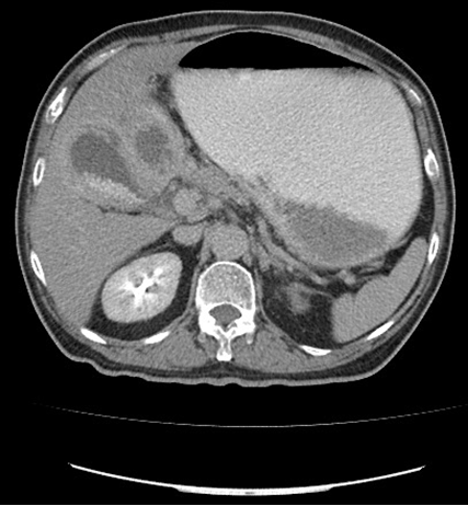
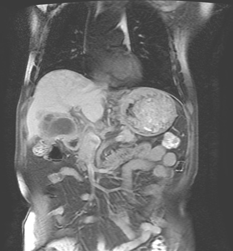
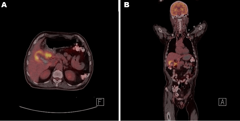
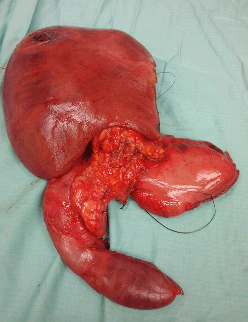
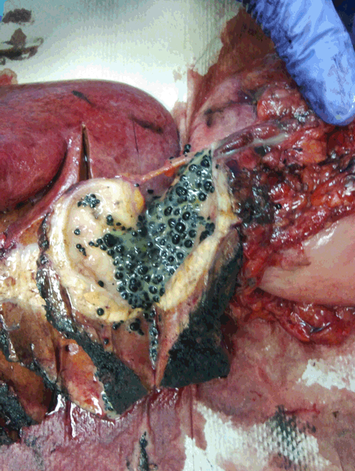
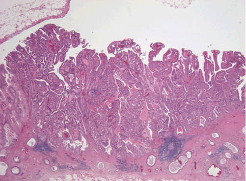
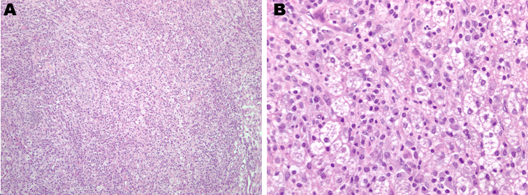
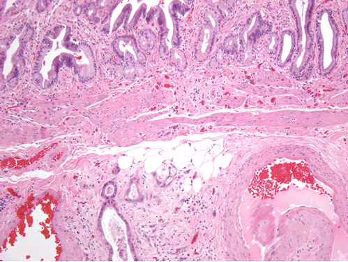
A 62-year-old male with a two-month history of right upper quadrant pain associated with nausea and vomiting causing 40 pounds weight loss. Empiric treatment with proton pump inhibitors did not achieve control of his symptomatology. The patient did not seek medical attention for all this time and he presented at the emergency department. Abdominal ultrasound showed features of acute cholecystitis characterized by pericholecystic fluid collection and thickness of the gallbladder wall. Contrast-enhanced abdominal computed tomography demonstrated extensive abnormal gallbladder wall thickening with direct invasion of the adjacent hepatic parenchyma and pylorus, with resulting gastric outlet obstruction (Figure 1). Subsequently, abdominal MRI scan confirmed the CT scan findings, and showed abnormal signal intensity and enhancement within the adjacent hepatic parenchyma, measuring up to 2.2 cm in thickness, invading the right hepatic duct as well compressing and encasing the pylorus (Figure 2). Positron emission tomography scan showed hypermetabolic activity around the gallbladder suggesting a neoplasm (Figure 3A-B). The patient underwent diagnostic laparoscopy to rule out distant metastasis. After conversion to open technique, intraoperative ultrasound, extended right hepatectomy with cholecystectomy, resection of extrahepatic bile duct and pancreaticoduodenectomy with Roux-Y intrahepatic biliary anastomosis were performed (Figure 4). Figure 5 shows a detailed aspect of the gallbladder wall. Pathology showed xanthogranulomatous cholecystitis forming a large mass, extending into liver, duodenum, and distal stomach without involvement of pancreas and ten unremarkable lymph nodes (Figure 6). Figure 7 shows the xanthogranulomatous cholecystitis characterized by mixed inflammatory cells with many foamy macrophages in different magnification. A pathologic picture of gallbladder carcinoma is shown in Figure 8 for comparison to the benign xanthogranulomatous cholecystitis. | ||||||
| ||||||
|
| ||||||
| ||||||
| ||||||
| ||||||
| ||||||
| ||||||
|
| ||||||
| ||||||
|
Discussion
| ||||||
|
Christensen et al. first described this rare entity as "fibro-xanthogranulomatous inflammation" of the gallbladder in the seventies [1]. It is characterized by acute on chronic cholecystitis with dense fibrosis and aggressive accumulation of xanthomas in areas of destructive inflammatory response. This rare disease, based on its aggressive inflammatory behavior, mimics a gallbladder malignancy with involvement of surrounding anatomical structures, and this characteristic leads to perform an unnecessary extensive surgical resection of the surrounding organs. Jain et al. recently reported a retrospective study comparing the clinical and radiographic findings between the xanthogranulomatous and gallbladder cancer. Despite the absence of difference in clinical symptomatology between the two groups, contrast enhancement CT scan of the abdomen and pelvis helped to identify some radiographic properties commonly founded in xanthogranulomatous cholecystitis compared to gallbladder carcinoma [2]. Gallbladder wall thickness was similar in both groups, but the presence of intramural hypo-attenuating nodules in about 60% of xanthogranulomatous cholecystitis group in the absence of disrupted mucosal lining and obstructive features of the bile duct system, implied the inflammatory component. In support of these radiographic findings, Yuan et al. reviewed the clinical role of contrast-enhanced ultrasound technique in differential diagnosis of xanthogranulomatous cholecystitis. A total of 60 patients (17 xanthogranulomatous cholecystitis and 43 gallbladder carcinoma) were enrolled in the study. An initial conventional ultrasound was performed in both groups and subsequently their data were compared with contrast-enhanced technique in searching for wall thickening characteristics, gallbladder stones and hypoechoic nodule frequencies. Contrast-enhanced ultrasound showed superior technique in analyzing the gallbladder wall thickening, gallbladder stones and hypoechoic nodules compared with conventional ultrasound. Particularly, a hypoenhancement time greater than 80 seconds, a diffuse thickening and hypoechoic nodules were highly suggesting features of xanthogranulomatous cholecystitis [3]. An ultrasound-guided fine needle aspiration (UGFNA) of gallbladder lesions should be always be part of our armamentarium in differential diagnosis of xanthogranulomatous cholecystitis. We did not perform a UGFNA in our patient. Rana et al. proved in their largest experience the paramount safeness, reliability and cost-effective of this technique in diagnosis gallbladder diseases. Their conclusions prompted them to suggest for all the providers in hepatobiliary specialty to perform a UGFNA in all cases with gallbladder mass upon presentation [4]. The premalignant condition of xanthogranulomatous cholecystitis is unknown. Ghosh et al. addressed the premalignant property of xanthogranulomatous cholecystitis [5]. Proliferating cell nuclear antigen (PCNA), beta-catenin and p53 expression were studied comparing xanthogranulomatous cholecystitis, gallbladder cancer, chronic cholecystitis and cholelithiasis as control group. p53 mutation and PCNA were present in 52% and 60% of gallbladder carcinoma and only in 3% and 11% of xanthogranulomatous cholecystitis. No evidence of this mutation was found in chronic cholecystitis or cholelithiasis. Beta-catenin expression was positive in all four analyzed groups. We supported the inflammatory component of xanthogranulomatous cholecystitis without evidence of premalignant condition. A new clinical scoring system has been proposed in differentiating xanthogranulomatous cholecystitis and gallbladder carcinoma [6]. The authors retrospectively reviewed their experience in treating almost 500 patients with gallbladder diseases focusing on clinical and imaging parameters. Patients with a long history of abdominal pain and gallbladder wall thickening, cholelithiasis and gallbladder submucosal hypoattenuated nodules compared with anorexia, weight loss are more prone to be diagnosed as xanthogranulomatous cholecystitis more than gallbladder malignancy. A scoring system closed to 11 to 13 helps to narrow the diagnosis towards xanthogranulomatous cholecystitis. | ||||||
|
Conclusion
| ||||||
|
Xanthogranulomatous cholecystitis is a rare entity leading to potentially unnecessary extensive surgical resection with possible associated increase morbidity and mortality. A defined role of scoring systems should be implemented in differentiating this rare disease from the more lethal gallbladder carcinoma. Identifying radiographic features and performing preoperative ultrasound-guided fine needle aspiration (UGFNA) of gallbladder masses and possible intraoperative biopsy through the liver parenchyma may assist the hepatopancreaticobiliary surgeon in intraoperative planning and decision-making. | ||||||
|
References
| ||||||
| ||||||

|
[HTML Abstract]
[PDF Full Text]
|
|
Author Contributions
Massimo Arcerito – Substantial contributions to conception and design, Acquisition of data, Analysis and interpretation of data, Drafting the article, Revising it critically for important intellectual content, Final approval of the version to be published John Moon – Analysis and interpretation of data, Revising it critically for important intellectual content, Final approval of the version to be published Kevin Tri Nguyen – Analysis and interpretation of data, Revising it critically for important intellectual content, Final approval of the version to be published |
|
Guarantor of submission
The corresponding author is the guarantor of submission. |
|
Source of support
None |
|
Conflict of interest
Authors declare no conflict of interest. |
|
Copyright
© 2017 Massimo Arcerito et al. This article is distributed under the terms of Creative Commons Attribution License which permits unrestricted use, distribution and reproduction in any medium provided the original author(s) and original publisher are properly credited. Please see the copyright policy on the journal website for more information. |
|
|