|
|
|
|
Case Report
| ||||||
| Dental implant placement in focal osteoporotic bone marrow defect: A case report | ||||||
| Hamdan S. Alghamdi | ||||||
|
Department of Periodontics and Community Dentistry, College of Dentistry, King Saud University, Riyadh, Saudi Arabia
| ||||||
| ||||||
|
[HTML Abstract]
[PDF Full Text]
[Print This Article] [Similar article in Pumed] [Similar article in Google Scholar] 
|
| How to cite this article |
| Alghamdi HS. Dental implant placement in focal osteoporotic bone marrow defect: A case report. Int J Case Rep Images 2017;8(12):817–821. |
|
ABSTRACT
| ||||||
|
Introduction: Dental implants are becoming the most efficient treatment option for missing teeth. These are because of their ability to osseointegrate directly into the jawbone and then support dental prosthesis. However, a challenged bone condition may prevent the achievement of an optimal implants placement and then cause clinical complications. One of these conditions could be the focal osteoporotic bone marrow defects (FOBMD). Keywords: Bone grafts, Dental implants, Focal osteoporotic defects, Primary stability | ||||||
|
INTRODUCTION
| ||||||
|
Nowadays, dental implants are becoming the most alternative treatment option for missing teeth. These are because of their ability to osseointegrate directly into the jawbone and then support dental prosthesis [1]. However, a challenged bone condition may prevent the achievement of an optimal implants placement and then cause clinical complications [2]. One of these conditions could be the focal osteoporotic bone marrow defects (FOBMD) [3]. However, in oral implantology, serious complications might occur in the presence of FOBMD, mainly in the posterior mandibular region of the jawbone. This would probably cause an incidence of implant fixture displacement inside the body of the mandible [4][5]. Such serious complication can occur immediately with the surgical insertion of dental implants or postoperatively because of a deficiency in the primary stability of implants [6][7]. Clinically, FOBMD is an unusual condition that appears as undefined radiolucent area in the jawbone [3][8]. It is usually localized, asymptomatic, and associated with previous history of extraction of teeth. They occur more commonly in the middle-aged female patients and show a predilection for the region of mandibular molars [9]. The radiographic appearance of FOBMD including scattered trabeculae may extend short distances into the defect with irregular borders [10]. The etiology of the FOBMD is still unknown. However, it may be related to an alteration in the repair process of bone trabeculae in the area of trauma or inflammation after teeth extraction [11]. This would also be related to ischemic changes in the bone marrow tissue, which stimulate the development of hematopoietic foci [8]. For the placement of dental implants, bone morphology as well as density plays an important role in the implant stability and then success outcome of implant treatment. Serious complications and accidents related to surgery can include the displacement or migration of implants into the body of jawbones because of poor surgical technique or anatomic variances (i.e., FOBMD). This paper describes a successful dental implant placement in a focal osteoporotic bone marrow defect located in the posterior left mandible of a 22-year-old female patient. | ||||||
|
CASE REPORT
| ||||||
|
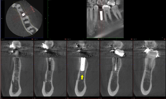
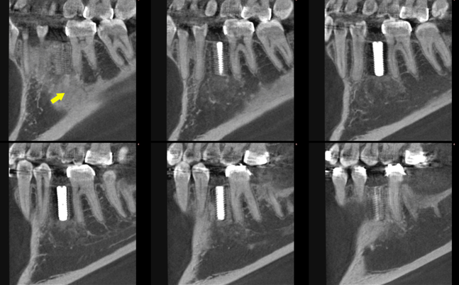
A 22-year-old female was referred to the dental implants clinic, with complaint of missing tooth in the left lower posterior region. Dental history revealed that #35 was badly decade, non-restorable, and then had been extracted five years ago. Intraoral examination showed healthy mucosa and there was not any sign of infection. Her past medical history was unremarkable. Panoramic radiography of the jaws showed radiolucent area with ill-defined borders associated to the region of #35 (Figure 1). This condition was asymptomatic and no expansion of the mandibular cortical bone was clinically detected. Thus, a provisional diagnosis of focal osteoporotic bone marrow defect (FOBMD) was considered as a differential diagnosis based on age, site, clinical and radiographic findings. The patient was given the option of possibility of dental implant treatment. Pros and cons of the procedure were explained to the patient and informed consent was obtained. Then, preoperative evaluation included clinical and radiographical assessments of implant size, position of implant, and anatomical landmarks were applied. Under local anesthesia (2% lignocaine with adrenaline 1:80,000), a crestal incision was placed in the edentulous #35 region. A full-thickness mucoperiosteal flap was elevated and reflected. Implant site was marked with round bur. A 2-mm pilot drill osteotomy was done to perforate the crestal cortical bone. Large FOBMD was founded isolated with several millimeters in diameter. The void was filled using deproteinized bovine bone grafts (Bio-Oss®, Osteohealth Co, Shirley, NY, USA) (Figure 2). Thereafter, Straumann® ITI Dental Implant System (Institut Straumann AG Waldenburg, Switzerland) was used. Implant fixture (ø 3.3 mm, Roxolid®, SLActive®) was immediately placed with a torque wrench with adequate primary stability. The selection of Straumann® implant was suitable for bone level treatment in combination with subgingival healing. It has a special anatomical design, which combines a cylindrical shape in its apical region and a conical shape in the coronal region, making this implant particularly suitable for implantation in challenged condition. Roxolid® Titanium-Zirconium alloy material is designed to offer more confidence when placing small diameter implants in the molars region. Also, SLActive® has unique properties of hydrophilicity and chemical activity, which can accelerates implant osseointegration [12]. Flap repositioned and sutured with simple interrupted sutures using resorbable Vicryl material size 4-0 (Vicryl®, Ethicon, Johnson & Johnson, Norderstedt, Germany). Primary closure was achieved. Immediate postoperative radiograph was taken to confirm position of implant fixture (Figure 3). Antibiotics and analgesics were prescribed for seven days. The patient was placed on regular maintenance protocol. Four months after placement, the implant was fully integrated and showed good healing (Figure 4) and (Figure 5). No clinical abnormality was noted. | ||||||
| ||||||
| ||||||
|
| ||||||
|
| ||||||
|
| ||||||
|
DISCUSSION
| ||||||
|
Focal osteoporotic bone marrow defect (FOBMD) has been reported as an unexpected radiolucency in the posterior mandible of the middleaged female patients [8][13]. Our case diagnosis is in consistent. Moreover, the present case shows the fact of the rigorous method for radiographic assessment of the patients and X-Ray prescription as an initial step for treatment planning. In addition, the use of CBCT examination should not be neglected, as it can be an accurate method for the pre-assessment during dental implant treatments [14]. The exact cause of FOBMD has not been confirmed [3]. However, it frequently occurs in an extraction area and might be related to alterations in socket healing and bone trabeculation [8]. Other possibilities might be related to some systemic conditions, e.g., hematologic disorders [11]. In this case, we could speculate that the FOBMD is associated with the impairment of alveolar bone healing after tooth extraction of #35. Also, the patient medical history was normal excluding a possible association with other medical conditions. Previous reports showed that ~70% FOBMD is occurred in the posterior mandible in adult women and only minority of the cases are reported in maxilla [15]. Most of the cases are asymptomatic and have no swelling, found incidentally on routine radiographs. Radiographic appearance of FOBMD shows isolated radiolucency with several millimeters to centimeters in diameter and ill-defined areas [10][16]. In addition, the radiolucent area of FOBMD is trabeculated randomly and separately distributed. Unlike other odontogenic lesions, FOBMD tend to respect the lamina dura of adjacent teeth without expanding the bone cortex to the corresponding area [17]. Clinically, serious complications in oral implantology might occur in presence of FOBMD. This would probably cause an incidence of implant fixture displacement inside the body of the mandible [4] [5][18]. The displacement of an implant can occur immediately or within a short period due to poor initial implant stability can result from the anatomical variances. And this because of the bone pattern of FOBMD provides low mechanical stability for dental implants at placement and should be carefully considered [6]. As previously reported, unforeseen accidents of implant displacement could occur especially in the posterior FOBMD, when could not be delineated on a panoramic radiograph [6][19]. Thus, careful radiographic evaluations may be necessary for female patients with extracted posterior teeth prior to dental implants placement. For management, it has been suggested that surgical manipulation of dental implants may improve the mechanical pattern of bone at an implant site [20]. As in this case, we recommend augmenting the FOBMD with bone grafting materials prior to implant insertion. For such surgical protocol, it was done similar to the method of immediate implant placement after teeth extraction [21]. Also, it has to be mentioned that the presence of active bone marrow (i.e. rich in stem cells) in the FOBMD would favor the biology of implants osseointegration [22]. Nonetheless, additional studies with more cases and long-term follow-up should be conducted. | ||||||
|
CONCLUSION
| ||||||
|
Focal osteoporotic bone marrow defect (FOBMD) is generally a rare condition that might be detected in posterior mandible of middle-aged women. When FOBMD presents with unusual radiographic findings, it requires more specific examination of the tissue. The presence of FOBMD does hinder the mechanical stability of dental implants, although it is not negatively affect implants osseointegration from biological point of view. Therefore, we recommend securing the initial stability of dental implants using bone grafting procedure simultaneous the implant placement. | ||||||
|
REFERENCES
| ||||||
| ||||||
|
[HTML Abstract]
[PDF Full Text]
|
|
Author Contributions
Hamdan S. Alghamdi – Substantial contributions to conception and design, Acquisition of data, Analysis and interpretation of data, Drafting the article, Revising it critically for important intellectual content, Final approval of the version to be published |
|
Guarantor of Submission
The corresponding author is the guarantor of submission. |
|
Source of Support
None |
|
Conflict of Interest
Author declares no conflict of interest. |
|
Copyright
© 2017 Hamdan S. Alghamdi. This article is distributed under the terms of Creative Commons Attribution License which permits unrestricted use, distribution and reproduction in any medium provided the original author(s) and original publisher are properly credited. Please see the copyright policy on the journal website for more information. |
|
ABOUT THE AUTHOR
| |||
| |||
|
|