|
|
|
|
Case Report
| ||||||
| The combined use of surgical rehearsal platform and BrainLab navigation for mandibular osteotomy in Nager syndrome | ||||||
| Tian Ran Zhu1, Justine C. Lee2 | ||||||
|
1MD, The Warren Alpert Medical School of Brown University, Providence, RI, USA
2MD, PhD, UCLA Division of Plastic and Reconstructive Surgery, Los Angeles, CA | ||||||
| ||||||
|
[HTML Abstract]
[PDF Full Text]
[Print This Article] [Similar article in Pumed] [Similar article in Google Scholar] 
|
| How to cite this article |
| Zhu TR, Lee JC. The combined use of surgical rehearsal platform and BrainLab navigation for mandibular osteotomy in Nager syndrome. Int J Case Rep Images 2017;8(10):717–720. |
|
ABSTRACT
| ||||||
|
Introduction: Nager syndrome is a rare genetic condition characterized by defects primarily of the face, arms, and hands. Children with Nager syndrome are frequently born with maxillary hypoplasia in conjunction with micrognathia and associated cleft lip/palate anomalies. These abnormalities severely restrict proper feeding, impair normal speech and language development, and contribute to life-threatening breathing problems. Surgical management options include mandibular osteotomy and distraction osteogenesis to correct the maxillofacial defects. Operative complications include damage to surrounding nerves and vessels, entry to the skull base, and recurrent temporomandibular joint (TMJ) ankylosis. Keywords: Brainlab, Craniofacial, Distraction osteogenesis, Mandibular osteotomy, Nager syndrome, Surgical planner | ||||||
|
INTRODUCTION
| ||||||
|
Nager syndrome is a congenital disorder of the first and second branchial arches and appendicular system that result in underdeveloped face, arms, and legs [1]. Nager and Reynier first coined the term acrofacial dysostosis in 1948 to distinguish Nager syndrome as a craniofacial malformation from mandibular dysostosis [2]. While the exact cause is unknown, most cases are sporadic with case reports of autosomal dominant and recessive pattern of inheritance that is associated with deletion in SF3B4 gene in the long arm of chromosome [1][2]. The resulting malformations of the branchial arches manifest as mandibular hypoplasia, malocclusion, micrognathia, cleft palate, and microtia [1][3][4]. These abnormalities frequently cause feeding problems in infants with Nager syndrome secondary to mandibular hypoplasia and palatal defects. In addition, micrognathia with subsequent glossoptosis can lead to life-threatening apnea and asphyxiation, necessitating distraction osteogenesis concomittant with mandibular osteotomy to advance the jaw both anteriorly and inferiorly to alleviate the soft-tissue obstruction of the airway [5][6][7][8]. Similar patterns of craniofacial anomalies have been observed in other genetic syndromes affecting children including Treacher Collins syndrome, Pierre Robbin syndrome, and Cleidocranial dysplasia. All of these syndromes share commonality in that specific genetic alterations affect key craniofacial developmental pathways leading to micrognathia, glossoptosis, and subsequent air obstruction [9][10]. Therefore, the goals of treatment for these children focuses on breathing and feeding and optimizing growth and nutrition. What differentiates Nager syndrome from these other aforementioned syndromes is involvement of distal limb buds that can result in deformed or absent thumbs, shortened or absent forearms, hammer toes, and leg and feet bone abnormalities [3][5][11]. Herein, we report a case of recurrent temporomandibular joint (TMJ) ankylosis in a child with Nager syndrome and demonstrate the efficacy of SuRgical PlannerTM surgical rehearsal platform (SRP) in conjunction with Brainlab intraoperative computed tomography navigation system to augment intraoperative visualization, enhance surgical proficiency and safety, and improve overall patient outcome. | ||||||
|
CASE REPORT
| ||||||
|
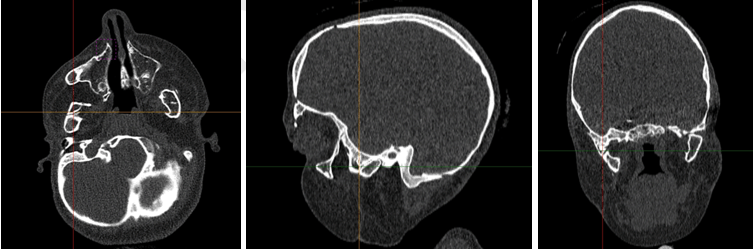
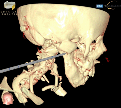
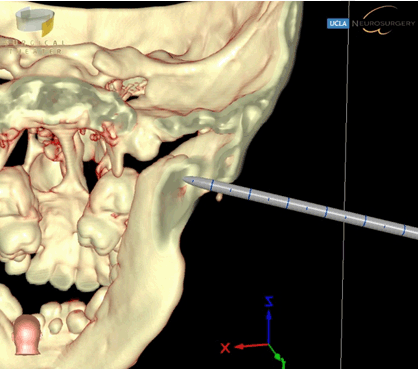
We present a case of a nine-year-old girl with Nager syndrome who was born with severe micrognathia, auricular atresia, high arched palate, bilateral radial deficiencies, and left club foot. At birth, she presented with significant respiratory distress secondary to mandibular hypoplasia and subsequent glossoptosis. Because of her difficult airway, intubation was attempted but failed, requiring emergent tracheostomy tube placement and mechanical ventilation. The patient was weaned off the ventilator prior to discharge. Since then she has not required mechanical ventilation. The relevant surgical history includes tracheostomy at birth, right index finger pollicization at age two, implantation bone anchored hearing aid at age five, and previous bilateral mandibular osteotomy at five years of age. Prior to her last surgery, the patient had reankylosis of her bilateral TMJ resulting in severe limited jaw opening that required repeat mandibular osteotomies (Figure 1). For the previous mandibular osteotomy, Brainlab intraoperative CT-guided navigation system was used to aid in the preoperative planning of localizing and assessing the extent of the TMJ fusion. The surgery was completed in nine hours with no complications. For most recent mandibular osteotomy, SuRgical PlannerTM was used in conjunction with Brainlab intraoperative CT-guided navigation system (Figure 2). This additional surgical guidance tool allowed us to rehearse the operation for TMJ resection and also assess the extent of our dissection, thereby accelerating operative efficiency (Figure 3). Additionally, based on principle of CT scanning Hounsfield unit, the SRP simulator can display or hide slices of tissue in real time, thereby allowing us to visualize surrounding skull base, vessel, and soft tissue anatomy as well as the location of our surgical probe to minimize skull base complications [12]. Overall, the operative time was four hours, a notable decrease from nine hours previously. There were similar scars noted from prior surgeries and no complications. The exact same sequence of surgery and placement of Matthews device was performed for these two operations to justify the comparison. | ||||||
| ||||||
| ||||||
|
| ||||||
|
DISCUSSION
| ||||||
|
Severe airway obstruction in Nager syndrome remains the major cause of morbidity and mortality. Respiratory instability secondary to micrognathia and glossoptosis frequently necessitates a tracheostomy, while a gastrostomy tube may be needed for adequate nutrition [6][13]. Later, mandibular osteotomy and distraction osteogenesis are necessary to address any mandibular and TMJ anomalies. Significant intraoperative risks surround the use of instrumentation to resect fused TMJ, in which the close vicinity to facial nerve branches, adjacent soft tissues, and the undersurface of skull base presents a notable challenge for craniofacial surgeons. Brainlab image guidance, a technology first developed for neurosurgery, has been applied to craniofacial surgery to address these concerns. Thus, by using Brainlab imaging in our operative planning we were able to design osteotomy lines, track the extent of bilateral TMJ and condylar resections, and identify the stylomastoid foramen to minimize damage to the facial nerve. Similarly, The SuRgical Planner™, a novel surgical rehearsal platform (SRP) developed by Surgical Theater LLC, approved by FDA in 2013, offers surgeons the opportunity to rehearse surgery prior to operation [14][15][16]. By using SRP, we rehearsed the sequence of utilizing the Pineapple bur and Kerrison rongeurs to resect bilaterally fused TMJ and coronoid processes prior to operation. Additionally, when combined with Brainlab 3D reconstruction of the patient’s craniofacial anatomy, SRP allowed us to visualize in real time the extent and anatomic spatial location of our resection, so we were able to further limit damage to local tissues, nerves, and vessels and avoid entry into the skull base. | ||||||
|
CONCLUSION
| ||||||
|
With the increased risk associated with surgical approaches to craniofacial reconstructions that closely border skull base and orbital floors, it is critical to advance training to minimize complications and improve surgical outcome. The trend to using 3D image guidance has advanced neurosurgery with improved operative time and patient safety. In addition, visual simulation technology further enhances resident training and provides an additional modality to guide operative decision making. We present this case to highlight the applicability of these innovations for mandibular osteotomy in Nager syndrome and other craniofacial operations. In particular, we demonstrate that the combined use of surgical rehearsal platform and Brainlab image guidance resulted in additional improvement in operative efficiency, enhanced real-time visualization of important anatomy, and minimized surgical complications. | ||||||
|
REFERENCES
| ||||||
| ||||||
|
[HTML Abstract]
[PDF Full Text]
|
|
Acknowledgements
We would like to acknowledge Patrick O’Neal, VR Clinical Specialist, Surgical Theater for helping with intraoperative set-up of Brainlab Surgical Navigation platform and for acquisition of images used in this case report. |
|
Author Contributions
Tian Ran Zhu – Substantial contributions to conception and design, Acquisition of data, Analysis and interpretation of data, Drafting the article, Revising it critically for important intellectual content, Final approval of the version to be published Justine C. Lee – Substantial contributions to conception and design, Acquisition of data, Analysis and interpretation of data, Revising it critically for important intellectual content, Final approval of the version to be published |
|
Guarantor
The corresponding author is the guarantor of submission. |
|
Source of support
None |
|
Conflict of interest
Authors declare no conflict of interest. |
|
Copyright
© 2017 Tian Ran Zhu et al. This article is distributed under the terms of Creative Commons Attribution License which permits unrestricted use, distribution and reproduction in any medium provided the original author(s) and original publisher are properly credited. Please see the copyright policy on the journal website for more information. |
|
|