| |
|
|
|
Case Series
| ||||||
| Respiratory failure in adults due to foreign body aspiration: A case series | ||||||
| Lycke R. Woittiez1,2, Elsbeth J. Wesselink1, Marcel A. de Leeuw1,3, Cornelis Slagt1,4 | ||||||
|
1Zaans Medical Center, Koningin Julianaplein 58, 1502 DV Zaandam, The Netherlands.
2Academic Medical Center, Department Internal Medicine, p.o. box 22660, 1100 DD Amsterdam. 3VU University Medical Center, Postbus 7057, 1007 MB Amsterdam, The Netherlands. 4Radboud University Medical Center, Department Anaesthesia, Pain and Palliative Medicine, Geert Grooteplein-Zuid 10, 6500 HB Nijmegen, The Netherlands. | ||||||
| ||||||
|
[HTML Abstract]
[PDF Full Text]
[Print This Article]
[Similar article in Pumed] [Similar article in Google Scholar] 
|
| How to cite this article |
| Woittiez LR, Wesselink EJ, de Leeuw MA, Slagt C. Respiratory failure in adults due to foreign body aspiration: A case series. Int J Case Rep Images 2016;7(6):422–426. |
|
Abstract
|
|
Introduction:
Foreign body aspiration (FBA) is rare in adults and its clinical presentation can be very diverse. Acute symptoms as dyspnea and choking are often immediately linked to FBA. However, mild or even asymptomatic chronic pulmonary symptoms can be presented as a result of FBA. Physical examination is usually nonspecific. Chest X-ray is often normal or shows nonspecific findings. Treatment and definite diagnosis can be accomplished using rigid or flexible bronchoscopy.
Case Series: We present two cases of foreign body aspiration. The first case was the aspiration of a broken tracheostomy tube leading to acute respiratory failure and the second case was the aspiration of a medication blister which initially presented as atypical chronic pulmonary symptoms but evolved to a medical emergency of acute respiratory failure. Conclusion: These two cases show the broad range of symptoms and findings associated with FBA. When patients present with nonspecific pulmonary findings, FBA should be included in the differential diagnosis. | |
|
Keywords:
Aspiration, Bronchoscopy, Chest X-ray, Foreign body, Pulmonary medicine, Respiratory failure
| |
|
Introduction
| ||||||
|
Foreign body aspiration (FBA) occurs frequently in children, but rarely in adults [1]. In different series where both children and adults with FBA were included, children represented 46–92% of the total study group [2][3]. Most adult patients who experience FBA have predisposing conditions resulting in a decreased consciousness, such as cerebrovascular accidents, intracranial hemorrhage or septic encephalopathy. Other possible risk factors are tracheostomy handling, emergency intubation, cranioencephalic trauma, intravenous drug abuse, alcohol or sedative use and dental and medical procedures [4]. Furthermore, the occurrence of FBA is dependent on the region where people live. In Islamic countries, aspiration of headscarf pins is quite common. How often FBA in adults occurs in the Netherlands is unknown. In this article we describe two cases of FBA in adults. The first patient presented with acute respiratory failure. The second patient presented with atypical chronic pulmonary symptoms which evolved to acute respiratory failure. | ||||||
|
Case Series
| ||||||
|
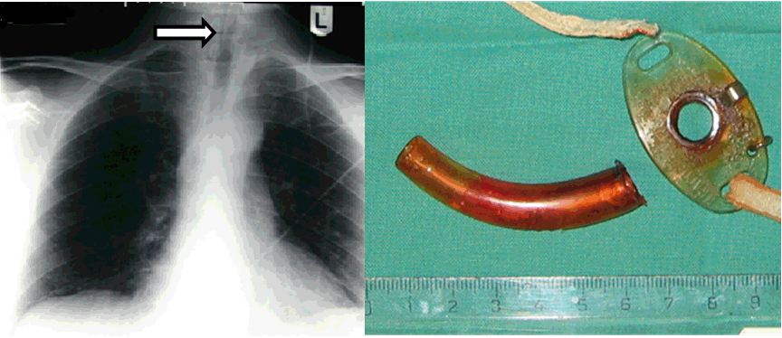
Case 1 The patient rapidly deteriorated. Therefore, an attempt was made to remove the barely visible tracheostomy tube which was trapped in the larynx under local anesthesia. During this attempt the tube dislocated to more distal airways, which caused an improvement in the vital signs. After dislocation, the patient was alert without experiencing any dyspnea. The pulmonologist performed a flexible bronchoscopy and retrieved the tube from the left main bronchus ( (Figure 1); right panel). After this procedure the patient remained in good condition. Case 2 | ||||||
|
| ||||||
|
| ||||||
| ||||||
|
Discussion
| ||||||
|
The above mentioned cases show that FBA in the adult patient can present in very different ways and the diagnosis can be challenging. Foreign Bodies Symptoms There can be a significant delay in the diagnosis as symptoms can be absent or atypical. The time from aspiration to clinical presentation is determined by the severity of the symptoms. In most studies, a minority of patients (19–53%) present within one week of aspiration [4] [6]. In 58–70% of the patients the delay in diagnosis was more than 1 month [1] [6]. Much longer delays (1–40 years) after aspiration have been described [4] [5] [6]. Twenty-five percent of the patients did not remember FBA, and only 22% remembered it on clinical suspicion [4]. In the geriatric population, only 30% could remember FBA at the first visit to the doctor [7]. Again, the physical examination is non-specific, clinical signs are absent in 39–87% of patients. Decreased breath sounds were noted in 13–47% of patients and respiratory distress was seen in only 5% [2]. Both our cases show that FBA can result in respiratory failure as a result of the aspiration itself or in a later stage due to dislocation of the FB. When respiratory failure develops and a FB is expected, rapid bronchoscopic removal is indicated. Radiology The sensitivity of computed tomography for diagnosing FBA ranges from 90–100%, its specificity from 75–100% [8]. The slice thickness of the CT scan has to be taken into account [7] [8]. Atypical findings, such as atelectasis (63%), hyperlucency (44%), thickened bronchial wall adjacent to the FB (44%), bronchiectasis (31%), pleural effusion (19%) and hilar lymphadenopathy (31%) can be found [5]. Virtual bronchoscopy, in which high resolution CT scan is used to depict the bronchi from an endoscopic viewpoint, has shown high sensitivity and specificity in the diagnosis of a foreign bodies in children. However, no studies were found for this indication in adult patients [9]. Treatment Bronchoscopy In the past, a rigid bronchoscopy was mostly used with a high success rate of 98%. However, for chronic aspiration flexible bronchoscopy is as effective as rigid bronchoscopy and causes fewer complications. Therefore, flexible bronchoscopy is now often used as first option. Flexible bronchoscopy can be performed under local anesthesia and has a success rate of 60–97% [1] [6]. Another advantage of flexible bronchoscopy is that it visualizes segmental airways to the third generation of branching, and rigid bronchoscopy only visualizes the trachea and proximal bronchi. Therefore, when the FB is impacted in distal airways, flexible bronchoscopy is the treatment of choice. Flexible bronchoscopy is also indicated in patients with cervicofacial trauma. Computed tomography scan can help distinguishing which technique should be used first [8]. When removal with flexible bronchoscopy is unsuccessful, a repeat procedure should be performed [4]. Usually a rigid bronchoscopy under general anesthesia [1]. Reasons for failure include entrapment of the FB in the bronchial wall, serious granulation with bronchial atresia or serious hemorrhage [6]. Complications after bronchoscopy are laryngeal edema, subcutaneous emphysema and pneumothorax [1]. The FB is usually located in the right lung, probably because the right main bronchus is more in line with the trachea [2] [5]. However, abnormalities on chest X-ray that are not right-sided should not lead to questioning the diagnosis since up to 25% of FB are located in the left bronchus and 6% in the trachea [4] [6]. Pathology Complications | ||||||
|
Conclusion
| ||||||
|
The presented cases show the different clinical presentations in patients presenting with foreign body aspiration (FBA), varying from chronic nonspecific to acute life-threatening clinical conditions. Importantly, chronic nonspecific presentations may evolve into acute life-threatening events. Maintaining the airway patency is essential in the acute setting. The diagnosis of FBA can be difficult, since history, physical examination and chest X-ray are often atypical. When suspicion is high, a bronchoscopy should be performed. Chest computed tomography scan can be helpful in distinguishing between flexible and rigid bronchoscopy. When patients present with nonspecific pulmonary findings, FBA should be included in the differential diagnosis. | ||||||
|
References
| ||||||
| ||||||
|
[HTML Abstract]
[PDF Full Text]
|
|
Author Contributions:
Lycke R. Woittiez – Substantial contributions to conception and design, Acquisition of data, Analysis and interpretation of data, Drafting the article, Revising it critically for important intellectual content, Final approval of the version to be published Elsbeth J. Wesselink – Analysis and interpretation of data, Revising it critically for important intellectual content, Final approval of the version to be published Marcel A. de Leeuw – Analysis and interpretation of data, Revising it critically for important intellectual content, Final approval of the version to be published Cornelis Slagt – Analysis and interpretation of data, Revising it critically for important intellectual content, Final approval of the version to be published |
|
Guarantor of submission
The corresponding author is the guarantor of submission. |
|
Source of support
None |
|
Conflict of interest
Authors declare no conflict of interest. |
|
Copyright
© 2016 Lycke R. Woittiez et al. This article is distributed under the terms of Creative Commons Attribution License which permits unrestricted use, distribution and reproduction in any medium provided the original author(s) and original publisher are properly credited. Please see the copyright policy on the journal website for more information. |
|
|
|
About The Authors
| |||
| |||
| |||
| |||
| |||