| |
|
|
|
Case Report
| ||||||
| Dabska tumor of the tongue: A clinicopathological study with confocal laser scanning microscopy | ||||||
| Gianfranco Favia1, Luisa Limongelli2, Angela Tempesta2, Eugenio Maiorano3 | ||||||
|
1MD, DDS, Ph.D Path, Ph.D Surg, Professor, Department of Interdisciplinary Medicine, Odontostomatology, Aldo Moro University, Bari, Italy.
2DMD, Department of Interdisciplinary Medicine, Odontostomatology, Aldo Moro University, Bari, Italy. 3MD, MS, Professor, Department of Emergency and Organ Transplantation, Pathological Anathomy, Aldo Moro University, Bari, Italy. | ||||||
| ||||||
|
[HTML Abstract]
[PDF Full Text]
[Print This Article]
[Similar article in Pumed] [Similar article in Google Scholar] 
|
| How to cite this article |
| Favia G, Limongelli L, Tempesta A, Maiorano E. Dabska tumor of the tongue: A clinicopathological study with confocal laser scanning microscopy. Int J Case Rep Images 2015;6(1):46–50. |
|
Abstract
|
|
Introduction:
Dabska tumor, or papillary intra-lymphatic angioendothelioma (PILA), first described in 1969 by Dabska, is a rare vascular low grade malignancy that usually affects the skin and subcutaneous tissues of infants.
Case Report: We report a case of PILA of the right tongue in a 65 years old male patient presenting as a white-bluish multinodular lesion, measuring 4x3 cm. After wide excision, histological examination was carried out with confocal laser scanning microscopy (CLSM). The histological features seen during optical examination were: thin-walled vascular spaces resembling a cavernous lymphangioma, and the formation of prominent intraluminal papillary tufts with hyaline cores lined by hobnail endothelial cells. The CLSM analysis highlighted muscle infiltration, and high fluorescence of intraluminal papillary core due to the presence of young thin collagen fibers devoid of cross-links. Thus, a final diagnosis of PILA was made. Conclusion: Confocal laser scanning microscopy analysis could surely facilitate the histological diagnosis, allowing the identification of some features of the tumor. | |
|
Keywords:
Benign vascular tumor, Confocal laser scanning microscope (CLSM), Dabska tumor, Papillary intra-lymphatic angioendothelioma (PILA)
| |
|
Introduction
| ||||||
|
Vascular tumors are very common and most frequently occur in the skin and oral cavity and they could be classified, according to WHO classification, in benign tumors, low grade and high grade malignant tumors. A rare low grade vascular malignant tumor is papillary intralymphatic angioendothelioma (PILA), also known as Dabska tumor, a locally aggressive, rarely metastasizing vascular lesion characterized by lymphatic-like channels and papillary endothelial proliferation. These tumors appear to be closely related to retiform hemangioendothelioma [1]. We report a case of Dabska tumor of the tongue emphasizing the importance of confocal laser scanning analysis for diagnosis and differential diagnosis. | ||||||
|
Case Report
| ||||||
|
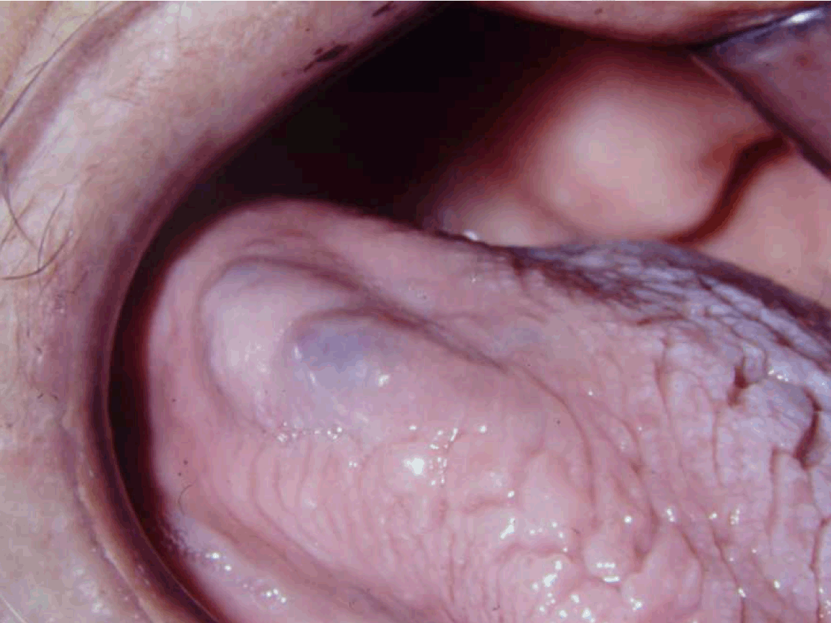
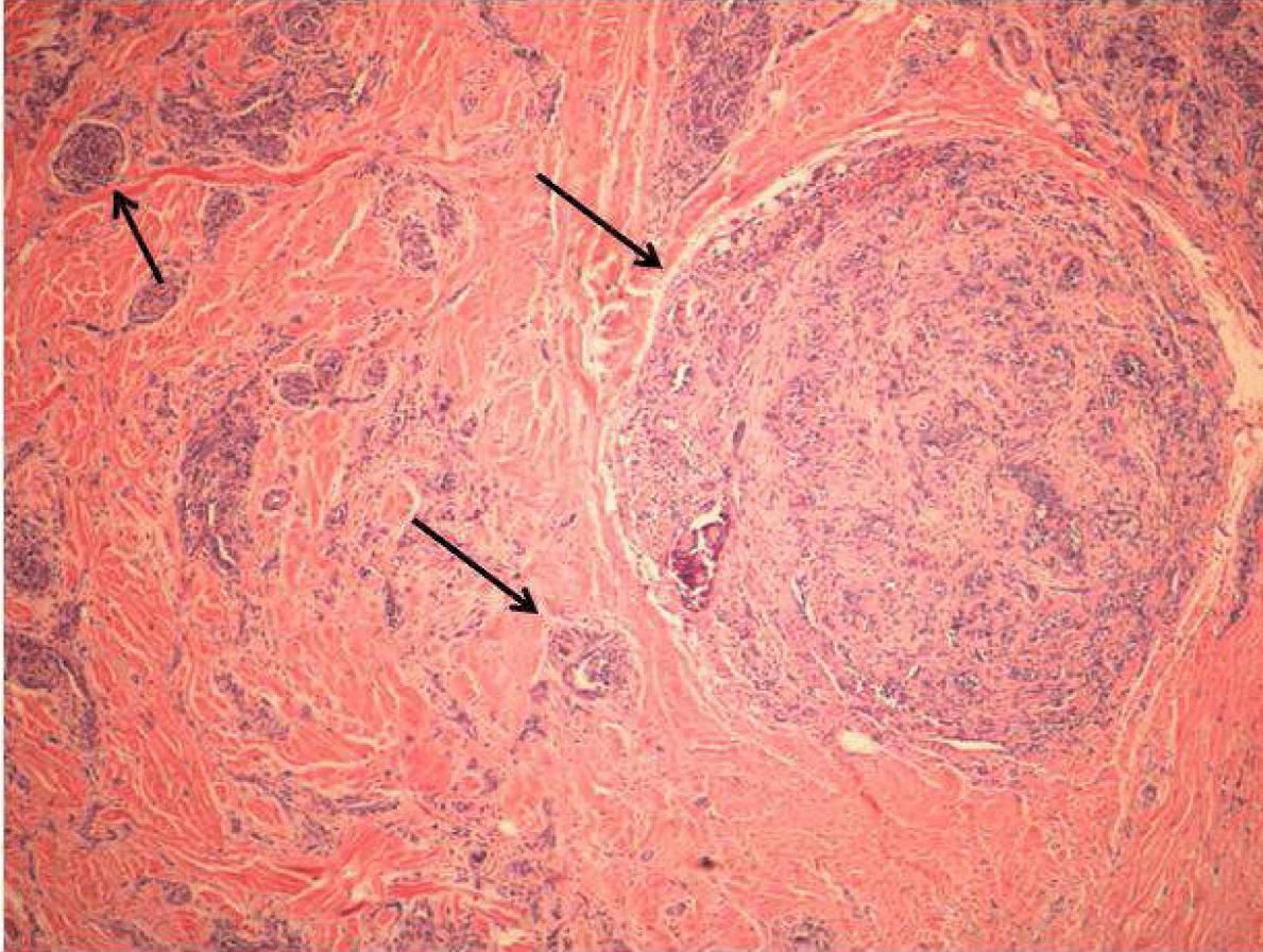
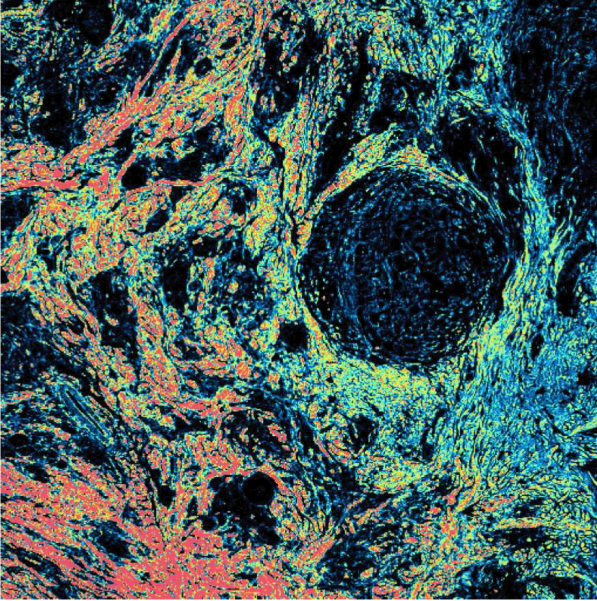
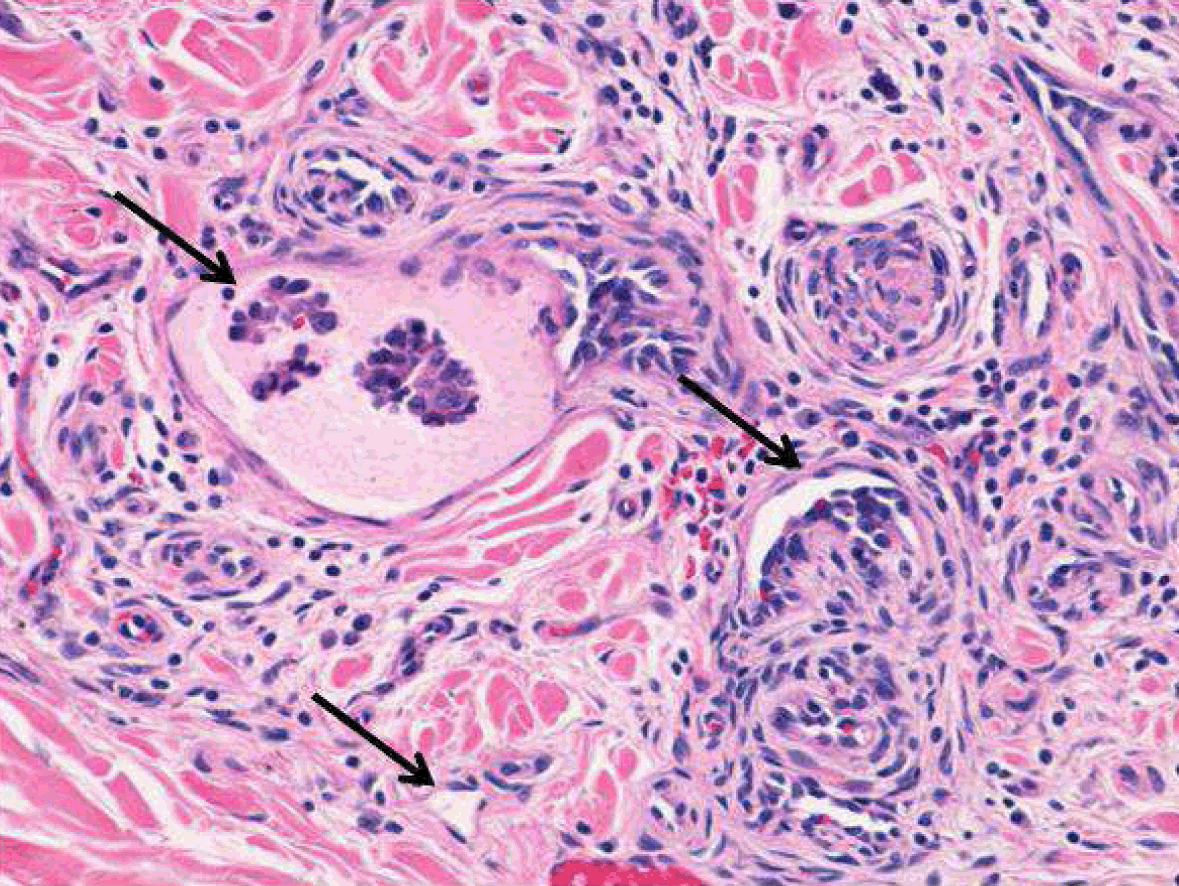
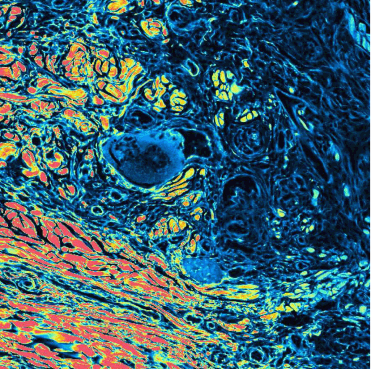
A 65-year-old male referred to the oral surgery unit of the University of Bari, presenting a multinodular painless exophytic mass on the right posterior margin of the tongue 4 cm in size. The patient's clinical history highlighted the rapid growth of the tongue nodule within two months on a pre-existing red plane lesion. The lesion was characterized by white or bluish overlying mucosa, and variable consistency in different areas, ranging from soft to soft-elastic (Figure 1). The regional lymph nodes were not enlarged. Computed tomography scan showed a lesion with endophytic pattern of growth, intermingling cystic and solid areas with irregular border, diffuse enhancement with contrast media and a depth extension of 2 cm. The clinical differential diagnosis had been made among benign soft tissues vascular tumor (such as hemangioma or lymphangioma), vascular low grade malignant tumor (Dabska tumor or endovascular papillary hemangioendothelioma), vascular high grade malignant tumor (angiosarcoma or Kaposi's sarcoma), vascular tumor-like lesions (intravascular papillary endothelial hyperplasia), cysts and cystic tumor of salivary gland (mucoepidermoid carcinoma), and cystic metastatic tumor. A definitive diagnosis could not be reached because of the clinical aspects of the lesion. Therefore, a fine-needle aspiration biopsy (FNAB) was performed. The specimen was formalin-fixed, paraffin-embedded, stained with hematoxylin-eosin, PAS, Van Gienson, and Picrosirius red and then sent to the Department of Pathological Anatomy of the University of Bari. The histological examination was carried out using a Nikon Eclipse E600 microscope (Nikon Corporation, Tokyo - Japan), equipped with Argon-ion and Helium-Neon lasers, emitting at 488 nm and 543 nm wavelengths, which allows both optical and confocal laser scanning analysis. The Nikon EZ C1 software (Nikon Corporation, ver. 2.10 Coord Automatisering) was used for bi-dimensional image processing. Under standard histological analysis, the tumor showed a submucosal location, with extension and involvement of superficial muscle layer of the tongue. It was composed by dilated, thin-walled vascular spaces resembling a cavernous lymphangioma and different size neoplastic nodules infiltrating the tongue intrinsic muscular tissues (Figure 2) and (Figure 3). The characteristic finding was the formation of prominent intraluminal papillary tufts with hyaline cores lined by hobnail endothelial cells. The endothelial cells had a pale or dark eosinophilic cytoplasm and round or ovoid nuclei. At high magnification it was possible to highlight, between atrophic remnants of intrinsic muscular tissues, ecstatic lymphatic vessels with endovascular papillary neoplastic proliferation associated with empty small and semilunar shaped vascular spaces, in more solid nodular worly shaped neoplastic cells. Absent or minimal cytological atypia and rare mitotic figures were seen. Peripherally, the tumor showed dilated anastomosing blood vessels. Confocal laser scanning microscopy (CLSM) analysis highlighted lack of fluorescence in muscle infiltration area, depending on the resumption of soft tissues collagen fibers. Moreover, CLSM showed the high fluorescence of intraluminal papillary core due to the presence of immature thin collagen fibers devoid of cross-links (Figure 4) and (Figure 5). Under general anesthesia, the surgeon performed radiofrequency wide local excision with intraoperative histological negative control of resection margins. No evidence of local recurrence or lymph nodes metastasis during the follow-up of three years. | ||||||
| ||||||
| ||||||
| ||||||
|
| ||||||
| ||||||
|
Discussion
| ||||||
|
Papillary intralymphatic angioendothelioma is a locally aggressive, rarely metastasizing vascular lesion characterized by lymphatic-like channels and papillary endothelial proliferation. This tumor is closely related to retiform hemangioendothelioma [1], also known as endovascular papillary angioendothelioma or Dabska tumor, it was described for the first time in 1969 by Maria Dabska, reporting six cases as childhood tumors in the skin and subcutis [2]. PILA has predilection for infants and children, although 25% of the cases occur in adulthood [3]. Male to female ratio is 1:1. PILAs appear as plaques or nodules with asymptomatic growth, are ill defined and usually involve the dermis and subcutaneous tissues. In literature, these tumors are thought to arise from different anatomical locations such as the spleen, tongue, testicles and bone [4][5]. Usually, they are diagnosed when they are 2–3 cm in size and they are more commonly diagnosed as hemangioma [6]. In this case report, Dabska tumor arise on the right posterior margin of the tongue presenting as multinodular painless exophytic mass 4 cm in size. The clinical diagnosis of this tumor is very difficult in view of its variable consistency and color, as well as its multinodular growth pattern. In this case here reported, the clinical differential diagnosis had been made among benign soft tissues vascular tumor, vascular low grade malignant tumor, vascular high grade malignant tumor, vascular tumor-like lesions, cysts and cystic tumor of salivary gland, and cystic metastatic tumor. Benign soft tissues vascular tumor, vascular tumor-like lesions and cysts of salivary gland are usually described as painless soft exophytic masses with bluish overlying mucosa, although generally in CT imaging they lack in endophytic pattern of growth, and solid areas with irregular border. Multinodular appearance, exophytic and endophytic patterns of growth, size, growth rate, depth extension and alternation of cystic and solid areas could prompt diagnoses of vascular low grade malignant tumor, vascular high grade malignant tumor, cystic tumor of salivary gland, or cystic metastasis, even though the lack of ulcers, pain, bleeding and lymph nodes involvement. Although PILAs may show different presentations depending on the analyzed area, FNAB or biopsy could be performed to achieve a certain diagnosis. Microscopically, PILAs are composed by endovascular papillary proliferations that project into dilated, thin-walled, lymphatic spaces [7]. At high-power view, they demonstrate a pathognomonic hobnail-like appearance [8] with scant pink cytoplasm and a prominent nucleus, with little or no cytological atypia [1]. Mitotic figures are rare [9]. Endothelial cells are immunoreactive for CD34, CD31, and factor VIII-related antigen [10]. Histologically, PILA should be distinguished from other lesions presenting endothelial papillary overgrowth and vasoproliferation such as intravascular papillary endothelial hyperplasia (IPEH) [9], hemangioma, epithelioid hemangioendothelioma [10], hemangiopericytoma, and angiosarcoma. Confocal laser scanning microscopy examination allow to evaluate the high fluorescence of intraluminal papillary core due to the presence of immature thin collagen fibers devoid of cross-links and lack of fluorescence in muscle infiltration area, depending on the resumption of soft tissues collagen fibers. Wide local excision is considered the treatment of choice for this low grade malignancy: the use of radiofrequency excision results in a reduction in bleeding and improved healing of tissues after surgery. Moreover, radiofrequency excision resets the postoperative edema, pain, and infection to zero. A tendency for local recurrence and lymph nodes metastasis was reported [1]. Nevertheless, a few cases of PILA have been reported thus far, and this suggests the need for a standardized clinicopathological approaches to achieve an accurate diagnosis and effective treatment, as clear-cut criteria for the differential diagnosis between PILA and PILA-like lesions are still lacking. CLSM analysis allows to achieve a certain histological diagnosis. | ||||||
|
Conclusion
| ||||||
|
Confocal laser scanning microscopy analysis could surely facilitate the histological diagnosis, allowing the identification of some features of the tumor. | ||||||
|
References
| ||||||
| ||||||
|
[HTML Abstract]
[PDF Full Text]
|
|
Author Contributions
Gianfranco Favia – Substantial contributions to conception and design, Acquisition of data, Analysis and interpretation of data, Drafting the article, Revising it critically for important intellectual content, Final approval of the version to be published Luisa Limongelli – Acquisition of data, Drafting the article, Revising it critically for important intellectual content, Final approval of the version to be published Angela Tempesta – Acquisition of data, Drafting the article, Revising it critically for important intellectual content, Final approval of the version to be published Eugenio Maiorano – Acquisition of data, Drafting the article, Revising it critically for important intellectual content, Final approval of the version to be published |
|
Guarantor of submission
The corresponding author is the guarantor of submission. |
|
Source of support
None |
|
Conflict of interest
Authors declare no conflict of interest. |
|
Copyright
© 2015 Gianfranco Favia et al. This article is distributed under the terms of Creative Commons Attribution License which permits unrestricted use, distribution and reproduction in any medium provided the original author(s) and original publisher are properly credited. Please see the copyright policy on the journal website for more information. |
|
|