  |

|
 |
|
Case Report
| ||||||
| Isolated neurocutaneous peripheral T cell lymphoma, NOS | ||||||
| Talal Hilal | ||||||
|
MD, Internal Medicine Resident, University of Kentucky, Lexington, KY, USA
| ||||||
| ||||||
|
[HTML Abstract]
[PDF Full Text]
[Print This Article]
[Similar article in Pumed] [Similar article in Google Scholar] 
|


| How to cite this article |
| Hilal T. Isolated neurocutaneous peripheral T cell lymphoma NOS. Int J Case Rep Images 2014;5(12):859–863. |
|
Abstract
|
|
Introduction:
Peripheral T cell lymphomas (PTCLs) are a heterogeneous group of diseases that are a relatively uncommon subtype of non-Hodgkin's lymphoma (NHL) with an overall poor prognosis. They are difficult to classify and targeted therapy does not exist. Patients are usually treated with B cell specific drugs that have not shown to be effective in most subtypes.
Case Report: A 52-year-old male presented with a one-week history of confusion. He was found to have diffuse subcutaneous lumps on his extremities, back, and thorax, and a large swelling over his left face. Imaging revealed a right frontal lobe mass. Biopsy of the mass confirmed PTCL. Further histopathological analysis and imaging for staging diagnosed the subtype as not otherwise specified (NOS). The patient received radiation therapy to the brain followed by systemic chemotherapy with cyclophosphamide, doxorubicin, vincristine, prednisone (CHOP). He responded by the end of the first cycle, but his long-term clinical course remains to be seen. Conclusion: Treatment for PTCL is a realm requiring further research with efforts focusing on the development of T cell specific drugs. | |
|
Keywords:
B-cell lymphoma, Lymphoma, Non-Hodgkin lymphoma, Peripheral T cell lymphoma
| |
|
Introduction
| ||||||
|
Peripheral T cell lymphomas (PTCLs) derive from post-thymic T cells and generally arise in lymphoid tissue "peripheral" to the thymus such lymph nodes, spleen, gastrointestinal tract and skin. They have a mature T cell phenotype and are grouped, along with mature natural killer lymphomas, according to clinical presentation as leukemic, nodal or extranodal [1]. This case describes an atypical presentation of PTCL, not otherwise specified (NOS), and reviews the classification and current treatment options for this uncommon disease. | ||||||
|
Case Report
| ||||||
|
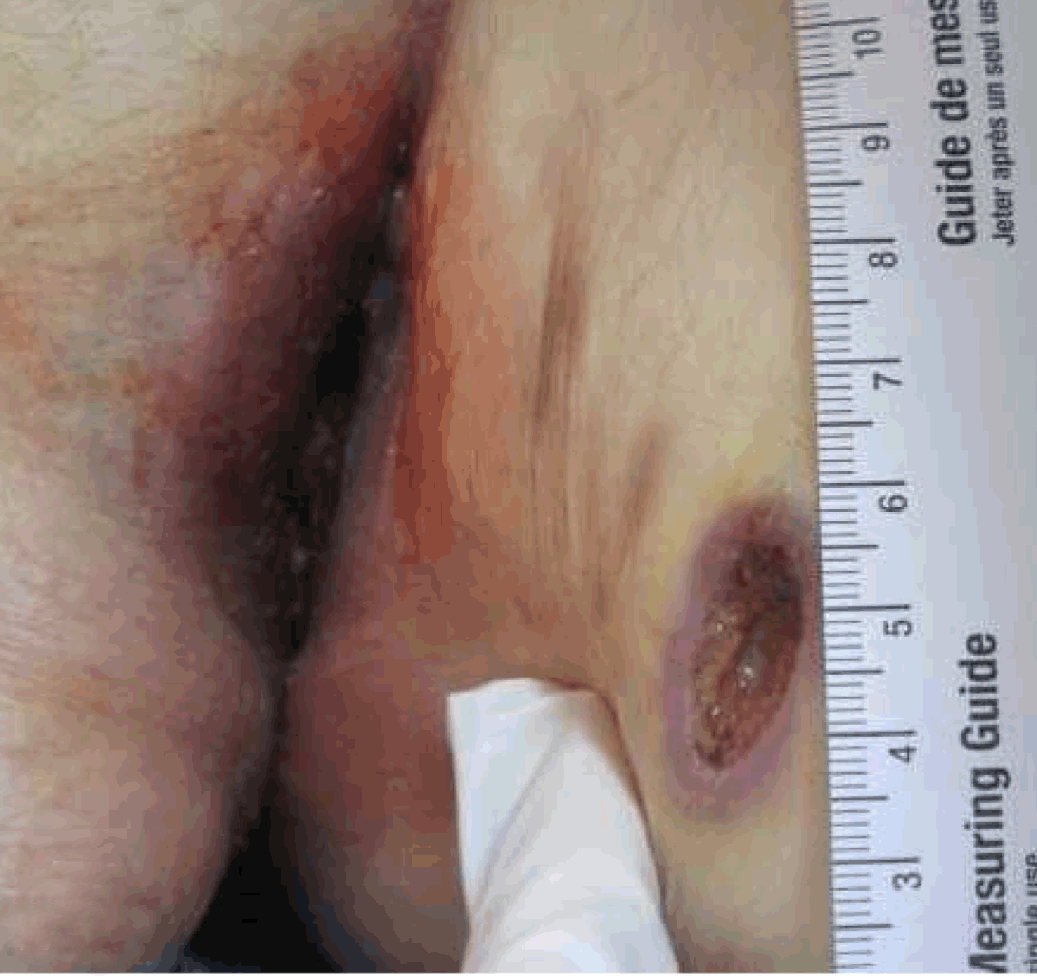
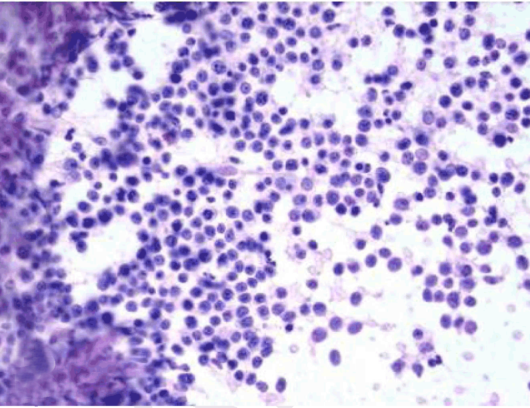
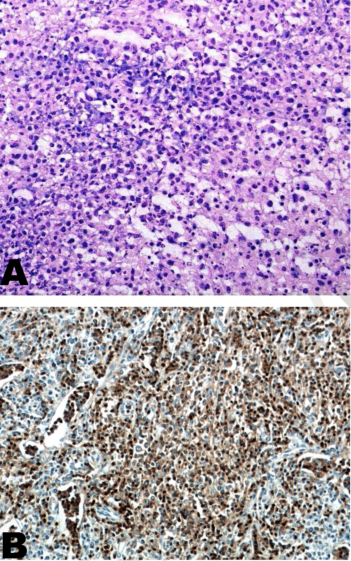
A 52-year-old male presented to the emergency department after being found confused at home. His family relayed that he had developed progressive left facial swelling and right scalp swelling over the past three months and had been becoming episodically confused over the past week. His past history was significant for poorly controlled type 2 diabetes mellitus, alcohol abuse and excision of a meningioma 15 years ago. On examination the patient was afebrile, his blood pressure 119/74 mmHg, heart rate 77 beats per minute, respiratory rate 18 breaths per minutes and his oxygen saturation was 97% on room air. He was drowsy and had delayed responses to questions. He had facial asymmetry with left sided swelling and facial droop. His scalp on the right side over the parietal area was swollen and mildly tender. Inspection of his oral cavity revealed a necrotic growth protruding from his soft palate. He had multiple diffuse violaceous lesions on his skin, some of which were ulcerated with central necrosis (Figure 1). He also had multiple firm, mildly tender nodules present on left forearm, right upper chest and several smaller nodules on upper back. Computed tomography (CT) scan revealed a right frontal mass. Further assessment with magnetic resonance imaging (MRI) of the brain showed a 4x3 cm enhancing mass in the right inferior frontal gyrus with hypercellullarity, hemorrhage and surrounding edema (Figure 2A-B). Initial assessment focused on histologic diagnosis and a biopsy of the soft palate growth and one of the skin lesions, both of which revealed features consistent with PTCL (Figure 3). Computed tomography scans of the chest, abdomen and pelvis showed no lymphadenopathy or evidence of primary neoplasm. Biopsy results of the brain mass were consistent with the same finding of PTCL (Figure 4A). Bone marrow biopsy and aspirate showed normal cellularity without involvement of lymphoma. Based on the pattern of involvement and histologic findings of CD3 (Figure 4B) and CD7 positivity with CD4, CD8, CD30, CD56 negativity, the patient's disease fit the subtype of PTCL, NOS. The patient initially received intavenous dexamethasone and emergent radiation therapy to the brain to decrease mass effect and risk for subfalcine herniation. He received a total of 10 days of radiation. During that period the lesions on his skin were ulcerating, the soft palate growth was causing pain and trouble swallowing and subcutaneous nodules were enlarging. We then decided to stop radiation therapy and start systemic chemotherapy using cyclophosphamide, doxorubicin, vincristine, and prednisone (CHOP). The patient responded within a few days and all his nodules disappeared by the end of the first of four cycles of therapy. | ||||||
| ||||||
| ||||||
| ||||||
| ||||||
|
Discussion
| ||||||
|
PTCLs are a group of lymphomas that fall under the category of mature T cell lymphomas, and are an uncommon subtype of non-Hodgkin's lymphoma (NHL). The International Peripheral T cell and Natural Killer/T cell lymphoma study reported that PTCLs accounted for only 5–10% of all NHL cases in Western countries and about 10–20% in Asian countries [2]. The disease tends to occur in adults with 40% of cases occurring between the ages of 55 and 74, and only about 5% of cases occurring after the age of 85. The incidence rates among all races in males and females are approximately 2.3 and 1.4 per 100,000 individuals, respectively, in contrast to 24 and 16.5 cases per 100,000 males and females for non-Hodgkin's lymphoma. The disease is almost twice as frequent in males than females [3] . The clinical presentation is variable and depends on the specific subtype of PTCL. Overall, patients present with more advanced disease and an increased incidence of B symptoms. Paraneoplastic features that are well described include eosinophilia, hemophagocytic syndrome, and autoimmune phenomena [4]. The incidence of brain metastasis in PTCLs is unknown with very few case reports demonstrating brain involvement at time of diagnosis and no cases that report isolated brain mass as the initial presentation of PTCL, NOS. Definitive diagnosis is based on examination of tissue biopsy specimen for histologic features supplemented by detailed immunohistochemistry, flow cytometry, cytogenetics, and molecular genetics [5]. Historically, PTCLs have been difficult to classify given the heterogeneity of the condition. The most widely used classification system currently is that of the World Health Organization (WHO) [6] (Table 1). The difficulty not only lies in diagnosing the subtype of PTCL, but also in treating it. Overall, the different subtypes all have a poor prognosis. The standard of care for all subtypes of PTCL has been combination chemotherapy using CHOP. This has been used based on trials that enrolled patients with diffuse large B cell lymphoma (DLBL) and has been shown to be effective only in the subtype anaplastic lymphoma kinase (ALK)-positive anaplastic large cell lymphoma (ALCL) [7] [8]. The outcomes of CHOP chemotherapy are variable. For PTCL, NOS, the reported complete response (CR) to treatment and overall survival (OS) is 50% and 30%, respectively [4]. Alternative first line therapies are currently under study with the focus being on the addition of a target-directed drug that takes on a role similar to that of rituximab in the treatment of mature B cell lymphomas. The addition of bortezomib, a selective proteosome 26S inhibitor, to CHOP has shown promising initial response rates in a phase II trial. The overall survival, however, was unchanged compared to CHOP alone [9] . Another agent, alemtuzumab, a anti-CD52 monoclonal antibody is currently being studied in phase III trials as an add-on to CHOP. The results have yet to be published. Another aspect of management is hematopoietic stem cell transplant (HSCT). Several studies have shown that first line high-dose chemotherapy with autologous HSCT had complete response rates ranging between 60–70% with the best studies showing a three-year survival ranging between 63–73%. Difficulties arise with toxicity and inability of patients to proceed with transplantation [10] [11]. Relapse is common with PTCLs and options at that point include salvage chemotherapy, autologous HSCT or allogeneic HSCT. Agents approved by the FDA for relapsed/refractory PTCL are pralatrexate, a folate analog, romidepsin, a histone deacytalse inhibitor and bretuximab vedotin (BV) used for ALCL. The first two showed response rates between 25–30%, a complete response between 10–15%. The overall survival for pralatrexate was 14 months and no overall survival was reported for romidepsin [12]. Autologous HSCT for relapsed disease showed poor results with a five-year overall survival <35% in most studies. Reduced intensity chemotherapy (RIC) followed by allogeneic HSCT shows promising results with problems arising from acute and chronic graft versus host disease (GvHD). Further prospective trials are warranted to address the role of RIC and allogeneic HSCT in PTCL, NOS [4]. | ||||||
| ||||||
|
| ||||||
|
Conclusion
| ||||||
|
Our understanding of the subcategory of peripheral T cell lymphomas (PTCLs) has improved over the years. What once was treated as a variant of mature B cell lymphomas is now seen under a different light. It is clear that patients who suffer from any of the PTCLs do worse than their mature B cell lymphoma counterparts. This understanding has led to further research into the molecular mechanisms that mediate disease. Clinical presentation is variable and histologic diagnosis is essential. Current efforts are focused on developing novel agents to target specific mutations that can alter the natural history of this poorly understood entity. | ||||||
|
Acknowledgements
| ||||||
|
Mr. Frank Davis for providing support with a concise literature review on the topic. Dr. Melissa Kesler, Dr. Janna Neltner and Zane Staubach, from the Department of Pathology their valuable help with the pathology slides. | ||||||
|
References
| ||||||
| ||||||


|
[HTML Abstract]
[PDF Full Text]
|
|
Author Contributions
Talal Hilal – Acquisition of data, Analysis and interpretation of data, Drafting the article, Final approval of the version to be published |
|
Guarantor of submission
The corresponding author is the guarantor of submission. |
|
Source of support
None |
|
Conflict of interest
Authors declare no conflict of interest. |
|
Copyright
© 2014 Talal Hilal. This article is distributed under the terms of Creative Commons Attribution License which permits unrestricted use, distribution and reproduction in any medium provided the original author(s) and original publisher are properly credited. Please see the copyright policy on the journal website for more information. |
|
|
|
About The Authors
| |||
| |||
