| |
|
|
|
Case Report
| ||||||
| Unusual imaging of pancreatic metastasis: A case report of tumor-to-tumor metastasis | ||||||
| Rossella Graziani1, Paola Spaggiari4, Silvia Carrara2, Giovanna Lo Bue1, Alessandro Zerbi3, Luca Balzarini1 | ||||||
|
1MD, Department of Radiology, Humanitas Clinical Reserch Hospital, Rozzano (Milan), Italy.
2MD, Department of Gastroenterology and Digestive Endoscopy, Humanitas Clinical Reserch Hospital, Rozzano (Milan), Italy. 3MD, Department of Surgery, Humanitas Clinical Reserch Hospital, Rozzano (Milan), Italy. 4Department of Pathology, Clinical Reserch Hospital, Rozzano (Milan), Italy. | ||||||
| ||||||
|
[HTML Abstract]
[PDF Full Text]
[Print This Article]
[Similar article in Pumed] [Similar article in Google Scholar] 
|
| How to cite this article |
| Graziani R, Spaggiari P, Carrara S, Lo Bue G, Zerbi A, Balzarini L. Unusual imaging of pancreatic metastasis: A case report of tumor-to-tumor metastasis. Int J Case Rep Images 2014;5(11):766–771. |
|
Abstract
|
|
Introduction:
Metastasis of one tumor to another tumor is a very rare and controversial phenomenon. Solitary renal cell carcinoma metastasis to a preexisting pancreatic endocrine tumor is distinctly uncommon. We report atypical imaging findings of pancreatic metastasis from renal cell carcinoma, due to tumor-to- tumor metastasis for presence of renal cell carcinoma metastasizing to a pancreatic endocrine tumor.
Case Report: A 78-year-old male suffering from mild anemia underwent to multidetector computed tomography scan showing renal cell carcinoma and solid-cystic pancreatic mass, both resectable, treated with right radical nephrectomy and spleno-distal pancreatectomy. Histopathology of the resected renal and pancreatic specimens confirmed a clear cells right renal cell carcinoma metastatic to endocrine neoplasm of pancreatic body-tail. We compared multidetector computed tomography scan findings and histopathological pancreatic specimen. The imaging finding of peripheral rim enhancement coincided in pancreatic pathologic specimen with presence of pancreatic endocrine tumor. The imaging finding of solid trabeculae inside the mass corresponded in pancreatic pathologic specimen to presence of pancreatic endocrine tumor mixed with lobules of typical renal carcinoma metastatic cells. Finally, the imaging finding of hypoenhancing central area of lesion coincided in pancreatic pathologic specimen with presence of large necrotic component. Conclusion: We describe an unusual multidetector computed tomography scan finding of renal cell carcinoma metastasizing to pancreatic endocrine tumor and emphasize the knowledge of rare phenomena of tumor-to-tumor metastasis. | |
|
Keywords:
Tumor-to-tumor metastasis, Pancreatic Endocrine Tumor, Renal cell carcinoma, Pancreatic metastasis
| |
|
Introduction
| ||||||
|
Renal cell carcinoma (RCC) metastases to the pancreas is especially rare. Their incidence in autopsy series has been reported as 1–3% in patients with primary RCC and their diagnosis is often radiological [1]. Early detection of RCC pancreatic metastases, frequently performed by multidetector computed tomography (MDCT) dut to the imaging pattern of hyperenhancing lesions allows for appropriate treatment and improved outcomes for metastatic disease [2] [3] [4]. Metastasis of one tumor to another tumor is a very rare phenomenon in which one tumor metastasizes into another tumor [5]. The aim of this case report is to describe atypical MDCT picture of pancreatic metastasis from RCC due to pancreatic endocrine tumor (PET) metastasized by renal cell carcinoma. We emphasize the knowledge of this rare phenomenon in order to avoid an incorrect imaging diagnosis and to planning a relevant treatment. | ||||||
|
Case Report
| ||||||
|
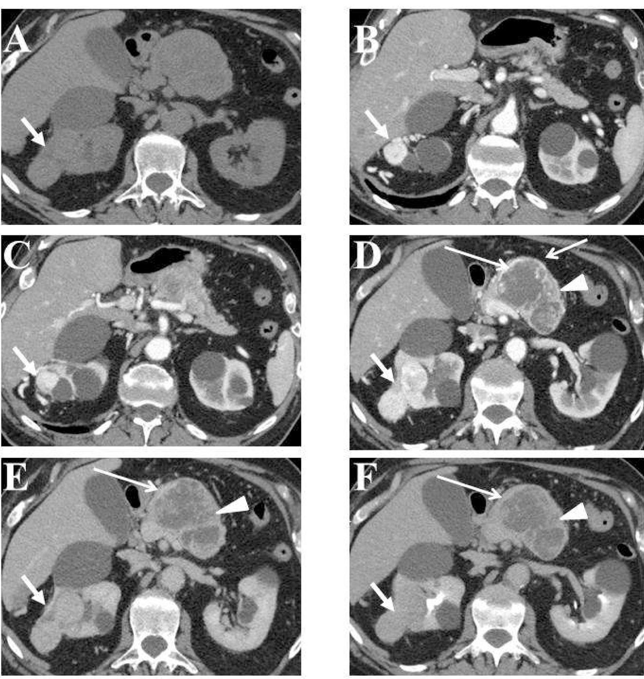
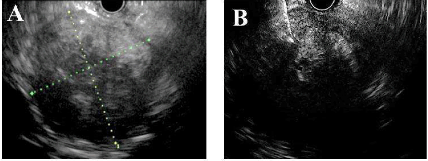
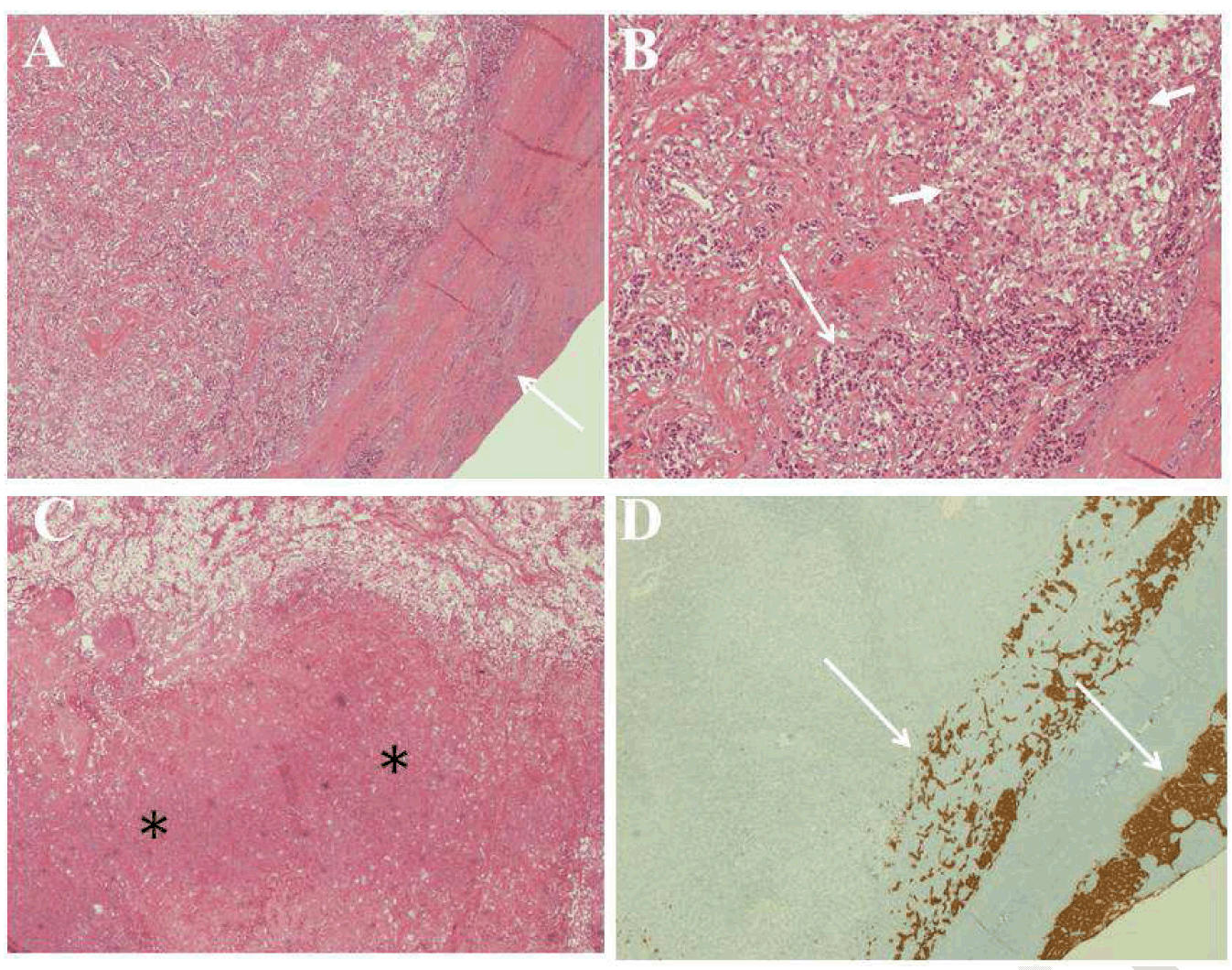
Asymptomatic 78-year-old male, non-smoker and non-drinker, with unremarkable past surgical history, was admitted to our hospital for occasional finding at check-up laboratory tests of persistent iron deficiency mild anemia during the last six months and ultrasound detection of pancreatic and renal masses. The physical examination was noncontributory. Laboratory investigations on admission showed a normal white blood cells count 10.0x10⁻⁹/L (reference range: 4.0–11.0x10⁻⁹/L) and a reduced serum level of hemoglobin 11.8 g/dL (reference range: 12–16 g/dL). His serum levels of lipase, amylase, CA19–9, liver enzymes and renal function tests were within the normal range. Moderate increased blood level of endocrine tumor markers was present with Chromogranin A of 115.87 U/L (reference range: 19–98 U/L) and NSE of 14.73 ng/mL (normal value inferior to 12.5 ng/mL). A 64-slice MDCT scan examination with quadriphasic study (pre-contrast enhanced, contrast enhanced pancreatic, venous and delayed phases) was performed. A contrast enhanced MDCT scan showed focal enlargement of pancreatic body due to the presence of large solid-cystic mass (Figure 1)), well-delimited but not encapsulated, measuring 80 mm in maximum diameter, mainly solid with some irregular, large, low-density central areas, suggesting presence of necrosis, and solid trabeculae inside central areas, hypoattenuating before contrast medium administration (Figure 1A). A thin peripheral rim enhancement was present during the pancreatic phase of contrast enhanced MDCT study (Figure 1D), showing wash-out in venous (Figure 1E) and late (Figure 1F) phases. The central areas and solid trabeculae of this mass remained hypoenhanced (Figure 1E-F) during all phases of MDCT study. The dilatation of upstream main pancreatic duct associated to parenchyma atrophy (Figure 1C) was present. There was no evidence of local invasion and peripancreatic vessels were preserved. A multinodular solid mass bulging from the upper pole of right kidney with maximum diameter of 60 mm was visible (Figure 1). This renal lesion was homogeneously hyperenhancing during pancreatic phase of examination (Figure 1B-D), showing wash-out during venous (Figure 1E) and late (Figure 1F) phases of MDCT study. Extra-renal involvement was absent and right renal vessels were preserved. There was no evidence of abdominal lymphadenopathy, free fluid or metastatic lesions in the liver and in left kidney. Computed tomography scan of the chest was normal. Endoscopic ultrasound (EUS) confirmed the presence of a well-demarcated solid-cystic mass of pancreatic body, hypoechoic with central fluid and hyperechoic areas, indicating intratumoral necrosis or hemorrhage (Figure 2A). Fine-needle aspiration biopsy endoscopic ultrasound guided (EUS-FNAB) of pancreatic lesion, performed using a 22-gauge needle (Figure 2B) revealed presence of malignant cells. Fine-needle aspiration ultrasound guided of one lesion bulging from the right renal upper pole revealed presence of malignant cells of RCC. The imaging findings were suggestive for presence of right kidney RCC associated with primary malignant lesion of pancreatic body without usual imaging pattern of pancreatic metastasis form renal cancer. All renal and pancreatic lesions were resectable. The patients underwent right radical nephrectomy and distal pancreatectomy. On gross pathologic examination, in the pancreatic specimen of resected body-tail, a well- circumscribed red and yellow variegated lesion measuring 6 cm in the greatest dimension was present. The lymph nodes identified separately were free of cancer, and the spleen was unremarkable. Histology of the pancreatic lesion showed two different cells population. In peripheral portion of the mass (Figure 3A) pancreatic endocrine tumor (PET) cells were exclusively observed, infiltrating even solid trabeculae inside pancreatic lesion, mixed with lobules of RCC composed of clear cells (Figure 3B). Large areas of necrosis were found in central portion of the mass, separated by solid trabeculae (Figure 3C). Immunohistochemistry confirmed the histologic picture: CD10 immunoperoxidase showed staining of RCC with no uptake of stain by PEN and synaptophysin immunoperoxidase of an adjacent section demonstrated of PEN only (Figure 3D), which was also positive at Chromogranin A and CD56 staining. PET resulted NET G1 according to WHO classification (2010), with Ki 67 of 1% and pT2N0 according TNM stage. This histological picture can be entirely consistent with a pancreatic endocrine neoplasm surrounding a well-defined nodule of metastatic RCC, representing an unusual case of RCC metastatic to a pancreatic endocrine neoplasm. On gross pathologic examination, in the renal specimen of resected right kidney a multinodular, circumscribed and exophytic lesion measuring 7 cm in the greatest dimension was present in the upper pole. Histopathology of the resected renal specimen confirmed a renal cell carcinoma of the kidney, composed mainly of clear cells. The TNM stage was pT3b, pNx, pM1. Adjuvant therapy were recommended after surgery but the patient declined. Follow-up with physical examination, laboratory tests, thoracic and abdominal MDCT scan were done every six months. The patient remain without evidence of disease 12 months from the original diagnosis. | ||||||
|
| ||||||
| ||||||
|
| ||||||
|
Discussion
| ||||||
|
Metastasis of one tumor to another tumor is a very rare phenomenon. The criteria for satisfying a true tumor-to-tumor metastasis are as follows [5] [6]:
This case report showed two distinct neoplasms and histologic evidence of encasement of an RCC by a PET. The comprehensive criteria that must be fulfilled for the diagnosis of a true tumor-to-tumor metastasis were present in our patient. Several authors have reported in literature lung cancer is the most common donor tumor, whereas RCC is the most common recipient [5] [6] [7] [8] [9] . The reason for tumor-to-tumor metastasis favoring specific tumors is still unknown. The RCC's rich vascularity, high content of glycogen and lipid, tendency to be localized without infiltration or metastasis could explain its favorable environment for receiving metastases from other cancers [6] [7] [9]. A solitary RCC metastasis to a preexisting pancreatic endocrine tumor (PET) is very uncommon. It is known in literature that PET are frequently hypervascular neoplasms. Matsukuma investigating 47 autopsy cases of lung cancer concomitant with other tumors found tumor-to-tumor metastasis in only one pancreatic endocrine microadenoma [7]. Cenkowski first described one case of RCC metastasizing to a preexisting PET, reporting MDCT and histopathologic findings [10]. In both cases, reported by Cenkowski and in our patient, MDCT findings of pancreatic lesion due to tumor-to-tumor metastasis from RCC are different from MDCT typical picture of pancreatic metastasis from RCC, which appears as enhancing lesions [1] [2], reflecting hypervascularity of the of primary tumor . We found a well-delimited solid-cystic mass, with MDCT peripheral rim enhancement, low-density central areas and solid trabeculae, both hypoehnancing after contrast medium administration during pancreatic phase of MDCT study. We assessed MDCT imaging findings and histopathological pancreatic specimen, comparing them. We have found that MDCT imaging finding of peripheral rim enhancement coincided in pancreatic pathologic specimen with presence of pancreatic endocrine tumor. The imaging finding of solid trabeculae inside the mass corresponded in pathologic specimen to the presence of pancreatic endocrine tumor mixed with lobules of typical renal carcinoma metastatic cells. Finally, the MDCT finding of hypoenhancing central area of lesion coincided in pathologic specimen with presence of large necrotic component. Early detection of metastases to the pancreas allows for appropriate treatment and improved outcomes of disease. In patients with pancreatic metastases from RCC, absence of extrapancreatic metastases and limited vascular involvement, 2 and 5 years survival rates of 78% and 65%, respectively after resection of pancreatic disease are reported [3] [4]. In our patients, radical nephrectomy and distal pancreatectomy were performed without complications. Clinical, laboratory, imaging follow-up after one year are all negative. However, the true prognosis of tumor-to-tumor metastasis remains unknown because this phenomenon is rare and most of the articles in literature about this disease are sporadic cases reports. | ||||||
|
Conclusion
| ||||||
|
A knowledge of rare phenomenon of RCC metastasizing to a preexisting pancreatic endocrine tumor is useful to avoid an incorrect diagnosis in the presence of unusual imaging findings in the pancreatic metastasis from renal cancer. On the basis of these reports, the mechanisms for RCC to pancreatic endocrine tumor specific metastasis, as well as correct treatment and prognosis of this rare disease may be elucidated in the future. | ||||||
|
References
| ||||||
| ||||||
|
[HTML Abstract]
[PDF Full Text]
|
|
Author Contributions
Rossella Graziani – Substantial contributions to conception and design, Acquisition of data, Analysis and interpretation of data, Drafting the article, Revising it critically for important intellectual content, Final approval of the version to be published Paola Spaggiari – Analysis and interpretation of data, Revising it critically for important intellectual content, Final approval of the version to be published Silvia Carrara – Analysis and interpretation of data, Revising it critically for important intellectual content, Final approval of the version to be published Giovanna Lo Bue – Analysis and interpretation of data, Revising it critically for important intellectual content, Final approval of the version to be published Alessandro Zerbi – Analysis and interpretation of data, Revising it critically for important intellectual content, Final approval of the version to be published Luca Balzarini – Analysis and interpretation of data, Revising it critically for important intellectual content, Final approval of the version to be published |
|
Guarantor of submission
The corresponding author is the guarantor of submission. |
|
Source of support
None |
|
Conflict of interest
Authors declare no conflict of interest. |
|
Copyright
© 2014 Rossella Graziani et al. This article is distributed under the terms of Creative Commons Attribution License which permits unrestricted use, distribution and reproduction in any medium provided the original author(s) and original publisher are properly credited. Please see the copyright policy on the journal website for more information. |
|
|
|
About The Authors
| |||
| |||
| |||
| |||
| |||
| |||
| |||