| |
|
|
|
Case Report
| ||||||
| Alobar holoprosencephaly with unfused thalami: A rare variety of holoprosencephaly | ||||||
| Abubakar A.1, Ibrahim S.M.2, Ahidjo A.1, Tahir A.1 | ||||||
|
1Department of Radiology, University of Maiduguri Teaching Hospital, Maiduguri, Nigeria.
2Department of Obstetrics and Gynecology University of Maiduguri Teaching Hospital, Maiduguri, Nigeria. | ||||||
| ||||||
|
[HTML Abstract]
[PDF Full Text]
[Print This Article]
[Similar article in Pumed] [Similar article in Google Scholar] 
|
| How to cite this article |
| Abubakar A, Ibrahim SM, Ahidjo A, Tahir A. Alobar holoprosencephaly with unfused thalami: A rare variety of holoprosencephaly. Int J Case Rep Images 2014;5(11):756–760. |
|
Abstract
|
|
Introduction:
Holoprosencephaly with unfused thalami is a rare malformation involving the forebrain and the face. The epidemiology of the disease is poorly known due to paucity of population based studies.
Case Report: A 32-year-old grand multipara at 27th week gestation found on routine ultrasound examination to have a single live fetus with the fetal head showing dilated single cerebral ventricle, with no evidence of anterior midline echo (falx, inter hemispheric cistern and septum pellucidum). The thalami appear relatively small but not fused with a thin midline linear echoic septum separating them. Two subsequent sonograms at 30th and 33rd weeks of pregnancy, including coronal sonograms of the fetal head, correctly identified a dilated single cerebral ventricle. There was no history of diabetes mellitus, hypertension or previously affected child. Pregnancy termination was done on the couple's request, because of the poor fetal prognosis. Postmortem clinical examination revealed a female newborn with normal body structure. The couple declined consent for autopsy. Conclusion: Alobar holoprosencephaly with unfused thalami is a rare and severe variety of holoprosencephaly with poorly understood aetiology and poor prognosis. | |
|
Keywords:
Alobar holoprosencephaly, Unfused thalami, Pregnancy termination
| |
|
Introduction
| ||||||
|
Holoprosencephaly, the most common malformation of the forebrain in humans, is a structural anomaly of the brain resulting from failed or incomplete forebrain division in the third to fourth weeks of gestation [1] [2]. Its incidence is estimated to be 1 in 16,000 live births and 1 in 250 spontaneous abortions and with a prevalence of 1:250 in embryos [2] and approximately 1:10,000 among live-born infants [3] [4] [5]. Holoprosencephaly with unfused thalami is very rare. The epidemiology of the disease is poorly known due to the paucity of population based studies [1] [6]. Environmental, mechanical, and genetic factors have been mentioned as possible causes [1] [6]. Such factors include chromosome aneuploidy, structural abnormality, autosomal recessive and dominant syndromes and maternal diabetes [4]. The role of maternal and paternal ages, parental consanguinity, maternal smoking and drinking habits is controversial [3]. The imaging study of choice in prenatal assessment of holoprosencephaly includes meticulous transvaginal or transabdominal ultrasound and Magnetic resonance imaging (MRI) scan. However, with continued refinement in ultrasonic imaging devices, it is now assuming an increasingly important role in the diagnosis [7] [8] . Three distinct types of holoprosencephaly have been documented in the literature, which include lobar, semilobar and alobar. The latter is the severest with characteristic intracranial findings of monoventricular cavity, fused thalami, and absence of midline structures such as the corpus callosum and falx cerebri [7]. However, the alobar variety with unfused thalami is rare, which informed this presentation. | ||||||
|
Case Report
| ||||||
|
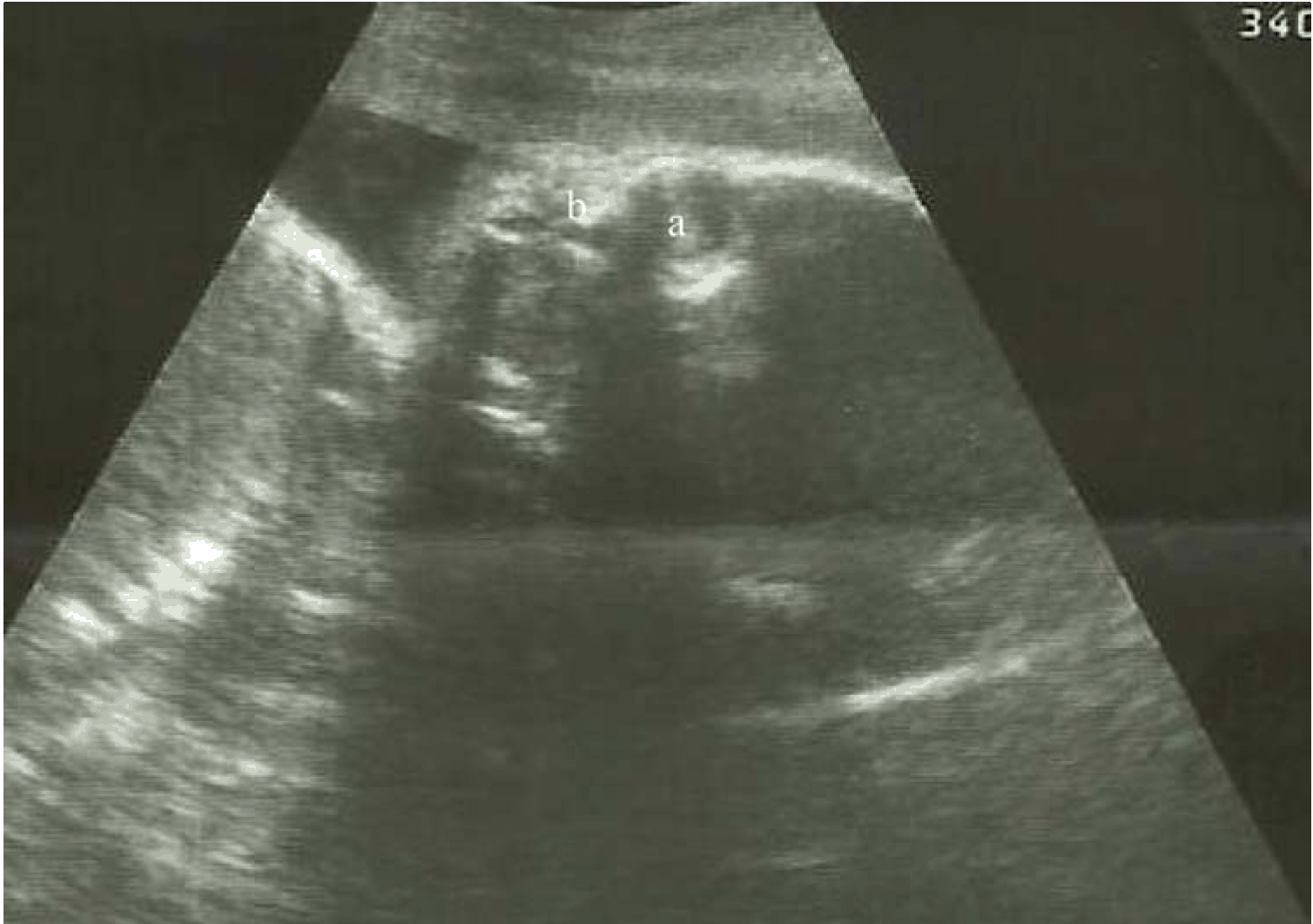
A 32-year-old gravida 6 para 5+0 female referred at 27th week gestation, for routine antenatal ultrasonography. There were no symptoms or signs suggestive of any clinical condition. The patient had no history of diabetes mellitus, hypertension or previously affected child. General physical examination was unremarkable. The uterus was gravid with symphysiofundal height of 29 cm. There was singleton fetus in cephalic presentation. The fetal heart sound was 110 beats per minute. Sonographic examination of the gravid uterus revealed a single live fetus. Axial sonograms of the fetal head (Figure 1) and (Figure 2) showed dilated single cerebral ventricle, with no evidence of anterior midline echo (falx, inter hemispheric cistern and septum pellucidum). The thalami appear relatively small but not fused; with a thin midline linear echoic septum separating them. It protrudes into the single ventricular cavity. The third ventricle was not visualized. The spine and cranium appeared to be well formed. Biparietal diameter, head circumference, abdominal circumference, and femur length were all consistent with the clinically estimated gestational age of 27 weeks. The interorbital distance was 1.3 cm; no evidence of facial anomalies was noted sonographically (Figure 3). The amniotic fluid index was normal. Two subsequent sonograms at 30 and 33 weeks of pregnancy, including coronal sonograms of the fetal head, correctly identified a dilated single cerebral ventricle. The couple requested for the termination of pregnancy because of the poor fetal prognosis. This was performed by administration of vaginal prostaglandin (misoprostol). Postmortem clinical examination revealed a female newborn with normal body structure. The couple declined consent for autopsy. | ||||||
| ||||||
| ||||||
| ||||||
|
Discussion
| ||||||
|
Alobar holoprosencephaly with unfused thalami is a very rare congenital malformation of intracranial and midfacial structures, which may be part of a Smith-Lemli-Opitz syndrome. It may be also an isolated finding or may occur in combination with other extra cephalic defects [8]. Although autopsy and maternal genetic testing were not conducted in the case cited above, prenatal history, detailed family history and focused physical examination of the parents and stillborn to identify microform of holoprosencephaly showed no definitive link to any syndrome or extra cerebral anomalies. The average gestational age at diagnosis is 21.9 weeks (range, 10.5–32.3 weeks) in most literature and in our patient the fetal age is about 27 weeks, which is relatively late, but this can be explained by delay in assessing the hospital by the patient [9]. The spectrum of prenatal cranial ultrasonographic anomalies in the three forms of holoprosencephaly include monoventricle with fused thalamus and corpus striatum, absent corpus callosum, fornix, and falx in alobar form. The features in semilobar include monoventricle with rudimentary occipital horns, falx, and interhemispheric fissure and fused thalamus and basal ganglia. The lobar form is characterized by separated lateral ventricle, absent septum pellucidum; the basal ganglia and thalamus may be fused or separated and the corpus callosum may or may not be present [7] [8] [10]. The prenatal axial fetal head sonogram of the index case showed dilated single cerebral ventricle, with no evidence of anterior midline echo (falx, interhemispheric cistern and septum pellucidum) which is consistent with the alobar holoprosencephaly. However, the thalami appear relatively small but not fused with a thin midline linear echoic septum separating them. This finding has only been reported in a few literatures [10] [11] [12]. Greene et al. proposed the use of two criteria for the prenatal sonographic diagnosis of alobar holoprosencephaly [10]. First, the intracranial criterion of a large central fluid collection in the fetal head, with no visible midline structures but with the presence of a mantle around the fluid collection and fusion of the thalami and corpus striatum. Second, sonographic facial abnormalities including hypotelorism, central clefts, facial asymmetry and abnormal orbits [10]. Similarly, Chervenak et al. considered that both hypotelorism and absence of the midline should be observed sonographically to diagnose holoprosencephaly with certainty [11]. The same criteria were used by Parant et al. [12]. In the present case, the first of these criteria was satisfied but no convincing evidence of the second criteria was noted. Demyers and other researchers have also found reasonable percentage of patients with a lobar prosencephaly with a normal face, which is in keeping with our patient [6] [13] [14]. Nerberg et al. had demonstrated that the absence of falx and fusion of the thalami are independent diagnostic features of alobar and semilobar holoprosencephaly irrespective of facial feature [[15]. Wenghoefer et al. found that exact prenatal assignation of holoprosencephaly into lobar, alobar, and semilobar was inconsistent in 41% of the cases and the ultrasound diagnosis was not confirmed in 19% of the cases [9]. The ongoing improvement of ultrasound equipment is not very likely to improve the ultrasound diagnosis but will allow for a reduction in the number of cases with a wrong assignment to the three groups. Distinguishing the alobar holoprosencephaly from other causes of large intracranial fluid collections such as semilobar holoprosencephaly, ventriculomegaly, hydranencephaly, and large Dandy-Walker cyst is very challenging but may not help in the fetal outcome because all of these malformations have a poor prognosis [9] [15] [16]. | ||||||
|
Conclusion
| ||||||
|
Alobar holoprosencephaly with unfused thalami is a rare and severe variety of holoprosencephaly with poorly understood aetiology and poor prognosis. During routine obstetric sonographic examination, attention should be paid to excluding possible malformations. | ||||||
|
References
| ||||||
| ||||||
|
[HTML Abstract]
[PDF Full Text]
|
|
Author Contributions
Abubakar A. – Substantial contributions to conception and design, Acquisition of data, Analysis and interpretation of data, Drafting the article, Revising it critically for important intellectual content, Final approval of the version to be published Ibrahim S.M. – Substantial contributions to conception and design, Revising it critically for important intellectual content, Final approval of the version to be published Ahidjo A. – Substantial contributions to conception and design, Revising it critically for important intellectual content, Final approval of the version to be published Tahir A. – Substantial contributions to conception and design, Revising it critically for important intellectual content, Final approval of the version to be published |
|
Guarantor of submission
The corresponding author is the guarantor of submission. |
|
Source of support
None |
|
Conflict of interest
Authors declare no conflict of interest. |
|
Copyright
© 2014 Abubakar A. et al. This article is distributed under the terms of Creative Commons Attribution License which permits unrestricted use, distribution and reproduction in any medium provided the original author(s) and original publisher are properly credited. Please see the copyright policy on the journal website for more information. |
|
|
|
About The Authors
| |||
| |||
| |||
| |||
| |||