| |
|
|
|
Case Series
| ||||||
| Proliferative vitreoretinopathy secondary to degenerative retinoschisis: A case series | ||||||
| Mansvkhlal Amer1, Safia Ahmed2, Carpi Olali3, Sohail Ahmed4, Gupta M5 | ||||||
|
1Junior Doctor in Ophthalmology, Pilgrim Hospital, Boston. United Kingdom.
2Junior Doctor in Ophthalmology, Royal Blackburn Hospital Blackburn, United Kingdom. 3Ophthalmic Surgeon, Pilgrim Hospital, Boston, United Kingdom, Special Interest: Glaucoma and Neuro-Ophthalmology. 4Consultant Ophthalmic Surgeon, Pilgrim Hospital, Boston, United Kingdom, Special Interest: Strabismus and Vitreoretinal Surgery. 5Consultant Ophthalmic Surgeon, Pilgrim Hospital, Boston, United Kingdom, Special Interest: Glaucoma and Cataract. | ||||||
| ||||||
|
[HTML Abstract]
[PDF Full Text]
[Print This Article]
[Similar article in Pumed] [Similar article in Google Scholar] 
|
| How to cite this article |
| Amer M, Ahmed S, Olali C, Ahmed S, Gupta M. Proliferative vitreoretinopathy secondary to degenerative retinoschisis: A case series. International Journal of Case Reports and Images 2014;5(5):329–333. |
|
Abstract
|
|
Introduction:
We report two cases proliferative vitreoretinopathy that occurred as complication of degenerative retinoschisis.
Case Series: Two patients; a male and a female with vitreo-retinal hemorrhage seen in our clinic were evaluated and retinoschisis found as the only cause of the bleeding. Both were treated with Argon barrier photocoagulation which arrested the hemorrhage and resolved the visual symptoms. Conclusion: It has been suggested that retinal ischemia occurs in retinoschisis and this triggers upregulation of growth factors and subsequent formation of new vessels. This hypothesis would need to be tested to ascertain the particular growth factors, that trigger this process and if other cytokines and interleukins also play a role as the process is a vitreoretinpathy. | |
|
Keywords:
Retinoschisis, Vitreous hemorrhage, Growth factors, Vitreoretinopathy
| |
|
Introduction
| ||||||
|
Degenerative retinoschisis rarely present with symptoms because of their peripheral location. [1] Sometimes, however, advanced cases may present with a large peripheral visual field defect corresponding to the area of retinoschisis and in some cases, it could be confused with retinal detachment by the referring individual. [1] [2] Patients who present with vitreous haemorrhage related symptoms on the other hand usually have vitreo-proliferative conditions such as diabetes mellitus, retinal vascular occlusion, hemoglobinpathies, coagulopathies or tumors. [3] [4] [5] We present two cases of retinoschisis who had ocular symptoms related to vitreous hemorrhage which occurred as a complication of the retinoschisis only and the treatment we instituted to arrest this complication. | ||||||
|
Case Series
| ||||||
|
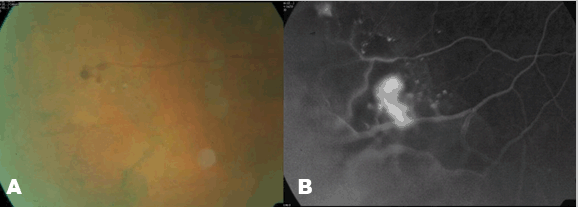
Case 1: A 58-year-old male presented with a week history of triangle-like floater in his right eye. His vision was otherwise normal and there was no medical history of significance. His corrected visual acuity in both eyes was normal at 20/15 in the right and 20/20 in the left eye. Dilated fundoscopy revealed what looked like three closely spaced macroaneurysms in the inferior-temporal periphery with retinoschises adjacent to the macroaneurysms in the right eye. (Figure 1A) The neovessels were located in the inner layer of the retinoschisis. Retinochisis was also found in the fellow eye located inferiorly, but there was no associated neovascularisation. He was kept on annual follow up, but two years after the initial diagnosis, developed proliferative vitreoretinopathy with new vessels and some fibrous tissue at the edge of the schises in the right eye. Fluorescein fundus angiography showed COATS- like disease. (Figure 1B) Clinically, there was progression of neovascularisation and glial tissue proliferation along the retinschises. There were some vascular anomalies noted in the inner layer of the retinoschisis, but no holes in either layer. A few months later, repeat retinal angiograms showed areas of non-perfusion in and around the retinoschisis with hyperflourescence of the new vessels. He had barrier argon laser photocoagulation and has since remained stable with normal vision and no vitreous hemorrhage. | ||||||
| ||||||
|
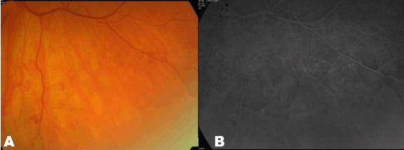
Case 2: A 60 year old female patient was referred because of bleeding in the lower mid-peripheral retina of the left eye noticed by an optician during routine visit. She had had no ocular problems but was on treatment for hypertension. Her vision with correction was normal at 20/25 in the right eye and 20/20 in the left eye. Dilated fundoscopy, revealed bilateral retinoschisis located inferior-temporally in the right eye and inferiorly in the left. There was evidence of neovascularization in the inner layer of the retinoschisis associated with small vitreous hemorrhage in the left eye. (Figure 2A) The fluorescein angiography showed hypoperfusion in the area of the retinoschisis in the left eye and hyperflourescence in the location of the neovessels. (Figure 2B) In order to prevent further vitreous hemorrhage, the patient underwent barrier argon laser photocoagulation. The patient has remained symptom free with no further vitreous hemorrhage in the last 18 months after the laser treatment. | ||||||
| ||||||
|
Discussion
| ||||||
|
Degenerative retinoschisis affects about 5–7% of the adult population and are usually incidental findings in clinical settings. [6] Indeed, in Bayer's [2] studies of 108 cases of retinoschisis, none had symptoms primarily due to the disease. Complications however, can sometimes result in retinal detachment or rarely vitreous hemorrhage producing symptoms. These are usually limited to the reticular (bullous and cyst in nerve fibre layer) form rather than the typical (flat and cyst in outer plexiform layer) form of the disease. Retinoschisis is responsible for less than 2.5% of all rhegmatogenous retinal detachments and the presence of holes in both outer and inner layers of the retina is the greatest risk factor as this can allow a large amount of fluid from the vitreous cavity to migrate into the potential space, resulting in the detachment. [7] [8] [9] Vitreous hemorrhage resulting from retinal neovascularisation as seen in our two cases is rare. Campo [10] and colleagues using retinal fluorescein fundus angiography demonstrated that the neovascularization was on the surface of the cyst and this accounted for the vitreous hemorrhage. They also suggested that the neovascularization in bullous retinoschisis may occur because of hypoxia induced by the combination of chronic retinal elevation and capillary non-perfusion. Growth factors are known to mediate various inflammatory and angiogenic processes in biologic systems and of particular importance is vascular endothelial growth factor (VEGF) because of its clinical relevance. In the eye, the potent angiogenic factor VEGF has since been recognized to be produced by many retinal cells such as endothelial cells, pericytes, retinal pigment epithelial cells, Muller cells and astrocytes. [11] [12] VEGF is upregulated in retinal ischaemic conditions such as diabetic retinopathy, retinal vein occlusion, rubeosis irides, and retinopathy of prematurtiy and is recognized as the main cytokine inducing the retinal neovascularization that results. [13] [14] Although it has five isoforms (121, 145, 165, 189, and 126), the 126 is the predominant. It has high affinity for the 180-kDa fms-like tyrosine kinase (also called FLt-1 or VEGFR1) and 200-kDa kinase insert domain-containing receptor (KDR) also known as fetal liver kinase Flk-1 or VEGFR2. KDR transduces the signals for endothelial proliferation and chemotaxis. [15] Hypoxia stimulates VEGF mRNA expression through binding of Hypoxia-inducible factor 1 alpha subunit (HIF - α) to consensus and ancillary hypoxia - response elements (HREs) in the VEGF promoter and once expressed, VEGF stimulates endothelial cell proliferation and neovascularisation via the mitogen-activated protein kinases (MAPK) dependent pathway. [16] [17] VEGF may also promote endothelial cell migration and vascular permeability with the resulting vessel leakage leading to interstitial edema and more hypoxia which in turn stimulates further VEGF production. [18] It is thought that VEGF promotes vascular leakage by several mechanism including endothelial cell fenestrations, damage to tight junctions and the upregulation of the expression of Intercellular Adhesion Molecule 1 (ICAM 1) [19] [20] (This protein is encoded by the ICAM 1 gene which encodes a cell surface glycoprotein that is typically expressed on endothelial and leukocyte-associated transmembrane protein). Kon and colleagues [21] demonstrated high levels of interleukin 6 (IL-6) and protein in patients who had vitrectomy as part of the retinal detachment surgical procedures and suggested that these may play a role in the pathobiology of proliferative vitreoretinopathy (PVR). The role of platelet derived growth factor C (PDGF-C) in pathological angiogenesis independent of VEGF was shown by Li and colleagues in their study. [22] PDGF-C as they reported may have effect on vascular cells, tissue stroma fibroblasts and macrophages among others and have therefore been described as pleiotropic and versatile with potential for angiogeneic therapy. It is known that in the presence of a retinal hole, the PVR could be explained as the vitreous humour contains a number of cytokines including TNFα, TGFβ2, PDGF and interleukins; which when in contact with the RPE could stimulate RPE migration (and proliferation) and angiogenesis. In our two cases, no hole was seen in either side of the retinoschisis and therefore this may not be the reason for the neovascularization. Rather, retinal ischemia as earlier suggested may be the trigger for the upregulation of the growth factors and subsequent formation of new vessels. This hypothesis would, however, need to be tested to ascertain the particular growth factor (s) that triggers this process and if other cytokines and interleukins also play a role as the process is a vitreoretinopathy. Also, in proliferative retinopathies, VEGF dependent angiogenesis can be induced by inflammation, alteration of shear stress on blood vessels, and glycosylation of proteins; mechanisms that are independent of retinal hypoxia. [23] [24] In retinoschisis, the splitting of the retina results in the retinal vessels attached to the inner layer floating freely in the vitreous cavity and being subjected to tractional forces, with eye movements. This could exhibit and alter the shear stress on these blood vessels and thereby result in upregulation of VEGF, with subsequent angiogenesis once the critical level of the cytokine is attained. Our two patients responded to Argon laser treatment. As demonstrated by Lip et al. [14] as well as Aiello et al. [25] VEGF levels reduced markedly following laser treatment in their studies of patients with proliferative diabetic retinopathy. The effect of argon laser treatment on the level of any mediator found in retinschisis related vitreoretinpathy would equally be a significant revelation. | ||||||
|
Conclusion
| ||||||
|
Retinal ischemia, hypoxia and subsequent neovascularization is a cascade mediated by cytokines of which VEGF is the most widely studied and the one with current clinical application in ophthalmology. It has been suggested that retinal ischemia occurs in retinoschisis and this triggers upregulation of growth factors and subsequent formation of new vessels. This hypothesis would need to be tested to ascertain the particular growth factors that trigger this process and if other cytokines and interleukins also play a role as the process is a vitreoretinpathy. Factors which can trigger vascular endothelial growth factor upregulation independent of hypoxia such as alteration of shear stress on blood vessels could also play a role in neovascularization in retinoschisis. | ||||||
|
References
| ||||||
| ||||||
|
[HTML Abstract]
[PDF Full Text]
|
|
Author Contributions:
Mansvkhlal Amer – Substantial contributions to conception and design, Acquisition of data, Analysis and interpretation of data, Drafting the article, Revising it critically for important intellectual content, Final approval of the version to be published Safia Ahmed – Substantial contributions to conception and design, Acquisition of data, Analysis and interpretation of data, Drafting the article, Revising it critically for important intellectual content, Final approval of the version to be published Carpi Olali – Substantial contributions to conception and design, Acquisition of data, Analysis and interpretation of data, Drafting the article, Revising it critically for important intellectual content, Final approval of the version to be published Sohail Ahmed – Substantial contributions to conception and design, Acquisition of data, Analysis and interpretation of data, Drafting the article, Revising it critically for important intellectual content, Final approval of the version to be published Gupta M – Substantial contributions to conception and design, Acquisition of data, Analysis and interpretation of data, Drafting the article, Revising it critically for important intellectual content, Final approval of the version to be published |
|
Guarantor of submission
The corresponding author is the guarantor of submission. |
|
Source of support
None |
|
Conflict of interest
Authors declare no conflict of interest. |
|
Copyright
© 2014 Mansvkhlal Amer et al. This article is distributed under the terms of Creative Commons Attribution License which permits unrestricted use, distribution and reproduction in any medium provided the original author(s) and original publisher are properly credited. Please see the copyright policy on the journal website for more information. |
|
|