|

|
 |
|
Case Report
| ||||||
| Retroperitoneal desmoid tumor with unusual appearance as a spontaneous ureteral rupture and rare association with desmoid tumor of the breast | ||||||
| Faouzi Mallat1, Wissem Hmida1, Sarra Mestiri2, Badreddine Sriha2, Moncef Mokni2, Faouzi Mosbah1 | ||||||
|
1Urology Department, Sahloul Hospital, Sousse.
2Pathology Department, Fahat Hached Hospital, Sousse. | ||||||
| ||||||
|
[HTML Abstract]
[PDF Full Text]
[Print This Article]
[Similar article in Pumed] [Similar article in Google Scholar] 
|


| How to cite this article |
| Mallat F, Hmida W, Mestiri S, Sriha B, Mokni M, Mosbah F. Retroperitoneal desmoid tumor with unusual appearance as a spontaneous ureteral rupture and rare association with desmoid tumor of the breast. International Journal of Case Reports and Images 2014;5(5):360–364. |
|
Abstract
|
|
Introduction:
Desmoid tumors of retroperitoneum and breast are rare. Their association is exceptional and their management is controversial. Spontaneous ureteral rupture as a complication and a circumstance of discovery of the retroperitoneal desmoid tumor is a rare entity and poses management difficulties. To the best of our knowledge, this case is the first to present this exceptional association, and to report the ureteral rupture as a circumstance of discovery of the retroperitoneal desmoid tumor.
Case Report: We present the first reported case, seen in a young girl without Gardner's syndrome, in which upper ureteral rupture was the primary symptom of an aggressive retroperitoneal desmoid tumor. She underwent laparotomy for septic peritonitis secondary to rupture of the right ureter, in which initially right ureterocutaneostomy was done, later right nephrectomy was made; for recurrent right ureteral rupture. Imatinib (Glivec) 200 mg was prescribed. However, due to intractable diarrhea, the target therapy was discontinued. At follow-up, left breast desmoid tumor was diagnosed by mammography, sonography and magnetic resonance imaging scan; and confirmed by sonographically guided core biopsy; and large excision was made. At the last follow-up, radiological exams did not show any significant change in size of the retroperitoneal masse or recurrent tumor in the breast or other sites. Conclusion: Desmoid tumors can occur synchronously or metachronously in many locations, even the absence of Gardner's syndrome. Their evolution is unpredictable and it may be complicated, in the case of retroperitoneal desmoid tumor, by spontaneous ureteral rupture that should be considered. | |
|
Keywords:
Retroperitoneal desmoid tumor, Spontaneous ureteral rupture, Breast desmoid tumor
| |
|
Introduction
|
|
Desmoid tumors are benign myofibroblastic neoplasms originating from the muscle aponeurosis and classified as deep fibromatoses. [1] They consist 3% of all soft tissue tumors and 0.03% of all neoplasms. [2] We present a very particular case of a young female patient, without Gardner's syndrome, presented with spontaneous rupture of the right ureter due to a retroperitoneal desmoid tumor. At follow-up a second location in the left breast was discovered to the best of our knowledge, these aspects were never reported in literature. |
|
Case Report
|
|
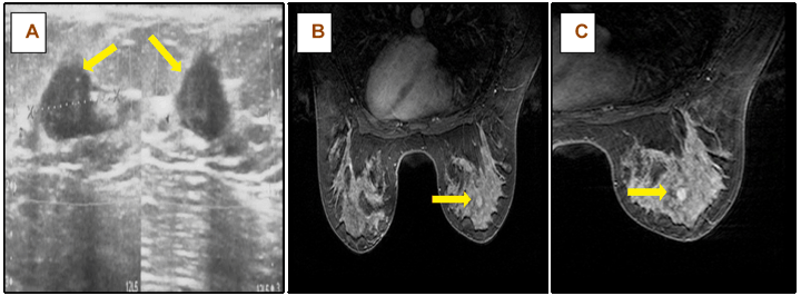
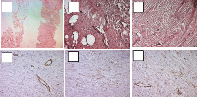
A 24-year-old female patient was initially admitted to the emergency department with persistent right for abdominal pain 48 hours. The patient had been suffering vague upper abdominal pain and discomfort for two years with fullness of upper abdomen part for the preceding one month; and had undergone two colonoscopic examinations in those two years, which did not reveal any colorectal polyps and hence excluded familial adenomatous polyposis. There was no history of weight loss, loss of appetite, abdominal trauma or oral contraceptive intake. No family history of similar ailment or malignancy was reported. An abdominal computed tomography (CT) scan (Figure 1) was performed which revealed large, hypodense and heterogeneously enhancing mass (138×98×112 mm) with an exophytic component arising from retroperitoneal space, extending up to the mesenteric vessels and encased them, compressing the right kidney, and also abutting the anterior abdominal wall. Both ureters were encased in the infiltrative tumor, with extravasation of radiocontrast from the right upper ureter at the L3 to L4 transverse process level leading to a 20-cm huge retroperitoneal urinoma over the right retroperitoneal space, extending from the perirenal space along the psoas muscle to the pelvic and peritoneal cavity. Based on the clinical and radiological findings, and after proper preoperative preparation, the patient was explored with a large median abdominal incision. After cleaning the peritoneum with a saline solution, a large exophytic mass arising from the retroperitoneum, abutting the abdominal cavity, was found. It was most densely adherent to the mesentere and encraised the mesenteric vessels. There was fibrosis near the region of suprahepatic inferior vena cava and no significant adherence of the mass with the anterior abdominal wall. The lesion was found to be completely adhered to many structures making total resection impossible. It had a petrous consistency when cut and partial resection of the lesion was carried out. At this site, the ureteric wall was hard, thickened, and adherent to the surrounding tissue; both ureters were encased in the infiltrative tumor and the right ureter was completely obstructed and ruptured 3 cm below the point where it crossed the common iliac artery. Right ureterocutaneostomy was performed. After 16 days, antegrade opacification performed through the ureterocutaneostomy catheter showed no extravasation of radiocontrast from the ureter. The percutaneous retroperitoneal drain tube was removed. The next day, she was discharged without complications. Histological and immunohistochemical findings confirmed the diagnosis of desmoid tumor. The evolution was characterized by a rapidly septic peritonitis and uroperitoneum secondary to recurrent rupture of ureterostomy one month later confirmed by the CT scan. The patient underwent laparotomy which includes a radical nephrectomy plus removal of the entire upper ureter, and the postoperative course was uneventful. Since that, a double-J catheter (6F, 24 cm) was placed in the left ureter for prevention. After the operation, imatinib (Glivec) 200 mg was prescribed. However, due to intractable diarrhea, the target therapy was discontinued and the patient refused any other treatment. After 10 months of follow-up, the patient presented with the chief complaint of a palpable lump in her left breast. Mammography revealed a 2-cm spiculated mass in the upper inner quadrant of the left breast. Sonography showed a suspicious hypoechoic mass with irregular margins. (Figure 2A) In magnetic resonance imaging (MRI) scan, the tumor was found to be of iso-intensity in the T1-weighted phase and of homogeneously low intensity in the T2-weighted phase and slow enhancement after contrast administration, and the marginal part showed very low intensity in both phases. (Figure 2B-C) Sonographically guided core biopsy was performed, and the histopathologic findings observed in this case were typical of desmoid tumor of the breast. (Figure 3) The patient underwent wide excision of the lesion. Currently, the patient is doing well. Follow-up CT scan, done at interval of three months, did not show any significant change in the size of retroperitoneal and mesenteric masse or left ureteral obstruction or other complications. There is no recurrent tumor in the breast or other sites. The patient changes the double-J catheter of the left ureter every six months and still refuses any medical or surgical treatment. |
|
|
|
|
|
|
|
Discussion
|
|
Desmoid tumors which belong to a family of fibroblastic proliferations that include a variety of fibromatoses, are benign tumors composed of fibrous elements. Desmoid tumors have an estimated incidence of 3.7 new cases per million people per year. [3] Desmoids may occur in the abdominal wall, the mesentery, or the retroperitoneum. Extra-abdominal desmoids may involve shoulder, thigh, buttock, or trunk whereas only a few cases describe a location within the breast. [3] [4] To the best of our knowledge, this is the first documented case of three locations of desmoid tumors in retroperitoneal space, mesentery and breast. Desmoids are the most common primary tumor of the mesentery. [5] Most of these tumors occur sporadically; however, patients with Gardner's syndrome are at higher risk than others. The incidence of abdominal wall and mesenteric desmoids in patients with Gardner syndrome ranges between 4–29%, and the tumors typically occur after abdominal surgery. [3] Desmoid tumors may be also associated with trauma and estrogen therapy. [5] In the present case, symptoms were sporadic, and the patient had no previous history of any of the above conditions. In addition, family history, upper gastrointestinal endoscopy, colonoscopy, and ophthalmoscopy were normal suggesting that our patient may be negative for the Gardner syndrome. Desmoids may occur in all age groups but they are typically seen in the third and fourth decades of life [3] [6] and have no significant racial or ethnic predilection. The difference in sex distribution is statistically insignificant, but there is a slightly higher incidence of this tumor in women than in men. [5] Symptoms of these fibromatosis depend on the site of the tumor. The signs are insidious and usually manifest when there is a large palpable tumor, or with abdominal discomfort or pain, nausea, vomiting, weight loss, and fever. [4] [5] [7] Desmoids can be locally aggressive and may invade contiguous structures. Some complications that have been reported and may lead to severe morbidity and mortality, [8] include small-bowel obstruction and hydronephrosis, [9] or ureteric obstruction, intestinal perforation, enterocutaneous fistula, and intestinal hemorrhage. However, we have not seen any previous case of spontaneous ureteral rupture in the literature, such as seen in the patient presented in this report, leading to septic peritonitis and uroperitoneum and aggressive surgical operations for management and a loss of the right kidney. On gross histopathologic examination, desmoid tumors are usually circumscribed lesions, but they may have irregular or infiltrating borders. On the cut surface, they are white and coarsely trabeculated, resembling scar tissue. Desmoids are usually larger than 5 cm when they are discovered, and they may be larger than 15 cm. In 10–15% of cases, desmoids are multiple. [10] Histologically, desmoid tumors are lesions composed of bland spindled or stellate fibroblastic cells embedded in a collagenous stroma, without evidence of muscular or neural differentiation and with little or no inflammatory component. The tumor may infiltrate adjacent viscera and tissues at the periphery. [5] The imaging appearance of these tumors is variable and depends on fibroblastic proliferation, fibrosis, collagen content, and vascularity. On sonography, desmoid tumors have variable echogenicity, appearing as masses of low, medium, or high echogenicity with smooth, defined margins. [6] On CT scan, most desmoid tumors appear as well-circumscribed homogeneous masses that may be isodense or hyperdense relative to muscle. Some cases of heterogeneous masses with infiltrative outer margins are seen. Desmoid tumors may enhance after injection of IV contrast material, [3] localizes the tumor and excludes metastasis. Malignant rhabdoid tumors (MRT) reveals the tumor's hypointensity on T1 and demonstrates variable signal intensity on T2 weighted imaging, depending on the accumulation of mucoid structures. [6] Therefore, a differentiation from other solid tumors is impossible using morphological criteria. [6] A multidisciplinary approach including surgery, chemotherapy, and radiation therapy is required for treatment. Complete resection is the therapy of choice for this type of tumors, but these tumors are often unresectable because of their aggressive and local infiltration, compression of surrounding structures and massive involvement of adjacent vital structures and sometimes a complete resection is only possible through organ transplant. The high recurrence rate even after complete surgical resection favors the use of nonsurgical therapy, such as nonsteroidal anti-inflammatory drugs and antiestrogens. [3] In our case due to extensive fibromatoses of the retroperitoneal space and the mesentery, excision was impossible, but wide excision of the breast lesion with clear margins was made without recurrence. Distant metastases have not been reported, [5] but in our present case, the second location in the breast seen 10 months after the diagnosis of the intraabdominal desmoid tumor But with the same Histological and immunohistochemical features can be a simple concomittent location not be diagnosed at the same time of the other lesions; or sporadic location appeared after the other locations. |
|
Conclusion
|
|
Spontaneous ureteral rupture as a complication and a circumstance of discovery of the retroperitoneal desmoid tumor is a rare condition. Its diagnosis is difficult and should be considered when presented with symptoms of acute abdominal pain or renal colic. Computed tomography scan remains quite valuable to confirm the diagnosis whereas the treatment is difficult. Desmoid tumors are benign neoplasms but are often unresectable because of their aggressive and local infiltration, compression of surrounding structures and massive involvement of adjacent vital structures; especially in the retroperitoneal space, as seen in our case. To the best of our knowledge, no cases of retroperitoneal and breast desmoid tumor association have previously been reported. Our purpose is to increase awareness of this condition so that clinicians will consider diagnosing it. This will facilitate prompt diagnosis and treatment. |
|
References
|
|


|
[HTML Abstract]
[PDF Full Text]
|
|
Author Contributions
Faouzi Mallat – Substantial contributions to conception and design, Acquisition of data, Drafting the article, Revising it critically for important intellrctual content, Final approval of the version to be published Wissem Hmida – Substantial contributions to conception and design, Acquisition of data, Drafting the article, Revising it critically for important intellrctual content, Final approval of the version to be published Sarra Mestiri – Substantial contributions to conception and design, Acquisition of data, Drafting the article, Final approval of the version to be published Badreddine Sriha – Substantial contributions to conception and design, Drafting the article, revising it critically for important intellrctual content, Final approval of the version to be published Moncef Mokni – Substantial contributions to conception and design, Drafting the article, Final approval of the version to be published Faouzi Mosbah – Substantial contributions to conception and design, Acquisition of data, Drafting the article, revising it critically for important intellrctual content, Final approval of the version to be published |
|
Guarantor of submission
The corresponding author is the guarantor of submission. |
|
Source of support
None |
|
Conflict of interest
Authors declare no conflict of interest. |
|
Copyright
© 2014 Faouzi Mallat et al. This article is distributed under the terms of Creative Commons Attribution License which permits unrestricted use, distribution and reproduction in any medium provided the original author(s) and original publisher are properly credited. Please see the copyright policy on the journal website for more information. |
|
|
|
About The Authors
| |||
| |||