  |

|
 |
|
Case Report
| ||||||
| Mid-aortic dysplastic syndrome as a rare cause of hypertension in young | ||||||
| Kaushik Saha1, Dipa Saha2, Parinita Ranjit3, Sujoy Sarkar3, Rabi Ranjan Sow Mondal4, Thiyagrajan G4 | ||||||
|
1MBBS, DCH, MD (Medicine), Assistant Professor, Department of General Medicine, Calcutta National Medical College, Kolkata.
2MBBS, MD (Physiology), Assistant Professor, Department of Physiology, College of Medicine & JNM Hospital, Kalyani, West Bengal. 3MBBS, MD (PGT), 3rd year post graduate trainee, Department of General Medicine, Calcutta National Medical College, Kolkata. 4MBBS, MD (PGT), 2nd year post graduate trainee, Department of General Medicine, Calcutta National Medical College, Kolkata. | ||||||
| ||||||
|
[HTML Abstract]
[PDF Full Text]
[Print This Article]
[Similar article in Pumed] [Similar article in Google Scholar] 
|


| How to cite this article: |
| Saha K, Saha D, Ranjit P, Sarkar S, Mondal RRS, Thiyagrajan G. Mid aortic dysplastic syndrome as a rare cause of hypertension in young. International Journal of Case Reports and Images 2013;4(10):563–566. |
|
Abstract
|
|
Introduction:
Mid-aortic syndrome (MAS), coarctation of abdominal aorta is a rare disease with only 200 reported cases. It is characterized by constriction of distal thoracic and/or abdominal aorta and its branches, therefore is also known as abdominal aortic coarctation. The MAS is characterized radiologically by severe narrowing of abdominal aorta and its branches and most of these patients usually die due to progressive severe hypertension before the age of 35–40 if left untreated.
Case Report: A 13-year-old boy was admitted with persistent headache and vomiting for one month and repeated generalized tonic clonic seizures for two days. His past history was unremarkable. The highlight of the clinical examination blood pressure was 240/150 mmHg in both the upper limbs, and all peripheral pulses were palpable. Blood pressure was similar in upper and lower limbs and a systolic bruit heard over the epigastrium. Ultrasonography showed a localized narrowing of a suprarenal segment of the abdominal aorta with an abrupt focal dilatation of the abdominal aorta approx 1.7 cm below origin of superior mesenteric artery. Magnetic resonance angiography of the aorta and its branches showed a distinct fusiform dilatation of abdominal aorta just below the origin of the superior mesenteric artery. Focal narrowing of abdominal aorta was noted just proximal to the dilatation. Conclusion: We diagnosed a case of mid-aortic coarctation with post stenotic dilatation associated with left renal artery stenosis leading to secondary hypertension in a teenage boy presenting with reversible posterior leukoencephalopathy and seizures. The boy was referred for vascular surgery but the patient refused any operative procedure. | |
|
Keywords:
Mid-aortic dysplastic syndrome (MAS), Secondary hypertension, Posterior leukoencephalopathy
| |
|
Introduction
| ||||||
|
The coarctation of the abdominal aorta, also known as middle aortic syndrome (MAS) or mid-aortic dysplastic syndrome, is a rare vascular pathology caused by localized or extended narrowing of the abdominal or distal descending thoracic aorta secondary either to a congenital anomaly in the development of the abdominal aorta or to one of several acquired conditions such as infection, obliterative panarteritis, neurofibromatosis, retroperitoneal fibrosis, fibromuscular dysplasia, mucopolysaccharidosis and Takayasu’s arteritis. [1] Most patients are young, with a mean age of 22 at diagnosis. [2] We report a case of a young boy admitted with headache and seizures who was found to have this rare cause of secondary hypertension. | ||||||
|
Case Report
| ||||||
|
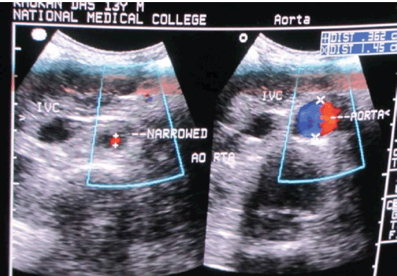
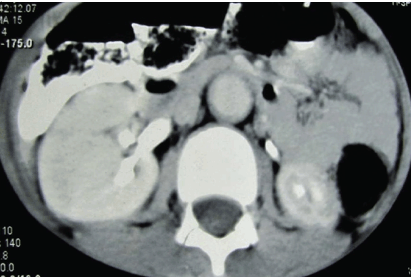
A 13-year-old boy was admitted with persistent headache and vomiting for one month and repeated generalized tonic clonic seizures for two days. He had no history of fever, chest pain, palpitations, claudication or visual disturbance. Enquiry about joint pain, photosensitivity, oral ulcers and recurrent infections were not contributory. His past history was unremarkable and his parents had no reason to worry about his milestones. The boy had good scholastic performance and had no difficulty in outdoor games. There was no history of contact with tuberculosis. Clinical assessment revealed an alert, conscious and co-operative boy, with corroborative apparent and chronological ages. Clinical examination of the patient showed blood pressure was 240/150 mmHg in both the upper limbs, and all peripheral pulses were palpable. Blood pressure was similar in upper and lower limbs and no radio radial or radio femoral delay was appreciated. Edema was absent and neck veins were not engorged. Systemic examination was unrewarding except for a systolic bruit heard over the epigastrium, which extended along the midline up to 1 cm above the umbilicus. Ophthalmoscopy was normal as well. Counts, baseline biochemistry (sugar, urea, creatinine, LFT, lipid profile, Na, K, TSH and FT4) and ECG were all within normal limits. X-ray of chest suggested borderline left ventricular hypertrophy and echocardiography with Doppler demonstrated eccentric hypertrophy of the left ventricle (left ventricular internal diameter 4.35 cm, LVIDs 2.98 cm, LVEF 60%). Moreover, a localized narrowing of a suprarenal segment of the abdominal aorta with a systolic pressure gradient around 40 mmHg, systolic pressure gradient 100 mmHg in celiac axis and superior mesenteric arteries. (Figure 1) Ultrasonography (USG) of abdomen with Color Doppler was done. Kidney sizes were 10.6 cm (right) and 8.5 cm (left) in the long axes. The right renal artery and interlobar arteries demonstrated normal spectral waveforms but the left renal artery could not be imaged properly. An abrupt focal dilatation of the abdominal aorta 1.7 cm (approx.) below the origin of superior mesenteric artery was observed. The dilated segment spanned 6.27 cm in the craniocaudal direction and measured 1.6 cm in diameter. A focal narrowing just before the dilatation was noted on USG. (Figure 2) In view of the seizures prior to admission, contrast enhanced computed tomography (CT) of brain was performed and bilateral symmetrical non-enhancing hypodense lesions involving the parieto-occipital areas suggestive of infarcts were noted. But repeat CT scan was normal and suggesting the diagnosis of reversible posterior leukoencephalopathy rather than infarct. Contrast enhanced computed tomography (CECT) abdomen corroborated the findings on USG and showed relatively small left kidney with poor enhancement patterns, suggestive of left renal artery stenosis. A focal dilatation of the abdominal aorta was also noticed on the CT scan. Tests for ANF (HEp2 method), RA factor and cANCA were all unrewarding. Magnetic resonance angiography of the aorta and its branches were performed. This showed a normally located aortic arch and the ascending aorta was seen to arise from the left ventricle with no evidence of any abnormal dilatation or flap. The arch of aorta and thoracic aorta were also normal. A distinct fusiform dilatation of abdominal aorta was seen just below the origin of the superior mesenteric artery. This dilated segment had a diameter of 19 mm and the craniocaudal extension of the dilated segment was about 63 mm. Focal narrowing of abdominal aorta as well as left renal artery was noted just proximal to the dilatation. The dilated segment extended below up to the division of aorta into the iliac vessels. (Figure 3) The boy was referred for vascular surgery but the patient refused any operative procedure. He was put on metoprolol (50 mg) twice daily and amlodipine (10 mg) daily and has been on irregular follow-up for the last 14 months, his last blood pressure record was 160/90 mmHg. | ||||||
| ||||||
| ||||||
|
| ||||||
|
Discussion
| ||||||
|
Coarctation of the abdominal aorta or mid-aortic dysplastic syndrome (MAS) is an extremely rare vascular defect in which congenital etiologies have described. Typical symptoms are hypertension in young and lower limb and/or visceral ischemia, which mostly occur later in life. [3] It is a rare condition that accounts for 0.5–2% of all aortic narrowing, most of which occur at the level of the proximal thoracic aorta. [4] Hypertension is the cardinal clinical feature in MAS and is present in more than 90% of the cases. Weak or absent femoral pulses may be appreciated and an audible bruit can typically be heard over the aorta. Most often, the patient is asymptomatic, but headache, vomiting, epistaxis, convulsions and other complication of severe hypertension are reported. Claudication and intestinal ischemia are present only in a minority of the patients, probably due to the gradual development of stenosis, which gives the body time to create effective collateral pathways. [5] From the embryological perspective, it has been suggested that a failure in fusion of the paired dorsal aortae during the fourth week of gestation may lead to MAS. Acquired conditions such as infection, obliterative panarteritis, neurofibromatosis, retroperitoneal fibrosis, fibromuscular dysplasia, mucopolysaccharidosis and Takayasu’s arteritis have been incriminated in MAS. In approximately 60% of cases, no etiology can be found. The renal arteries are involved in about 90% of the cases, and the coeliac axis and superior mesenteric artery in 35–50%, while the inferior mesenteric artery is almost never affected. A common histopathological finding in idiopathic MAS is fibroplasia of the intima and variable distortions of the internal elastic lamina with a lack of inflammatory changes that characteristically distinguish it from Takayasu’s arteritis. [6] The natural history of untreated symptomatic MAS is invariably death before the fourth decade due to complications secondary to severe hypertension such as cerebral hemorrhage or heart failure. Surgical correction remains the only definitive treatment when technically feasible. However, with newer and more effective antihypertensive drugs, surgery can be postponed until the patient reaches a more appropriate age. It is probably best to wait until full adult growth and adult vascular size are reached. [7] | ||||||
|
Conclusion
| ||||||
|
Middle aortic syndrome is a rare cause of uncontrolled hypertension with poor outcome if left untreated. We diagnosed a case of mid aortic coarctation with post stenotic dilatation associated with left renal artery stenosis leading to secondary hypertension in a teenage boy presenting with reversible posterior leukoencephalopathy and seizures. Although we have diagnosed this case early but definitive surgical treatment was denied by patient. | ||||||
|
References
| ||||||
| ||||||


|
[HTML Abstract]
[PDF Full Text]
|
|
Author Contributions
Kaushik Saha – Conception and design, Acquisition of data, Analysis and interpretation of data, Drafting the article, Critical revision of the article, Final approval of the version to be published Dipa Saha – Acquisition of data, Analysis and interpretation of data, Drafting the article, Critical revision of the article, Final approval of the version to be published Parinita Ranjit – Conception and design, Acquisition of data, Analysis and interpretation of data, Drafting the article, Critical revision of the article, Final approval of the version to be published Sujoy Sarkar – Conception and design, Acquisition of data, Analysis and interpretation of data, Drafting the article, Critical revision of the article, Final approval of the version to be published Rabi Ranjan Sow Mondal – Conception and design, Acquisition of data, Analysis and interpretation of data, Drafting the article, Final approval of the version to be published Thiyagrajan G – Conception and design, Acquisition of data, Analysis and interpretation of data, Drafting the article, Critical revision of the article, Final approval of the version to be published |
|
Guarantor of submission
The corresponding author is the guarantor of submission. |
|
Source of support
None |
|
Conflict of interest
Authors declare no conflict of interest. |
|
Copyright
© Kaushik Saha et al. 2013; This article is distributed the terms of Creative Commons Attribution License which permits unrestricted use, distribution and reproduction in any means provided the original authors and original publisher are properly credited. (Please see Copyright Policy for more information.) |
|
|