 |

|
 |
|
Case Series
| ||||||
| Chondroid lipoma, a rare lipomatous tumor: Report of two cases | ||||||
| Mohamed A Shawarby1, Maissa N El-Maraghy2, Ragaa A Salem2, Nafissa M El-Badawy2, Tarek M El-Sharkawy3, Tarek M Hashem3, Ahmad S Kamel3 | ||||||
|
1College of Medicine, University of Dammam, Dammam, Saudi Arabia.
2College of Medicine, Ain-Shams University, Cairo, EGYPT. 3College of Medicine, University of Dammam, Dammam, Saudi Arabia. | ||||||
| ||||||
|
[HTML Abstract]
[PDF Full Text]
[Print This Article]
[Similar article in Pumed] [Similar article in Google Scholar] 
|


| How to cite this article: |
| Shawarby MA, El-Maraghy MN, Salem RA, El-Badawy NM, El-Sharkawy TM, Hashem TM, Kamel AS. Chondroid lipoma, a rare lipomatous tumor: Report of two cases. International Journal of Case Reports and Images 2013;4(10):527–531. |
|
Abstract
|
|
Introduction:
Chondroid lipoma is a rare, benign lipomatous tumor that may be mistaken histologically for a liposarcoma or chondrosarcoma. Herein, two cases of chondroid lipoma are reported in two Egyptian females aged 38 and 51 years and literature about this rare tumor briefly reviewed with discussion of diagnostic criteria that may help distinguish it from other tumors with overlapping histologic features.
Case Report: The tumor was subcutaneous in both cases. One was located in the right knee area, the other in an infra-mammary location on the right side of the chest. Grossly, both neoplasms were encapsulated and exhibited a nodular, myxoid cut surface. Histologically, they consisted of an abundant myxoid and chondroid stroma interspersed by small round cells with eosinophilic or vacuolated cytoplasm, signet ring lipoblast-like cells, adipocytes and foci of mature adipose tissue. Immunohistochemically, diffuse reactivity of the neoplastic cells for vimentin and variable reactivity for S-100 protein were present in both cases. One case also showed focal immunoreactivity for CD68. The Ki67 proliferative index was less than 5%. The common differentials like myxoid liposarcoma and extraskeletal myxoid chondrosarcoma were ruled out based mainly on pure histologic criteria. Both patients underwent simple curative excision and were tumor free for at least one and three years after surgery, respectively. Conclusion: Because chondroid lipoma is easily misdiagnosed as a sarcoma of either adipose tissue or cartilage, a high level of suspicion by the pathologist and familiarity with its features are of practical importance to avoid misdiagnosis and overtreatment. | |
|
Keywords:
Chondroid, Myxoid, Liposarcoma, Chondrosarcoma, Lipoma
| |
|
Introduction
| ||||||
|
Chondroid lipoma is a rare, benign lipomatous tumor that may be mistaken histologically for a sarcoma of either adipose tissue or cartilage. [1] Because it is easily misdiagnosed as a malignant tumor, a high level of suspicion by the pathologist and familiarity with its features are of practical importance to avoid an overtreatment as the tumor does not recur or metastasize, and simple excision is curative. [2] Herein, we report two cases of chondroid lipoma followed by a brief literature review about this rare tumor with discussion of diagnostic criteria that may help distinguish it from other tumors with overlapping histologic features. | ||||||
|
Case Series
| ||||||
|
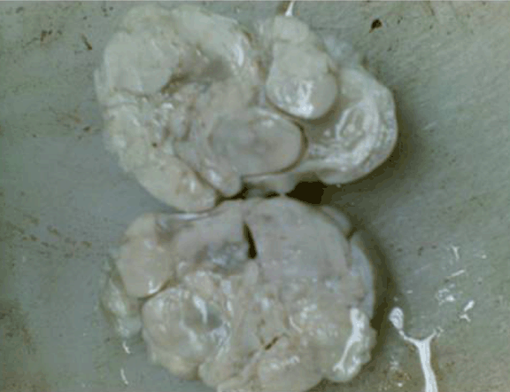
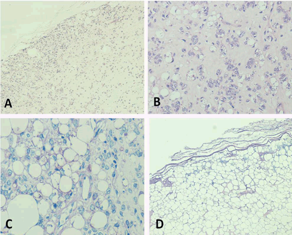
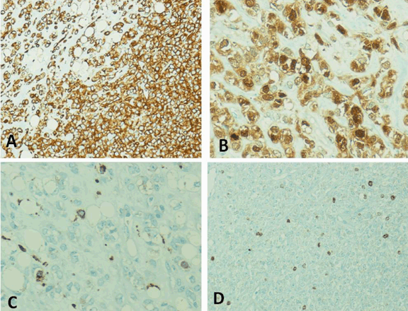
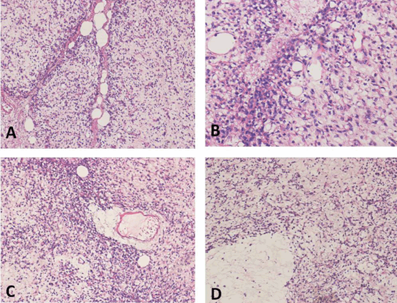
Case 1: A 38-year-old Egyptian female presented with a painless subcutaneous swelling in the right knee area. Her past medical history was unremarkable. On examination, the swelling was well defined and non-tender. The overlying skin was normal. Excision was done and the specimen submitted for pathological examination. Gross: An encapsulated, well circumscribed tumor received fragmented as two grayish-white pieces measuring 12x8x4 cm, and 8x5x4 cm. The cut surface was nodular with myxoid areas. (Figure 1) Microscopic: Sections revealed an encapsulated, lobulated tumor of varying cellularity, composed of strands, clusters and sheets of small round cells with eosinophilic or vacuolated cytoplasm in an abundant chondroid and myxoid stroma interspersed by scattered adipocytes and foci of mature adipose tissue. (Figure 2) Glycogen could be demonstrated in the cytoplasm of rare cells. There was no significant nuclear pleomorphism and only very occasional mitoses were seen. Immunohistochemically, there was strong diffuse cytoplasmic reactivity for vimentin. Most cells also showed strong cytoplasmic and nuclear reactivity for S-100 protein. Few scattered cells showed positive cytoplasmic staining for CD68. Positive nuclear staining for Ki67 was noted in only less than 5% of cells. (Figure 3) There was no reactivity for CK, EMA, SMA or HMB45. The initial impression was myxoid liposarcoma. However, the final diagnosis after intradepartmental consultation was chondroid lipoma, mainly based on the lack of the typical ‘chicken wire’ vasculature seen in myxoid liposarcoma and the presence of a chondroid matrix and mature adipose tissue. After three years of surgery, the patient was tumor free. | ||||||
| ||||||
|
| ||||||
|
| ||||||
|
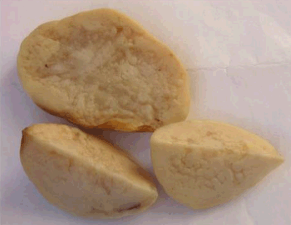
Case 2: A 51-year-old Egyptian female presented with a painless subcutaneous inframammary mass on the right side of the chest. Past medical history was unremarkable. The mass was well defined and non-tender, and the overlying skin was normal. It was excised and submitted for pathological study. Gross: An encapsulated, well circumscribed grayish-tan mass with a myxoid, nodular cut-surface, measuring 8x6x5 cm. (Figure 4) Microscopic: Sections revealed a distinctly lobulated tumor with a thin capsule, composed of an abundant myxoid to chondroid stroma interspersed by small round cells with eosinophilic cytoplasm, mainly located at the periphery of the lobules along with scattered signet ring lipoblast-like cells and more mature adipocytes. Adipocytes were also focally seen around blood vessels and occasionally formed islands within the tumor lobules. (Figure 5) There was no significant nuclear pleomorphism and mitoses could not be demonstrated. Immunohistochemically, there was strong diffuse cytoplasmic reactivity for vimentin. Occasional cells also showed nuclear reactivity for S-100 protein and Ki67. There was no reactivity for CD68, CK, EMA, SMA or HMB45. The initial impression was extraskeletal myxoid chondrosarcoma. However, the final diagnosis after intra-departmental consultation was chondroid lipoma, mainly based on the presence of adipocytes and mature adipose tissue within the tumor lobules. After one year of surgery, the patient was tumor free. | ||||||
| ||||||
|
| ||||||
|
Discussion
| ||||||
|
Chondroid lipoma is a rare, benign lipomatous tumor that may be confused histologically with a liposarcoma or chondrosarcoma. Meis and Enzinger were the first to recognize chondroid lipoma as a distinct entity when they reported a series of 20 cases in 1993. [1] An identical tumor had, however, been reported in 1986 by Chan et al. as an example of ‘an extraskeletal chondroma with lipoblast-like cells’. [3] The vast majority of chondroid lipomas occur in females and are seen between ages of 14 and 70 years (median of 36 years). [1] Both of our patients were female and the mean age was 44.5 years. They are usually located in the subcutaneous tissue, superficial muscular fascia, or skeletal muscles, most frequently in the proximal extremity and limb girdle, then in decreasing frequency in the leg, trunk, head and neck region, foot, and, finally, hand. [4] [5] The tumor mass in both of our case was subcutaneous with one located in the knee area, the other in the trunk in a infra-mammary location. Chondroid lipomas are well-circumscribed, may be encapsulated, lobulated tumors. They range in size from 1.5–11 cm, but are usually around 4 cm. The cut surface is smooth and yellow. [6] Both of our cases were well circumscribed, encapsulated and lobulated but one of them was distinctly larger than the sizes reported in literature (20x13x8 cm in aggregate). Microscopically, the tumor is composed of a varying admixture of eosinophilic, multivacuolated cells arranged in strands, nests and sheets, together with mature adipocytes. These are present in a myxoid and chondroid stroma. Mature adipose tissue may be interspersed within the tumor. Fibrous bands often divide the tumor into lobules. Identical histologic features were noted in both of our cases. Multivacuolated cells contain fat and often glycogen (demonstrated by periodic acid–Schiff and Oil-red-O stains) and may resemble chondroblasts, lipoblasts or hibernoma cells. The nuclei have irregular contours and lack pleomorphism and mitotic activity. [1] One of our cases showed intracytoplasmic glycogen within the neoplastic cells and nuclei did not show significant pleomorphism or mitotic activity in either case. Immunohistochemically, the neoplastic cells stain for vimentin, S-100 and CD68 and some tumors may be keratin positive. Ki67 immunoreactivity is typically very low and detected only in the more primitive cell population. [4] [7] Both of our cases showed strong immunoreactivity for vimentin and variable reactivity for S-100. However, only one case showed CD68 positivity. The Ki67 proliferation index was very low in both cases confirming the very low mitotic activity observed in H&E stained sections. Ultrastructurally, the cells of chondroid lipoma exhibit a spectrum of differentiation, ranging from primitive cells sharing features of prelipoblasts and chondroblasts, to lipoblasts and preadipocytes, to mature adipocytes. The myxohyaline matrix has ultrastructural features of cartilage. Numerous mitochondria and lysosomes are absent, indicating that chondroid lipoma is neither a fibromatous lesion nor a lipogranuloma. [8] Cytogenetic analysis of chondroid lipoma revealed a balanced translocation t (11, 16) (q13; p12-13) distinct from the known translocation involving 16p11 in myxoid and round-cell liposarcoma. The 11q13 breakpoint was previously noted in hibernomas, raising the possibility of a common genetic deregulation. [9] [10] The cellularity of chondroid lipomas and their myxochondroid matrix have caused them to be histologically confused with myxoid liposarcoma or extraskeletal myxoid chondrosarcoma. The histologic differential diagnosis also includes mixed tumors, soft tissue chondroma, cartilagenous metaplasia in a lipoma [11] and parachordoma. [12] Myxoid liposarcoma may contain areas of mature fat and vacuolated and eosinophilic cells in a myxoid stroma similar to chondroid lipoma. However, chondroid lipoma lacks the distinct delicate branching vasculature present in myxoid liposarcoma and is usually less cellular. Myxoid liposarcoma also lacks the chondroid matrix seen in chondroid lipoma. [11] Extraskeletal myxoid chondrosarcoma is composed of cords of eosinophilic cells that are smaller than those in chondroid lipoma. The tumor is more lobulated and there is absence of adipocytes and mature adipose tissue. [11] Mixed tumors of the dermis or deep soft tissue are composed of nests and cords of eosinophilic cells in a myxochondroid stroma, but they are not as vacuolated as the cells in chondroid lipoma, and do not demonstrate adipocytic differentiation. Epithelial differentiation manifested by glands and myoepithelial cells are also usually present in mixed tumors. Soft tissue chondroma usually has true hyaline cartilage, does not contain adipose tissue and arises in the soft tissues of the hand and feet. In cartilaginous, metaplasia in a lipoma (chondrolipoma), true hyaline cartilage is present. [11] Parachordoma does not contain mature fat and is EMA and Keratin positive. [12] | ||||||
|
Conclusion
| ||||||
|
Because chondroid lipoma is easily misdiagnosed as a sarcoma of either adipose tissue or cartilage, a high level of suspicion by the pathologist and familiarity with its features are of practical importance to avoid an overtreatment as the tumor does not recur or metastasize, and simple excision is curative. [2] | ||||||
|
Acknowledgements
| ||||||
|
The authors acknowledge the services of Mr Shakir Ahmed and Mrs Maria Rosario Lazaro from the histopathology laboratory of the University of Dammam, Saudi Arabia for conducting the histology and immuunohistochemistry work. | ||||||
|
References
| ||||||
| ||||||

|
[HTML Abstract]
[PDF Full Text]
|
|
Author Contributions:
Mohamed A Shawarby – Substantial contributions to conception and design, Acquisition of data, Analysis and interpretation of data, Drafting the article, Revising it critically for important intellectual content, Final approval of the version to be published Maissa N El-Maraghy – Acquisition of data, Revising it critically for important intellectual content, Final approval of the version to be published Ragaa A Salem – Acquisition of data, Revising it critically for important intellectual content, Final approval of the version to be published Nafissa M El-Badawy – Acquisition of data, Revising it critically for important intellectual content, Final approval of the version to be published Tarek M El-Sharkawy – Acquisition of data, Revising it critically for important intellectual content, Final approval of the version to be published Tarek M Hashem – Acquisition of data, Revising it critically for important intellectual content, Final approval of the version to be published Ahmad S Kamel – Acquisition of data, Revising it critically for important intellectual content, Final approval of the version to be published |
|
Guarantor of submission
The corresponding author is the guarantor of submission. |
|
Source of support
None |
|
Conflict of interest
Authors declare no conflict of interest. |
|
Copyright
© Mohamed A Shawarby et al. 2013; This article is distributed the terms of Creative Commons attribution 3.0 License which permits unrestricted use, distribution and reproduction in any means provided the original authors and original publisher are properly credited. (Please see Copyright Policy for more information.) |
|
|