  |

|
 |
|
Case Report
| ||||||
| Goldenhar syndrome presenting as limbal dermoid cyst: A case report | ||||||
| Sajid Ansari1, Kanchan Dhungel2, Kaleem Ahmad2, Mukesh Kumar Gupta2, Md. Farid Amanullah3, Pranav Kumar Santhalia4, Raj Kumar Rauniyar5 | ||||||
|
1Senior Resident, MD, Department of Radiodiagnosis and imaging, BP Koirala Institute of Health Sciences, Dharan, Nepal.
2Associate Professor, MD, Department of Radiodiagnosis and imaging, BP Koirala Institute of Health Sciences, Dharan, Nepal. 3Senior Resident, MS, Department of Orthopaedics, BP Koirala Institute of Health Sciences, Dharan, Nepal. 4Junior Resident, MD, Department of Radiodiagnosis and imaging, BP Koirala Institute of Health Sciences, Dharan, Nepal. 5Professor, MD, Department of Radiodiagnosis and imaging, BP Koirala Institute of Health Sciences, Dharan, Nepal. | ||||||
| ||||||
|
[HTML Abstract]
[PDF Full Text]
[Print This Article]
[Similar article in Pumed] [Similar article in Google Scholar] 
|


| How to cite this article: |
| Ansari S, Dhungel K, Ahmad K, Gupta MK, Amanullah MF, Santhalia PK, Rauniyar RK. Goldenhar syndrome presenting as limbal dermoid cyst: A case report with clinical and radiological findings. International Journal of Case Reports and Images 2013;4(7):384–387. |
|
Abstract
|
|
Introduction:
Goldenhar syndrome is a rare congenital disorder which manifests as ocular, auricular, dental and vertebral and several other systemic abnormalities. The abnormalities in the organs developing from first and second branchial arches during blastogenesis cause morphological anomalies in this disorder.
Case Report: We report a case of Goldenhar syndrome in a 12-year-old Asian girl with limbal dermoid cyst, lateral canthal tag, preauricular appendage and malocclusion of teeth. Computed tomography scan and grayscale ultrasonography of orbit revealed dermoid cysts. Multidisciplinary treatment approach was advised; dermoid cyst was excised surgically and dental consultation was advised for malocclusion. Conclusion: This case has been presented to increase the awareness about this rare entity, to highlight the importance of typical clinical and radiological findings and its association with various systemic conditions. Multidisciplinary treatment approach and long-term regular follow-up is important to monitor the growth and development of patients. | |
|
Keywords:
Goldenhar syndrome, Dermoid cyst, Preauricular appendages, Malocclusion
| |
|
Introduction
| ||||||
|
Goldenhar syndrome is a rare congenital disorder with occurrence of about 1 per 5800 births and male:female ratio of 3:2. In 1952, Goldenhar was first to describe this rare disorder. It comprises various anomalies such as periauricular appendages, limbal dermoid and auricular malformations. Goldenhar syndrome is also known as oculo-auriculo-vertebral dysplasia and hemifacial microsomia. [1] The abnormalities in the organs developing from first and second branchial arches during blastogenesis cause morphological anomalies. It has a multifactorial etiopathology that includes nutritional and environmental factors. [2] | ||||||
|
Case Report
| ||||||
|
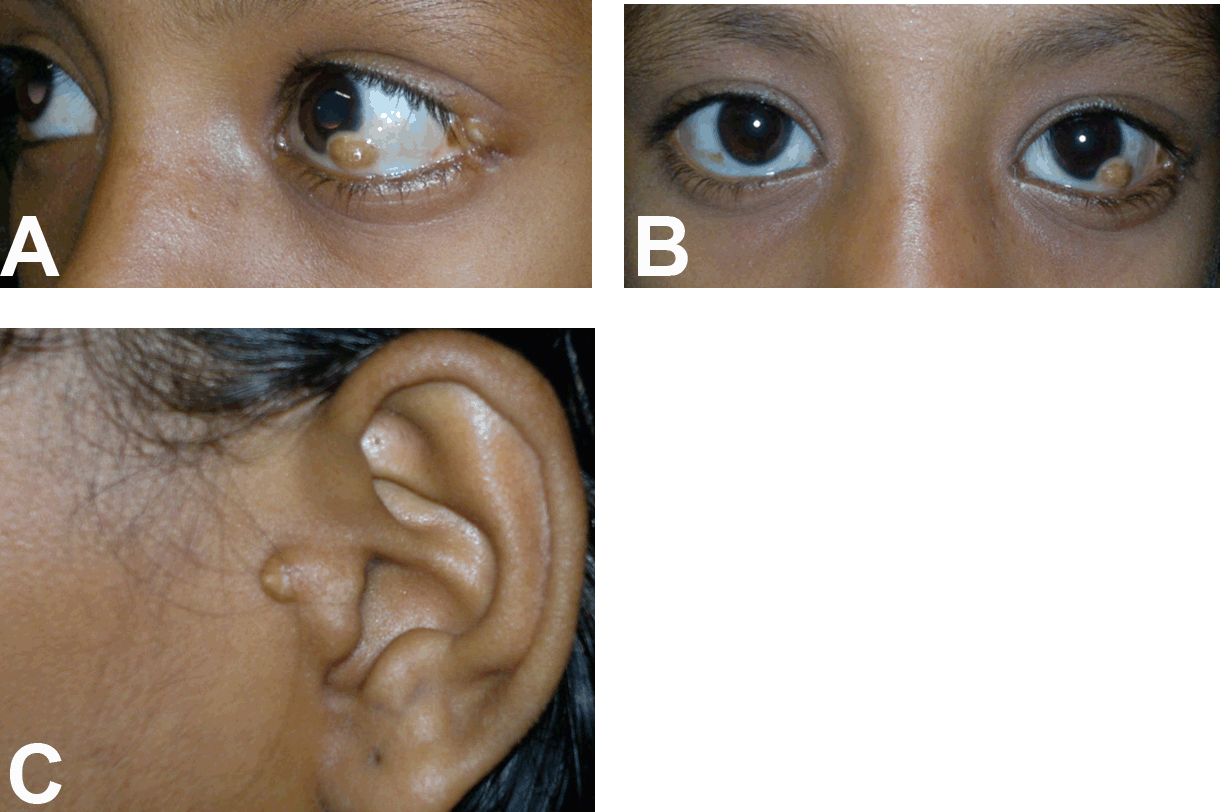
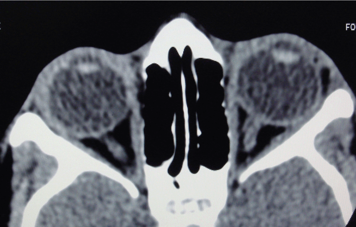
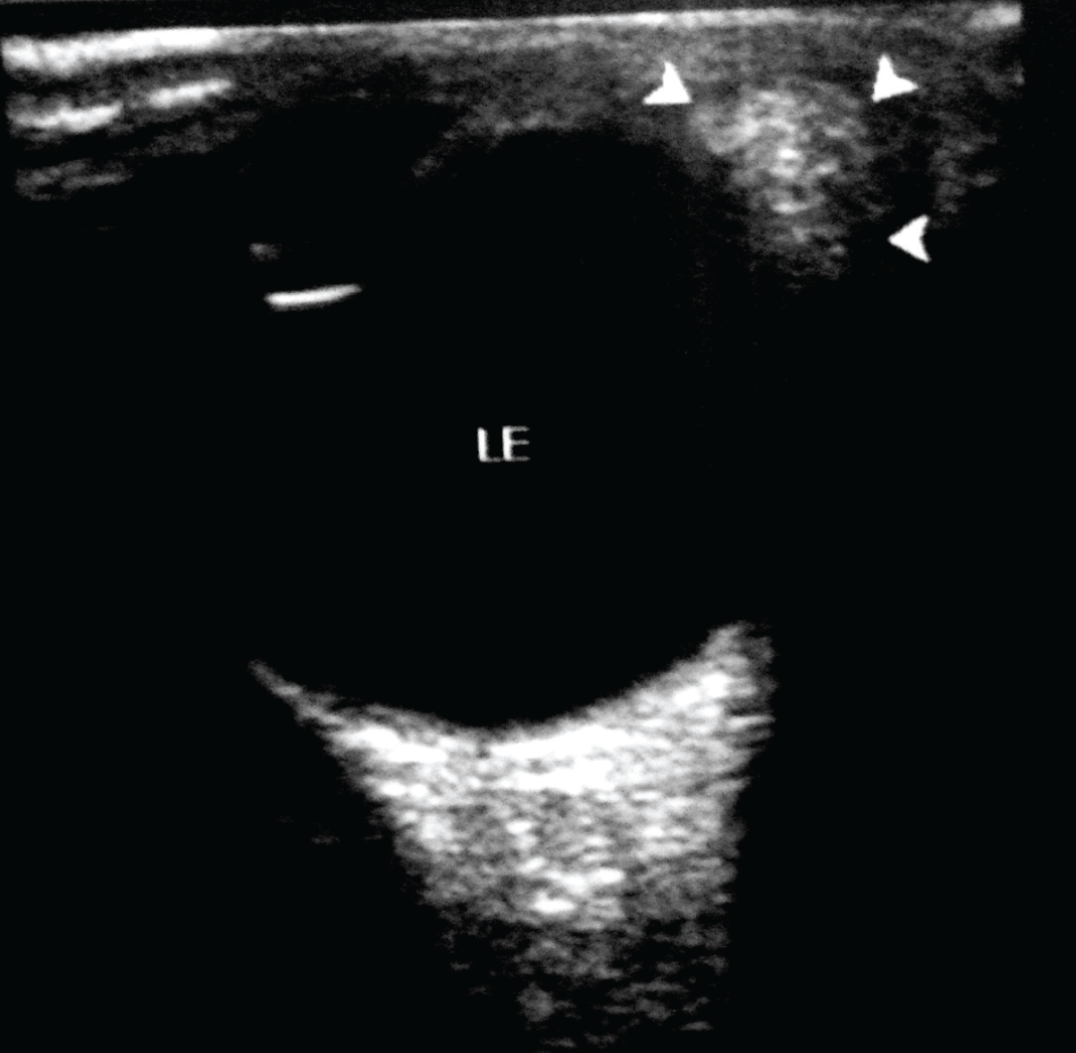
A 12-year-old Asian girl presented to our department for computed tomography (CT) scan of orbits for evaluation of ocular dermoid cyst. The child was born to non-consanguineous parents with uneventful prenatal and antenatal period. There was no relevant family history. Her mental status was normal and there was no evidence of developmental delay. Left limbal dermoid cyst measuring 0.5x0.5 cm with hair follicles was seen at 5 o’clock position since birth, (Figure 1A-B) which is painless and not increasing in size. A dermal tag was seen at the lateral canthus of the left upper eyelid. (Figure 1A) There was no redness or discharge from the eyes and ocular motility was normal bilaterally. Her visual acuity was normal (6/6). An accessory preauricular tag was seen on the left side. (Figure 1C) Audiological examination shows no evidence of hearing impairment. Intraoral examination revealed malocclusion of lower dentition. (Figure 2) Cardiovascular system and central nervous system were normal. Hematological investigation showed hemoglobin 11.9 g/dL, total leukocyte count (TLC) 8500/μL, neutrophils 38%, lymphocytes 48%, eosinophils 15%, monocytes 5% and platelet counts 274x103/μL. Routine and microscopic urine investigation were within the normal limits. No abnormality was seen on plain radiograph (antero-posterior and lateral views) of cervical and thoracic spine. The CT scan of orbits showed a hypodense lesion of fat attenuation measuring 0.8x0.5 cm on the lateral aspect of the left orbit abutting the lateral margin of the globe, suggestive of dermoid cyst. (Figure 3) Another small similar lesion measuring 0.5x0.5 cm seen at the left limbal region (suggestive of dermoid cyst). Grayscale ultrasonography (USG) of left orbit showed a small echogenic lesion at the limbus. (Figure 4) Clinical and radiological features were suggestive of Goldenhar syndrome. Pediatric consultation was also done to rule out any systemic associations. She was referred to dentist for needful correction of the maloccluded teeth. Surgical excision of the dermoid cysts was done. Postoperative period was uneventful. | ||||||
| ||||||
| ||||||
|
| ||||||
| ||||||
|
Discussion
| ||||||
|
Goldenhar syndrome has characteristic features of epibulbar dermoids, auricular abnormalities, preauricular appendages and fistulas, dacryocystitis, hypoplasia of the malar bones, mandible and zygomatic arch. It is also associated with macrostomia, micrognathia, cleft palate, bifid tongue and malocclusion, vertebral abnormalities, facial muscle hypoplasia, neurological, visceral, cardiac and genitourinary abnormalities. [2] [3] [4] [5] [6] In 40% cases significant hearing loss have been reported. [3] The presence of anomalies of the ear and of appendages on the ear is sufficient for the diagnosis. In our patient, preauricular appendage was seen but there was no hearing loss. Epibulbar dermoid is found in 30–60% cases with ocular manifestation of Goldenhar syndrome. [7] Dermoid cysts are usually observed on the inferotemporal or superotemporal aspect of the limbus. Astigmatism is a rare manifestation of the disease due to the corneal and scleral invasion by the lesions. In our patient, the dermoid was seen on the inferotemporal aspect of the limbus. In this disorder, the delayed development of teeth can be seen rarely; malocclusion was observed in our patient. Malformations are usually bilateral in about 10–33% of cases, with one side of the body affected more leading to asymmetry. In most of the cases, right sided involvement is more severely affected than the left. [8] Cardiovascular abnormalities are observed in range of 5–58% of cases. However, there were no cardiovascular abnormalities in our patient. Sporadic autosomal dominant and autosomal recessive modes of inheritance have been seen in most of the cases along with association of Trisomy of 7 and 22. [9] Goldenhar syndrome has also been found in children born to pregnant women who were exposed to various teratogenic agents like retinoic acid, primidone and thalidomide. [10] In our case there was no history of maternal drug intake, any febrile illness or diabetes during pregnancy. Prognosis of this disease is good in otherwise uncomplicated cases without any systemic associations. Multidisciplinary treatment approach is required which varies with age and systemic associations. Ocular dermoid cysts can be excised surgically. The ophthalmologist should focus on visual consequences, early treatment and meticulous follow-up of the patients. Reconstructive oral and facial surgeries are required for malocclusion and bony deformities. | ||||||
|
Conclusion
| ||||||
|
This case has been presented to increase the awareness about this rare entity, to highlight the importance of typical clinical and radiological findings and its association with various systemic conditions. Multidisciplinary treatment approach and long-term meticulous follow-up is important to monitor the growth and the development of patients. | ||||||
|
References
| ||||||
| ||||||


|
[HTML Abstract]
[PDF Full Text]
|
|
Author Contributions
Sajid Ansari – Substantial contributions to conception and design, Acquisition of data, Analysis and interpretation of data, Drafting the article, Final approval of the version to be published Kanchan Dhungel – Substantial contributions to conception and design, Acquisition of data, Drafting the article, Revising it critically for important intellectual content, Final approval of the version to be published Kaleem Ahmad – Analysis and interpretation of data, Revising it critically for important intellectual content, Final approval of the version to be published Mukesh Kumar Gupta – Substantial contributions to conception and design, Revising it critically for important intellectual content, Final approval of the version to be published Md. Farid Amanullah – Substantial contributions to conception and design, Drafting the article, Revising it critically for important intellectual content, Final approval of the version to be published Pranav Kumar Santhalia – Substantial contributions to conception and design, Acquisition of data, Analysis and interpretation of data, Revising it critically for important intellectual content Raj Kumar Rauniyar – Substantial contributions to conception and design, Acquisition of data, Analysis and interpretation of data, Final approval of the version to be published |
|
Guarantor of submission
The corresponding author is the guarantor of submission. |
|
Source of support
None |
|
Conflict of interest
Authors declare no conflict of interest. |
|
Copyright
© Sajid Ansari et al. 2013; This article is distributed the terms of Creative Commons Attribution License which permits unrestricted use, distribution and reproduction in any means provided the original authors and original publisher are properly credited. (Please see Copyright Policy for more information.) |
|
|