
 |

|
 |
|
Case Series
| ||||||
| Granular tumors of the central nervous system: A case series | ||||||
| Janese Trimaldi1, Nicole D Riddle2, Jeremy W Bowers3, Harry R van Loveren4, Kondi Wong1 | ||||||
|
1Department of Pathology and Cell Biology, University of South Florida College of Medicine, Tampa, FL, USA.
2Department of Pathology, University of Texas Health Science Center, San Antonio, TX, USA. 3Department of Pathology, Moffitt Cancer Center, Tampa, FL, USA. 4Department of Neurosurgery, University of South Florida College of Medicine, Tampa, FL, USA. | ||||||
| ||||||
|
[HTML Abstract]
[PDF Full Text]
[Print This Article]
[Similar article in Pumed] [Similar article in Google Scholar] 
|


| How to cite this article: |
| Trimaldi J, Riddle ND, Bowers JW, Loveren HRv, Wong K. Granular tumors of the central nervous system: A case series. International Journal of Case Reports and Images 2013;4(1):1–6. |
|
Abstract
|
|
Introduction:
Granular cell tumors of the central nervous system are rare tumors. To date, eight cases arising from cranial nerves have been reported. Granular cell tumors have also been found arising from the neurohypophysis and its stalk. Due to their rarity and histological similarity to other central nervous system (CNS) tumors with a granular appearance, they often pose a diagnostic conundrum. The differential diagnosis is surprisingly diverse and includes granular cell astrocytoma, infundibular granular cell tumor, spindle cell oncocytoma of the adenohypophysis, granular and oncocytic variants of pituitary adenoma, meningioma, pituicytoma and intrasellar schwannoma. Distinguishing between the CNS tumors with granular features is important because some tumors have an increased recurrence risk or a poor prognosis.
Case Report: To highlight the histological features of granular lesions of the central nervous system, including the immunohistochemical profile and electron microscopic depiction, we review two cases each with a similar granular histology and a different final diagnosis. Conclusion: Thorough online literature search revealed several cases of granular cell lesions of the CNS, however, oftentimes the diagnosis is difficult to come by and the differential is long. Conclusion: Granular cell tumor and its variants, though uncommon, must be included in the differential diagnosis of CNS lesions. | |
|
Key Words:
Granular, Tumors, Central nervous system (CNS)
| |
|
Introduction
| ||||||
|
Granular cell lesions of the central nervous system (CNS) are rather uncommon and may pose a diagnostic challenge. Granular histology may be present as the main characteristic of a neoplasm or as a variation in a number of benign and malignant lesions. The differential diagnosis must always include a true granular cell tumor when granular morphology is identified, especially when it encompasses a majority of the lesion. The differential diagnosis of a CNS granular cell tumor is widely varied and includes infundibular granular cell tumor, granular/oncocytic variants of pituitary adenoma, meningioma, pituicytoma and intrasellar schwannoma, as well as spindle cell oncocytoma of the adenohypophysis, which may be prone to recur and grow despite adjuvant therapy, and granular cell astrocytoma, which has a poor prognosis. | ||||||
|
Case Report
| ||||||
|
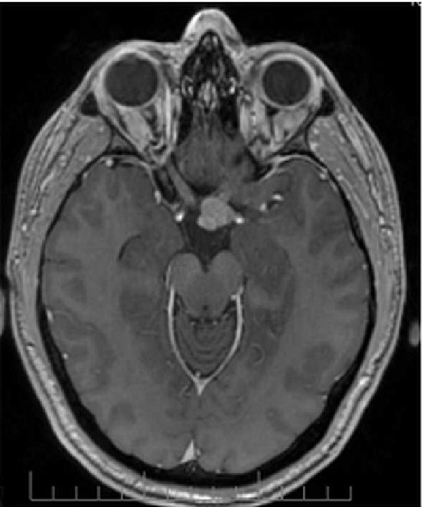
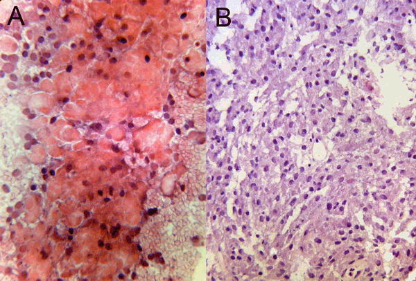
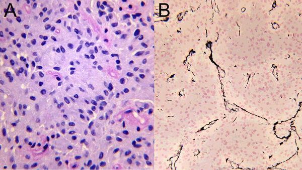
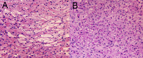
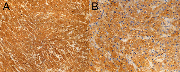
Case 1: A 37-year-old male presented with a history of depression, fatigue, and decreased libido for two years. Magnetic resonance imaging (MRI) was performed revealing a left-sided pituitary mass that was enlarging the sella with suprasellar extension. Post-contrast images demonstrated an intensely enhancing mass in the right middle cranial fossa that was hypointense on T2-weighted images and isointense to adjacent gray matter on T1-weighted images (Figure 1). It was centered in Meckel's cave with involvement of the cisternal segment of the trigeminal nerve. There was distal extension of the mass along the inferior alveolar branch of V3 into the mandibular foramen. The right side of the pituitary gland appeared to be uninvolved. Surgical removal was performed through a transnasal endoscopic approach and yielded a 1.4x0.4x0.1 cm tumor. Frozen section analysis revealed a granular epithelial cell neoplasm. The differential diagnosis at that time included: pituitary adenoma, xanthoma, infundibular granular cell tumor, langerhans cell histiocytosis, hemangioblastoma and granular cell astrocytoma. Routine histological examination with hematoxylin and eosin (H&E) stained sections revealed diffuse, densely granulated, eosinophilic cells (Figure 2). Special staining for PAS was also positive (Figure 3). Immunohistochemical studies showed the tumor cells to be weakly positive for prolactin and strongly synaptophysin positive. The tumor cells were non-reactive for CD68, inhibin, S100, chromogranin; as well as pituitary markers such as ACTH, FSH, LH, GH, and TSH. Pancytokeratin and GFAP were also negative. Complete disruption of the native lobular reticular architecture was evident on reticulin stain, characteristic of a pituitary adenoma. A diagnosis of pituitary adenoma with granular cell morphology, and evidence of prolactin expression was given. | ||||||
| ||||||
|
| ||||||
| ||||||
| ||||||
|
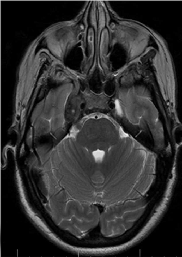
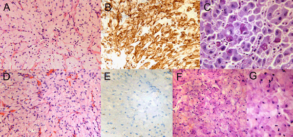
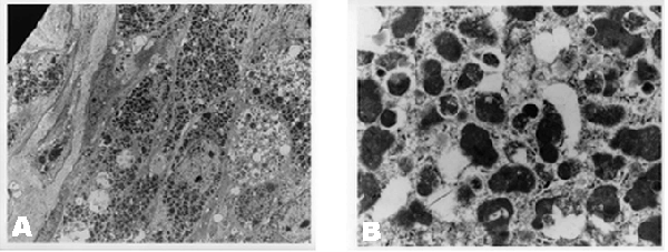
Case 2: A 54-year-old woman with a history of Bell's palsy one year prior with recurrence nine months later, presented with numbness primarily in the third division of the right trigeminal nerve. MRI scan showed a tumor homogeneously enhancing in the gasserion ganglion with an extension into the foramen ovale (Figure 4). A right temporal craniotomy and zygomatic osteotomy was performed, and the foramen ovale was opened for resection of a 6.1x4.3x1.2 cm tan-brown mass with multi-nodular areas. Frozen section analysis suggested a possible ganglioneuroma. Following histological evaluation a diagnosis of granular cell tumor of the right trigeminal ganglion was rendered. Permanent section analysis revealed peripheral nerve and entrapped ganglion cells (trigeminal ganglion) infiltrated by round, spindled and epithelioid cells with abundant, granular, PAS-positive cytoplasm consistent with granular cell tumor (Figure 5). The tumor cells were strongly positive for CD 68 with abundant PAS positive cytoplasmic granules and strong reactivity to S100 (Figure 6) (Figure 7). They were non-reactive for: synaptophysin, smooth muscle actin, muscle specific actin, melan-A, myogenin, GFAP, smooth muscle myosin heavy, and desmin. Ki67 demonstrated moderate proliferative activity as well as moderate amounts of reticulin collagen and neuron specific enolase (not shown). Electron microscopy demonstrated tumor cell cytoplasm filled and expanded with dark, granular lysosomes, characteristic of granular cell tumor (Figure 8). | ||||||
| ||||||
| ||||||
| ||||||
| ||||||
|
| ||||||
|
| ||||||
|
Discussion
| ||||||
|
A number of granular or similarly oncocytic tumors have remarkably similar histomorphology on initial staining with hematoxylin and eosin. Differing intracranial locations may be helpful at times for diagnosis, but at other times they may be misleading. Tumors containing granular cells can present in two forms: (i) as a "pure" form composed entirely of granular cells ("granular cell tumor"), (ii) or as a focal change that occurs in a neoplasm of a recognizable cell type. [1] In the latter form, one theory considers granular cell change to be a degenerative phenomenon. [2] The hypothesis is that the granular cell phenotype seems to be not specific of a certain tumor type, but rather a peculiar change characterized by an increase in intracellular lysosomes. Reported CNS tumors with granular cell or oncocytic change include: astrocytoma, medulloblastoma, ganglioglioma, glioblastoma, meningioma, schwannoma, ependymoma, oligodendroglioma, neurofibroma, anterior and posterior pituitary. [2] [3] [4] [5] [6] [7] Spindle cell oncocytoma, granular cell astrocytoma, and pituicytoma are distinct entities and possibly the infundibular granular cell tumor as well. Morphology on hematoxylin and eosin stain and special stains alongside tumor immunophenotype are a valuable aide in pathologic evaluation and distinguishing the different tumor types. There are distinguishing characteristics of different tumor types. Granular cell astrocytomas are GFAP reactive in more than 95% cases [19] and have a poor prognosis. Spindle cell oncocytomas of the adenohypophysis have S100 reactivity, but are negative for neuroglial markers and CD68 because they are filled with mitochondria, not phagolysosomes. Spindle cell oncocytomas are reactive for TTF-1 (thyroid transcription factor), but negative for thyroglobulin. Meningiomas are generally EMA (epithelial membrane antigen) reactive, at least focally, with a lobular reticulin pattern and meningeal pattern on ultrastructure. Granular/oncocytic pituitary adenomas lose the native lobular reticulin architecture of the pituitary gland and are reactive for neuroendocrine markers. Intrasellar schwannoma may have granular cytoplasm, but can be distinguished by the dense pericellular basal lamina on reticulin staining, diffuse S100 staining and non reactivity for other neuroglial markers. Electron microscopy shows a profuse basal laminar or basement membrane pattern with long spaced collagen or "Luse bodies". [20] Conventional granular cell tumors are similar and differ by primary location and possibly cell of origin. Eight cases of cranial nerve granular cell tumors have been reported in literature arising from the following nerves: vagus, oculomotor, abducens, optic, facial; and an additional three arising from the trigeminal nerve. [8] [9] [10] [11] [12] [13] [14] [15] Of the four arising from the trigeminal ganglion, two were reported as confined to Meckel's cave, while one reported the tumor originating from the brain stem and extending into Meckel's cave. [13] [14] [15] All patients experienced symptoms of trigeminal neuralgia. All masses were composed of clusters or nests of cells, most polyhedral, with round nuclei, separated by connective tissue ranging from dense strands of collagen to delicate reticulin fibers. Tumor in our case was non-reactive for synaptophysin and GFAP, helping to rule out this entity in our differential diagnoses. Granular cell change occurs as a cell accumulates lysosomes and phagosomes. There are numerous types of cytoplasmic accumulations that result in distinctive cell morphologies which correlate with the type of material that is being increased. Often a certain type of accumulated material favor a certain cell type. For example, clear cell change is usually due to glycogen accumulation, which is easily seen with a PAS stain with diastase digestion. Epithelial cells are the primary type of cell involved. Cytoplasmic vacuolization that leads to indentation of the nuclear membrane is due to lipid containing vacuoles, and is usually seen in sebaceous gland cells, adrenal cortex fasciculata cells, lipoblasts or adipose tissue neoplasms. In addition to phagosomes and lysosomes, other types of material such as neurosecretory granules or mast cell granules can lead to a granular appearance of the cytoplasm. In granular cell tumors, toluidine blue semithin sections of epon embedded tissue show cytoplasm filled with densely staining, but variegated vacuolar material that prove to be degenerated phagolysomes on electron microscopy. [16] Granular cell tumors may arise in the neurohypophysis or infundibulum of the pituitary gland. They have also been referred to as choristomas (microscopic nodules in the infundibulum) or myoblastomas. They show a preference for the stalk, are usually suprasellar and composed of nests of large, eosinophilic cells with abundant granular cytoplasm due to high lysosomal content. Most commonly, they are an incidental finding, usually at autopsy, composed of small, round, eosinophilic, infundibular nodules. Their origin is presumed to be the pituicyte-a specialized type of glial cell or modified astrocyte of the neurohypophysis. [1] Electron microscopy shows pituicytes to possess variable amounts of electron dense lysosomes, identical to those found in granular cell tumors. Hence, a newly described tumor, the pituicytoma, may possibly be related. Isolated or clusters of granular cells are a normal part of the histology of the neurohypophysis. It is rare for these microscopic nodules to enlarge enough to become symptomatic. When they do, they are usually suprasellar, discrete, and show intense heterogenous or homogenous enhancement with contrast. Common symptoms, including visual acuity deficits, or rarely, endocrine deficiency are usually the result of stalk compression. They show a predilection for adult females. Grossly, the tumor is lobulated, soft but rubbery (firmer than an adenoma) and very vascular, with a gray-yellow cut surface that usually lacks necrosis, cystic degeneration or invasion of surrounding structures. The tumor cells are polygonal, with small nuclei containing evenly dispersed chromatin and inconspicuous nucleoli, low mitosis/proliferation, and can grow in sheets, nodules, or in a spindled/fascicular pattern. The abundant cytoplasm contains granules that are diastase resistant with PAS staining. "Atypical" tumors show increased mitosis (up to 5/10 HPF, and Ki67 index of 7%), nuclear pleomorphism, prominent nucleoli and multi-nucleation. The significance of this difference is unknown. Giant cell tumors (GCTs) are positive for: CD68, S100, alpha-1-antitrypsin, alpha-1-antichymotrypsin, and cathepsin B; and negative for neurofilament proteins, cytokeratins, chromogranin A, synaptophysin, desmin, smooth muscle actin and pituitary hormones. Though most are negative for glial neural filament protein (GFAP), some variable reactivity has been reported. A presumed lineage from pituicytes may explain the conflicting results of IHC since there are different types of pituicytes. Electron microscopy demonstrates phagolysosomes with unevenly distributed electron dense material and membranous debris. Neurosecretory granules are absent. Most are slow growing, non-invasive and clinically benign. [17] Granular cell astrocytomas (GCA) are infiltrative neoplasms and ill-defined. They are considered an aggressive variant (WHO grade 3) despite the fact that they are cytologically bland with few mitosis. The majority are found in cerebral hemispheres. Since they are associated with high grade astrocytomas, they display ring enhancement on imaging. Their cytoplasm, like other granular cell neoplasms, contains PAS positive, diastase resistant granules that often create an eccentric nucleus. Some tumor cells display a clear central cytoplasmic area and the granules accumulate at the periphery beneath the cell membrane. Granular cell astrocytomas are positive for CD68, EMA, and S100 in the majority of cases. GFAP is positive in over 95% of tumors. [18] [19] [21] The malignant nuclei are larger and courser than normal astrocytes, but are not as hyperchromatic or pleomorphic as in glioblastoma (WHO grade 4). The granular cell component can either be mixed with an otherwise typical diffuse astrocytoma or be the exclusive cell type in a lesion. Diagnosis may be particularly difficult in the diffuse exclusive cases and they are often mistaken for non-neoplastic processes such as infarcts or demyelinating diseases or GCTs of the infundibulum. Making this distinction, however, is paramount, as GCA's have a poor prognosis, whereas, granular cell tumor of the neurohypophysis is a benign, slowly progressive tumor without an invasive growth pattern. | ||||||
|
Conclusion
| ||||||
|
Granular cell lesions of the central nervous system are very rare. However, granular histology may be seen in a vast array of benign and malignant lesions. Histomorphology on hematoxylin and eosin stain and special stains with tumor immunophenotype can be a helpful aid in distinguishing the various tumor types. The differential of a granular cell tumor must always be included when granular cell morphology is present. Also, a granular cell astrocytoma must be ruled out due to prognostic implications. | ||||||
|
References
| ||||||
| ||||||

|
[HTML Abstract]
[PDF Full Text]
|
|
Author Contributions:
Janese Trimaldi – Conception and design, Acquisition of data, Drafting the article, Final approval of the version to be published Nicole D Riddle – Conception and design, Acquisition of data, Analysis and interpretation of data, Drafting the article, Final approval of the version to be published Jeremy Bowers – Conception and design, Acquisition of data, Critical revision of the article, Final approval of the version to be published Harry Van Loveren – Acquisition of data, Drafting the article, Critical revision of the article, Final approval of the version to be published Kondi Wong – Conception and design, Analysis and interpretation of data, Critical revision of the article, Final approval of the version to be published |
|
Guarantor of submission:
The corresponding author is the guarantor of submission. |
|
Source of support:
None |
|
Conflict of interest:
Authors declare no conflict of interest. |
|
Copyright:
© Janese Trimaldi et al. 2013; This article is distributed the terms of Creative Commons Attribution License which permits unrestricted use, distribution and reproduction in any means provided the original authors and original publisher are properly credited. (Please see Copyright Policy for more information.) |
|
|