  |

|
 |
|
Clinical Image
| ||||||
| Paraduodenal hernia presenting as acute intestinal obstruction on computed tomography scan | ||||||
| Susannah Margaret Flexer1, David Scullion2 | ||||||
|
1Core Surgical Trainee, General Surgery Department, Harrogate District Hospital.
2Consultant Radiologist, Department of Radiology, Harrogate District Hospital. | ||||||
| ||||||
|
[HTML Abstract]
[PDF Full Text]
[Print This Article]
[Similar article in Pumed] [Similar article in Google Scholar] 
|


| How to cite this article: |
| Flexer SM, Scullion D. Paraduodenal hernia presenting as acute intestinal obstruction on computed tomography scan. International Journal of Case Reports and Images 2012;3(11):60–61. |
|
Case Report
|
|
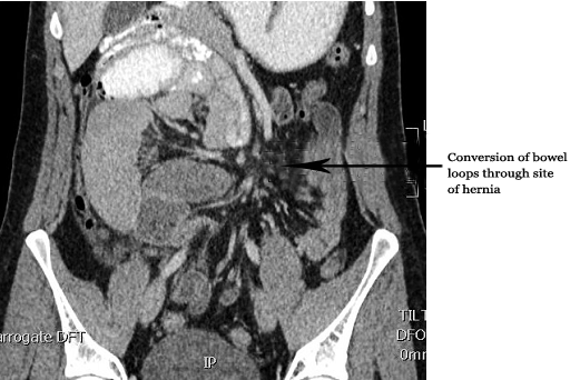
A 24-year-old male presented with a four-year history of intermittent colicky abdominal pain. These episodes were associated with abdominal distension and constipation and had previously resolved spontaneously. Over the four-year period a series of investigation were performed by his general practitioner, including baseline blood tests, serology for coeliac disease, Esophagogastroduodenoscopy (OGD) and colonoscopy. All investigations were normal. He had undergone an appendectomy for appendicitis. He presented acutely to hospital with symptoms and signs suggestive of mechanical bowel obstruction. Abdominal X-ray (AXR) and computed tomography (CT) scan were performed and they showed a cluster of abnormally dilated small bowel loops (Figures 1 and 2), with a mass effect on the ascending colon. The configuration of bowel loops seen on imaging suggested a closed loop obstruction secondary to an internal hernia. The patient proceeded to theatre for a laparotomy, findings at laparotomy were acute small bowel obstruction secondary to a right sided paraduodenal hernia. This was repaired and the patient recovered well. |
|
|
|
|
|
|
|
Discussion
|
|
Paraduodenal hernias are rare congenital anomaly arising from an error of midgut rotation. They are said to be the most common type of internal hernia. Both right and left paraduodenal hernias occur. [1] Right paraduodenal hernias occurs when bowel herniates through Waldeyer's fossa (a defect in the jejunal mesentery). [2] Left paraduodenal hernias can be considered truly congenital and occur when bowel prolapses through Landzert fossa, an aperture behind the fourth part of the duodenum. Signs and symptoms are variable. Intermittent episodes of self-limiting bowel obstruction may be associated with chronically incarcerated hernias. As in this case the patient had intermittent episodes of abdominal pain and distension. Complications of internal hernias include small bowel obstruction, ischemia, infarction and perforation. Differential diagnoses of internal hernias include foreign body impaction, intestinal volvulus, adhesions and tumor. [3] The treatment of a paraduodenal hernia is prompt surgical repair. |
|
Conclusion
|
|
Internal hernias are rare and can be a cause of recurrent episodes of abdominal symptoms. Critical review of imaging is needed to diagnose internal hernias. Prompt treatment by surgical repair is required to prevent complications of acute small bowel obstruction and the halt further recurrence of symptomatic episodes. |
|
References
|
|


|
[HTML Abstract]
[PDF Full Text]
|
|
Author Contributions:
Susannah Flexer – Conception and design, Acquisition of data, Analysis and interpretation of data, Drafting the article, Critical revision of the article, Final approval of the version to be published David Scullion – Analysis and interpretation of data, Critical revision of the article, Final approval of the version to be published |
|
Guarantor of submission:
The corresponding author is the guarantor of submission. |
|
Source of support:
None |
|
Conflict of interest:
Authors declare no conflict of interest. |
|
Copyright:
© Susannah Flexer et al. 2012; This article is distributed the terms of Creative Commons Attribution License which permits unrestricted use, distribution and reproduction in any means provided the original authors and original publisher are properly credited. (Please see Copyright Policy for more information.) |
|
|