|
|
|
|
Case Report
| ||||||
| Inguinal dermoid cyst masquerading as irriducible inguinal hernia: A case report | ||||||
| Soumen Das1, Utpal De1, Dilip Das1, Sudip Sarkar1 | ||||||
|
1Department of surgery, Ipgmer & sskm hospital, Kolkata, West Bengal, India.
| ||||||
| ||||||
|
[HTML Abstract]
[PDF Full Text]
[Print This Article]
[Similar article in Pumed] [Similar article in Google Scholar] 
|
| How to cite this article: |
| Das S, De U, Das D, Sarkar S. Inguinal dermoid cyst masquerading as irriducible inguinal hernia: A case report. International Journal of Case Reports and Images 2012;3(11):47–49. |
|
Abstract
|
|
Introduction:
Dermoid cysts are common developmental anomalies occurring along embryonic fusion lines. Occurrence of this in inguinal region is rare and imposes diagnostic challenge as it masquerades hernia.
Case Report: A 48-year-old male patient presented with irreducible left inguinal hernia. Exploration of the left inguinal canal revealed a cyst (10x7 cm) in the floor of the inguinal canal separated from the cord structures. The cyst was opened and foul smelling muddy paste like material along with a few hairs came out. Cyst was completely excised. The histopathology was consistent with a dermoid cyst. The patient is doing well at one year follow-up. Conclusion: Inguinal dermoid cyst mimicking irreducible hernia is rare but possible entity. If such cyst is encountered during hernia operation, complete excision is to be contemplated. | |
|
Key Words:
Dermoid Cyst, Inguinal Canal, Hernia
| |
|
Introduction
| ||||||
|
Dermoid cysts are developmental lesions occurring along the line of embryonic fusion. [1] Common sites include supraorbital region of forehead and midline. [1] Dermoid cyst of anterior abdominal wall is rare. Literature review revealed only five cases of inguinal dermoid till date. [1] [2] [3] [4] [5] [6] [7] We report a case with review of literature. | ||||||
|
Case Report
| ||||||
|
A 48-year-old male patient presented with a swelling in left groin since last 12 years. The swelling was progressive in nature, increased in size on straining, coughing and decreased on lying down. It was associated with occasional pain which subsided on medication. Physical examination revealed mild pallor with a left sided inguinoscrotal swelling. The swelling extended from mid inguinal region to root of left scrotum. It was pyriform shaped and measured 10 cm in its longitudinal axis. We could not get above the swelling. Cough impulse was present. The swelling was doughy on palpation. It was irreducible. Abdominal and per-rectal examinations were within normal limits. A clinical diagnosis of Irreducible left inguinal hernia was made and an elective inguinal hernioplasty was planned. Laboratory investigations reveled a hemoglobin- 9 g/dL, WBC count- 11,000/mm3, DLC-N63L30E7, 63%, L 30%, E 7%), blood glucose 110 g/dL, urea 20 mg/dL, serum creatinine 0.8 mg/dL. Chest X-ray and ECG were normal. Exploration of the left inguinal canal revealed a cyst (10x7 cm) in the floor of the inguinal canal separated Exploration of the left inguinal canal revealed a cyst (10x7 cm) in the floor of the inguinal canal separated from the cord structures. The cyst extended from the deep ring above to upper pole of left testis below. Cord structures were separated from cyst. No direct or indirect sac could be detected. The cyst was opened and foul smelling muddy paste like material along with a few hairs (Figure 1) came out. Cyst was completely excised and sent for histopathology. The postoperative recovery was uneventful and the patient was discharged on 10th post operative day after stitch removal. Histopathology revealed a thin-walled cystic lining composed of keratinized squamous epithelial cells. Underlying layers contained blood vessels, hair follicles, ecrine and apocrine glands. The above findings were consistent with a dermoid cyst. The patient is doing well at one year follow-up. | ||||||
| ||||||
|
Discussion
| ||||||
|
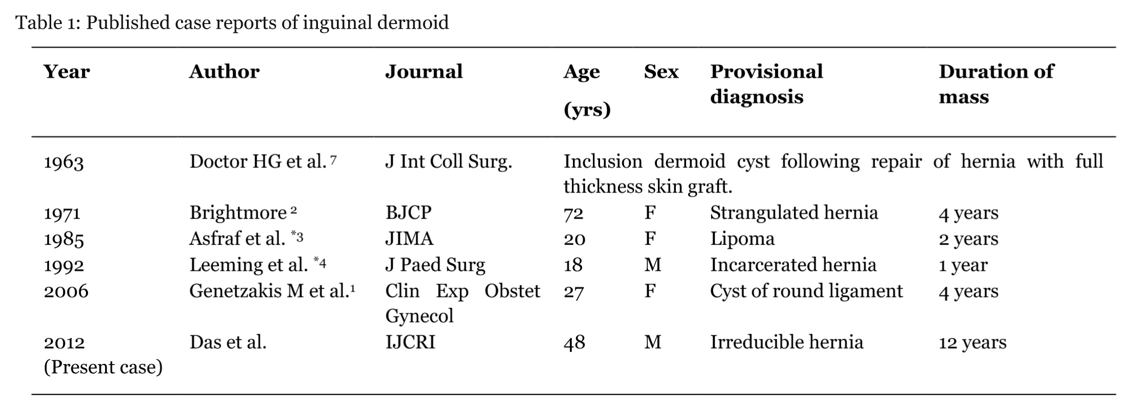
Hernia is the commonest inguinal swelling. Other common swellings include undescended testes, lipoma or hydrocele of spermatid cord. Rare inguinal swellings include preperitoneal lipoma, supernumerary pectineus bursa, haemorrhage into internal internal oblique muscle, round ligament angioma, pedunculated uterine fibromyoma, inguinal endometriosis and thrombophlebitis. [5] Dermoid cyst as a cause of inguinal swelling is rare. A search of English medical databases, using key words dermoid cyst and inguinal mass, revealed five case reports of inguinal dermoid till date (Table 1). [2] [3] [4] Of the five patients three were female [2] [3] and two male. [4] Four patients were below 30 years [3] [4] of age and the fifth patient was 72 years of age. [2] The duration of the swelling varied from 1 to 4 years. The swellings were provisionally diagnosed as inguinal hernia, [2] [4] lipoma [3] and cyst of the round ligament respectively. Dermoid cyst was revealed peroperatively and confirmed histopthologically. A fifth patient had dermo-plastic repair of inguinal hernia and later developed inclusion dermoid. [7]This procedure is obsolete and as such does not need further elaboration Dermoid cysts may be teratomatous or non teratomatous benign malformations. [3] [4] [5] Non teratomatous dermoids are common in the inguinal canal. Grossly the excised tumour may be mistaken for a sebaceous cyst or epidermoids. The microscopic presence of skin along with its appendages and sebaceous gland differentiates them from epidermoids and sebaceous cyst which have stratified squmaous epithelium surrounded by fibrous tissue forming their wall. [2] [3] [4] The absence of tissues foreign to the part differentiates it from a true dermoid found elsewhere. The contents of the cyst have been reported as tan-colored keratin resulting from accumulation of stratum corneum. [4] Diagnosis is often mistaken clinically as irreducible inguinal hernia. Complications of the cyst which resembles obstructed or incarcerated inguinal hernia include inflammation and hemorrhage. [4] The cyst may sometimes lead to compression of adjacent organs causing retention of urine and bowel obstruction. [4] The possibility of malignant degeneration exists especially in women with dermoid cysts arising from round ligament. [6] Tumor markers like alpha fetoprotein and beta chorionic gonadotropin estimation are helpful to monitor treatment and recurrence. [6] Surgical excision is the treatment of choice. [1] [2] [3] [4] [5] [6] | ||||||
| ||||||
|
| ||||||
|
Conclusion
| ||||||
|
Inguinal dermoid cyst is rare. If encountered complete excision is to be contemplated in order to avoid complications like-inflammation, hemorrhage and rarely malignant degeneration. | ||||||
|
References
| ||||||
| ||||||
|
[HTML Abstract]
[PDF Full Text]
|
|
Author Contributions:
Soumen Das – Substantial contributions to conception and design, Acquisition of data, Drafting the article, revising it critically for important intellectual content, Final approval of the version to be published Utpal De – Substantial contributions to conception and design, Acquisition of data, Drafting the article, revising it critically for important intellectual content, Final approval of the version to be published Dilip Das – Substantial contributions to conception and design, Acquisition of data, Drafting the article, revising it critically for important intellectual content, Final approval of the version to be published Sudip Sarkar – Substantial contributions to conception and design, Acquisition of data, Drafting the article, revising it critically for important intellectual content, Final approval of the version to be published |
|
Guarantor of submission:
The corresponding author is the guarantor of submission. |
|
Source of support:
None |
|
Conflict of interest:
Authors declare no conflict of interest. |
|
Copyright:
© Soumen Das et al. 2012; This article is distributed the terms of Creative Commons Attribution License which permits unrestricted use, distribution and reproduction in any means provided the original authors and original publisher are properly credited. (Please see Copyright Policy for more information.) |
|
|