| |
|
|
|
Case Report
| ||||||
| Primary duodenal adenocarcinoma of the fourth portion diagnosed using double-balloon enteroscopy and surgically resected: A case report | ||||||
| Shingo Kawano1, Koichi Sato1, Hiroshi Maekawa1, Mutsumi Sakurada1, Hajime Orita1, Ryo Wada2 | ||||||
|
1Department of Surgery, Juntendo Shizuoka Hospital, Juntendo University School of Medicine, 1129 Nagaoka, Izunokuni-shi, Shizuoka 410-2295, Japan.
2Department of Pathology, Juntendo Shizuoka Hospital, Juntendo University School of Medicine, 1129 Nagaoka, Izunokuni-shi, Shizuoka 410-2295, Japan. | ||||||
| ||||||
|
[HTML Abstract]
[PDF Full Text]
[Print This Article]
[Similar article in Pumed] [Similar article in Google Scholar] 
|
| How to cite this article: |
| Kawano S, Sato K, Maekawa H, Sakurada M, Orita H, Wada R. Primary duodenal adenocarcinoma of the fourth portion diagnosed using double-balloon enteroscopy and surgically resected: A case report. International Journal of Case Reports and Images 2012;3(11):35–39. |
|
Abstract
|
|
Introduction:
Primary duodenal adenocarcinoma is extremely rare. If this carcinoma occurs in fourth portion, it can now be diagnosed by recent developments in enteroscopy.
Case Report: We report a rare case of primary duodenal adenocarcinoma of the fourth portion diagnosed by double-balloon enteroscopy and resected surgically. A 57-year-old man was anemic. PET-CT revealed accumulation in the fourth portion of the duodenum. Double-balloon enteroscope showed circular tumor of the fourth portion of the duodenum, and biopsy disclosed poorly differentiated adenocarcinoma. Partial duodenectomy and partial colonectomy were performed. The marginal artery of the transverse colon was invaded. Histological examination disclosed that the tumor was poorly differentiated adenocarcinoma and two lymph node metastases were seen. Conclusion: Primary duodenal adenocarcinoma of fourth portion can be diagnosed by double-ballon enteroscopy and treated by surgical resection. | |
|
Key Words:
Double-balloon entroscopy, Primary duodenal adenocarcinoma, The fourth portion
| |
|
Introduction
| ||||||
|
Primary duodenal adenocarcinoma is extremely rare, accounting for 0.3v0.4% of all gastrointestinal cancers. [1] It is too difficult to anatomically diagnose primary duodenal adenocarcinoma of the fourth portion. However, primary duodenal adenocarcinoma of the fourth portion can be diagnosed by recent developments in enteroscopy. Here we report a case of duodenal adenocarcinoma of the fourth portion diagnosed by double-balloon enteroscopy and resected surgically, and discuss it based on a review of literature. | ||||||
|
Case Report
| ||||||
|
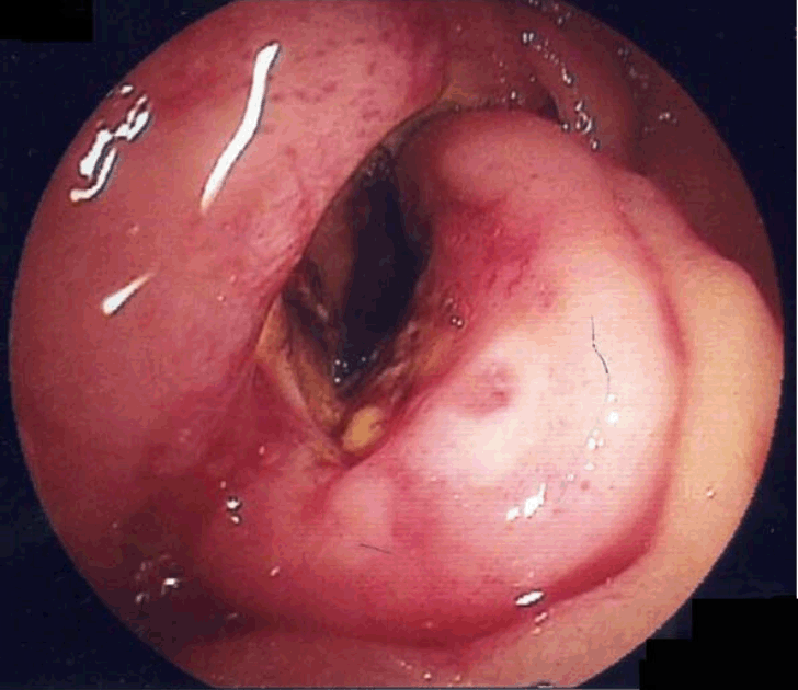
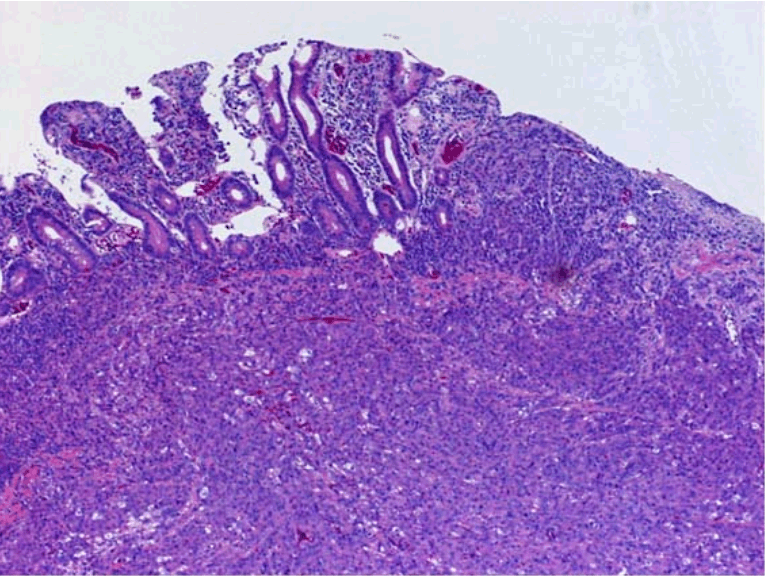
A 57-year-old Japanese male was admitted to our hospital in December 2009 for evaluation of severe anemia. Past medical history included a gastric ulcer when he was 50-years-old. His family history was noncontributory. Physical examination on admission revealed a body height of 175 cm, weight of 63 kg, blood pressure of 112/61 mmHg, a regular pulse of 85/min, and body temperature of 36.8oC. There was no sign of lymphadenopathy. His abdomen was soft and flat, and no abdominal, liver, or spleen masses were palpable. There were no abnormal laboratory findings, except evidence of anemia (hemoglobin was 6.6 g/dL) and an inflammatory reaction (white blood cell count was 8900/µg, and C-reactive protein was 3.7 mg/dL). Tumor markers, carcinoembryonic antigen (CEA) and carbohydrate antigen (CA19-9), were within normal ranges. Abdominal computed tomography (CT) scan indicated a circumferential, thick, and unequally enhanced wall in the fourth portion of the duodenum, and showed no lymph node swelling or ascites. Upper gastrointestinal series demonstrated a 7-cm 'apple core' sign in the fourth portion of the duodenum (Figure 1). Double-balloon enteroscopy showed a circumferential tumor with ulceration in the fourth portion of the duodenum (Figure 2). The enteroscope could not pass the lesion. Biopsy specimen from the lesion disclosed poorly differentiated adenocarcinoma. Positron Emission Tomography (PET) revealed accumulation in the fourth portion of the duodenum (Figure 3). Based on these findings, the patient was diagnosed with primary duodenal adenocarcinoma of the fourth portion and underwent partial duodenectomy and partial colectomy because the marginal artery of the transverse colon was invaded by the tumor on January 22nd, 2010. The resected tumor had deep ulceration with a round wall and measured 100×80 mm in size (Figure 4). Histological examination disclosed that the tumor was poorly differentiated adenocarcinoma which invaded into the subserosal layer and two lymph node metastases were seen (Figure 5). The patient had an uneventful postoperative course, and was discharged from hospital 40 days postoperatively. Combined chemotherapy of TS-1 and cisplatin was prescribed as neoadjuvant chemotherapy. TS-1 (80 mg/m2) was administered 21 days, followed by 14 days rest as one course. Cisplatin (60 mg/m2) was administered on the eight day. Six courses of this adjuvant chemotherapy were administered. The patient is alive with no recurrence for one year. | ||||||
| ||||||
| ||||||
| ||||||
| ||||||
| ||||||
|
Discussion
| ||||||
|
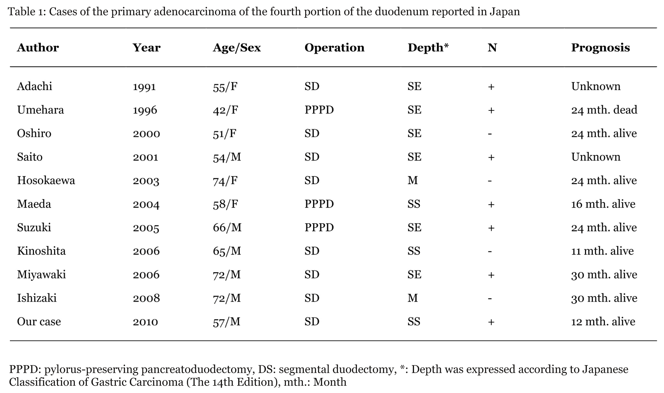
Primary duodenal adenocarcinoma is extremely rare. Most primary duodenal adenocarcinomas are in the first and second portions, with 20% in the third portion, and 10% in the fourth portion. [2] Recently, reports of primary duodenal adenocarcinoma have been increasing because of progresses in gastric enteroscopy. [3] [4] However, it is impossible to diagnose primary duodenal adenocarcinoma of the third and fourth portions by gastric enteroscopy. There are still many cases to be diagnosed by the upper gastrointestinal series. In this case, primary duodenal adenocarcinoma of the fourth portion was diagnosed by double-balloon enteroscopy. In Japan, there are no case reports of primary duodenal adenocarcinoma of the fourth portion diagnosed by double-balloon enteroscopy. In Brazil, there is only one case report. [5] In Japan, there are eleven case reports of primary duodenal adenocarcinoma of the fourth portion resected surgically, including our case [5] [6] [7] [8] [9] [10] [11] [12] [13] [14] [15] [16] (Table 1). The median age was 60.5 years (range, 42–74 years). Six cases were men. Two cases invaded into the mucosal layer, three cases into the subserosal layer, and six cases went beyond the serosal layer. Seven cases had metastasis of the lymph nodes. According to these reports, there are two kinds of surgery, three cases of pylorus preserving pancreatoduodenectomy and partial duodenectomy and eight cases of partial duodenectomy with lymphadenectomy. In Kaklamanos's report, there was no significant difference in prognosis between pancreatoduodenectomy and partial duodenectomy in sixty-three cases of primary duodenal adenocarcinoma. [16] In Lowell's report, the seven cases of primary duodenal adenocarcinoma of the third or fourth portions are all alive after five years, except for those who died of another disease. [17] Five of seven cases underwent partial duodenectomy. There are many reports that partial duodenectomy is better in the case of primary duodenal adenocarcinoma in the third or fourth portions. Consequently, partial duodenectomy was thought to be a better surgery for primary distal duodenal adenocarcinoma. However, in Suzuki's report, a case of primary duodenal adenocarcinoma of the fourth portion underwent pancreatoduodenectomy. [12] There were metastases of the lymph nodes in No. 13 and No. 14 according to the Japanese classification of gastric carcinoma (The 14th Edition). That report insisted on the necessity of pancreatoduodenectomy with lymphadenectomy. Our case underwent partial duodenectomy with lymphadenectomy. There was no recurrence for one year in spite of advanced carcinoma and metastasis of the lymph nodes. | ||||||
| ||||||
|
| ||||||
|
Conclusion
| ||||||
|
We reported an extremely rare case of primary duodenal adenocarcinoma of the fourth portion diagnosed by double-balloon enteroscopy and resected surgically. | ||||||
|
References
| ||||||
| ||||||
|
[HTML Abstract]
[PDF Full Text]
|
|
Author Contributions:
Shingo Kawano – Substantial contributions to conception and design, Acquisition of data, Analysis and interpretation of data, Drafting the article, Revising it critically for important intellectual content, Final approval of the version to be published Koichi Sato – Acquisition of data, Revising it critically for important intellectual content, Final approval of the version to be published Hiroshi Maekawa – Acquisition of data, Revising it critically for important intellectual content, Final approval of the version to be published Mutsumi Sakurada – Acquisition of data, Revising it critically for important intellectual content, Final approval of the version to be published Hajime Orita – Acquisition of data, Revising it critically for important intellectual content, Final approval of the version to be published Ryo Wada – Acquisition of data, Revising it critically for important intellectual content, Final approval of the version to be published |
|
Guarantor of submission:
The corresponding author is the guarantor of submission. |
|
Source of support:
None |
|
Conflict of interest:
Authors declare no conflict of interest. |
|
Copyright:
© Shingo Kawano et al. 2012; This article is distributed the terms of Creative Commons Attribution License which permits unrestricted use, distribution and reproduction in any means provided the original authors and original publisher are properly credited. (Please see Copyright Policy for more information.) |
|
|