|
|
|
|
Case Report
| ||||||
| An uncommon cause of apparent life-threatening event | ||||||
| Muhammad Waseem1, Dee Soontharothai1, Heidi Pinkert1, Evelyn Erickson2, Michael Trotman1 | ||||||
|
1Department of Emergency Medicine, Lincoln Medical & Mental Health Center, Bronx, NY, United States.
2Department of Pediatrics, Lincoln Medical & Mental Health Center, Bronx, NY, United States. | ||||||
| ||||||
|
[HTML Abstract]
[PDF Full Text]
[Print This Article]
[Similar article in Pumed] [Similar article in Google Scholar] 
|
| How to cite this article: |
| Waseem M, Soontharothai D, Pinkert H, Erickson E, Trotman M. An uncommon cause of apparent life-threatening event. International Journal of Case Reports and Images 2012;3(11):17–20. |
|
Abstract
|
|
Introduction:
An apparent life threatening event (ALTE) in infancy is a common reason for presentation to the emergency department (ED). We report a case of a breast-fed infant who presented with an ALTE secondary to hypocalcemia from undiagnosed rickets.
Case Report: A 9-month-old girl was brought to the ED because she stopped breathing. The parents reported that she stopped breathing for more than 20 seconds and was gasping for air. During the event, she became unresponsive and her eyes rolled back. There was a history of cough and nasal congestion with subjective fever for two days. Further questioning revealed that over the last few months, she had had episodes of 'gasping', and 'strange noisy breathing sounds.' Her mother reported an 'odd cough' for several months. The baby had been exclusively breast-fed and was noted by her mother to be feeding well. No solid foods had been introduced to the patient. The past medical history was significant for developmental delay. The patient was born full-term via normal spontaneous vaginal delivery. The pregnancy, however, had been complicated by oligohydramnios. The parents reported no family history of vitamin D deficiency, rickets or seizure disorder. A chest radiograph showed flaring of the costochondral junctions consistent with rickets. Her vitamin D 25-OH level was 5 ng/mL (20–100) and D3 level was less than 5 ng/ml. Conclusion: Hypocalcemia is a difficult to diagnose in infants because there may present with a wide range of non-specific clinical symptoms, or they may be asymptomatic. It is important for the emergency physician to recognize the association of hypocalcemia with ALTE in infants. Rickets is a re-emerging health problem in infants and children. Physicians should therefore maintain a high index of suspicion for hypocalcemia especially in a breast-fed infant. We suggest including serum calcium level as part of the evaluation of ALTE. | |
|
Key Words:
Rickets, Hypocalcemia, Seizures; ALTE
| |
|
Introduction
| ||||||
|
An apparent life threatening event (ALTE) is a common reason for presentation to the emergency department (ED). Although the occurrence of seizures due to hypocalcemia in early infancy is well known, [1] its association with an acute ALTE is rare. [2] We report a case of a breast-fed infant who presented with ALTE secondary to hypocalcemia from undiagnosed rickets. | ||||||
|
Case Report
| ||||||
|
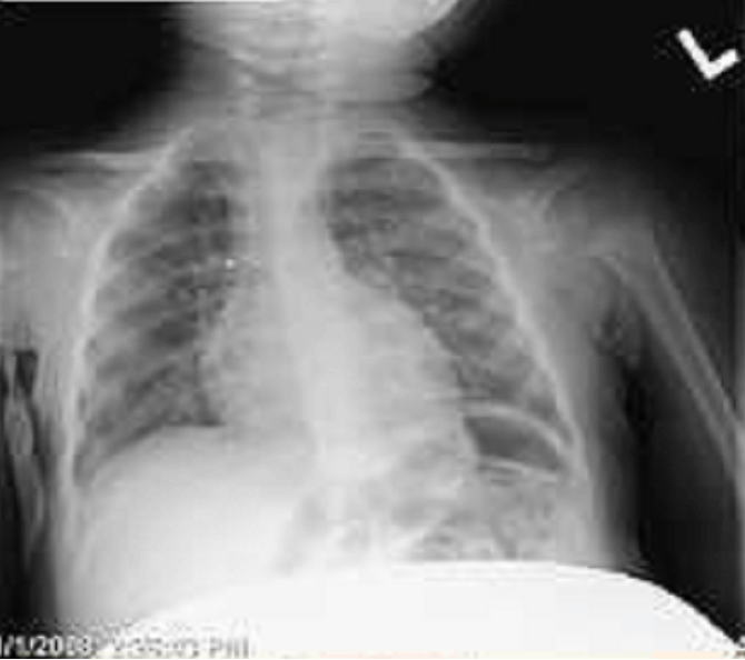
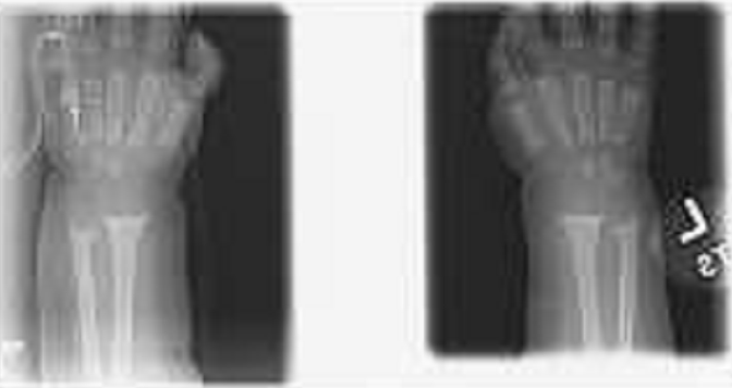
A 9-month-old female baby was brought to the ED because she stopped breathing. Parents reported that she stopped breathing for more than 20 seconds, and was gasping for air. During the event, she became unresponsive and her eyes rolled back. There was a prior history of cough and nasal congestion with subjective fever for two days. Further questioning revealed that over the last few months, she had episodes of 'gasping', and strange noisy breathing sounds. Her mother reported an 'odd cough' for several months. Although at nine months this infant should have been introduced to solid foods, this baby had been exclusively breast-fed and was noted by her mother to be feeding well. Her past medical history was significant for developmental delay. The patient was born full-term, via normal spontaneous vaginal delivery. The pregnancy, however, had been complicated by oligohydramnios. The parents reported no family history of vitamin D deficiency, rickets or seizure disorder. On arrival, the patient was alert and awake. The vital signs on arrival were as follows: temperature 102.8°F, heart rate 155/minute, respiratory rate 34/minute, and oxygen saturation 100% on room air. The growth parameters were as follows: weight 9.4 kg (75%), length 73 cm (75%), and head circumference 47 cm (95%). During the physical examination in the ED, however, the patient had a brief episode of seizure-like activity. The episode lasted a few seconds with desaturation of 88–90% on room air. Mother also reported similar episodes at home. She had no signs of trauma. The neck was supple, and pupils were 3 mm and equally responsive to light with normal extra-ocular movements. Tympanic membranes were normal, and the throat was unremarkable. Clear rhinorrhea was present. She had increased work of breathing with obvious retractions, and an increased respiratory rate with frequent stridor, but no rales or wheezing present. The heart sounds were normal. The abdominal exam was unremarkable. The neurological exam revealed normal tone and reflexes, and a normal sensory examination. No nystagmus was present. The infant was moving all extremities. No rashes or skin lesions were noted. The remainder of her physical examination was unremarkable. Laboratory investigations revealed a hemoglobin level of 10.7 g/L, a platelet count of 330,000/mm3, and a WBC count of 15,700 /mm3 with 64% neutrophils, 34% lymphocytes, and 0.9% monocytes. The serum electrolytes were as follows: sodium 137 mEq/L, potassium 4.3 mEq/L, chloride 103 mEq/L, bicarbonate 14 mg/dL, blood urea nitrogen 6 mg/dL, creatinine 0.4 mg/dL, glucose 113 mg/dL, and total calcium 4.9 mg/dL. Her magnesium was 1.9 mg/dL. Repeat total calcium was 6.5 mg/dL, and phosphate 3.2 mg/dL (Nasal secretions for respiratory syncitial virus, and influenza A and B antigens were negative. A chest radiograph showed bilateral pulmonary opacities consistent with pneumonia and flaring of the costochondral junctions consistent with rickets (Figure 1). Findings on radiographs of her wrists also supported the diagnosis of rickets (Figure 2). Her vitamin D 25-OH level was 5 ng/mL (20–100) and D3 level was less than 5 ng/mL. The patient responded to intravenous calcium replacement with rapid resolution of symptoms. Her calcium level returned to normal and she was discharged home in a stable condition. | ||||||
| ||||||
| ||||||
|
Discussion
| ||||||
|
An ALTE is defined as 'an episode that is frightening to the observer and is characterized by some combination of apnea, color change, change in muscle tone, and choking'. [3] It accounts for 0.6–0.8% of emergency department visits in infants. [4] Hypocalcemia is a difficult to diagnose in infants because there may be a wide range of non-specific clinical symptoms, or patients may be asymptomatic. A high index of suspicion is needed to diagnose hypocalcemia and it is important for the emergency physician to recognize hypocalcemia as a cause of ALTE in infants. These patients may present with non-specific symptoms of weakness, feeding problems, facial spasms, jitteriness or seizures. Other symptoms of hypocalcemia may include lethargy, irritability, and vomiting. Commonly, infants present with hypocalcemic tetany or seizures, whereas older children present with failure to thrive or skeletal abnormalities. Laryngospasm also can occur during seizure-like activity but may itself be a symptom of hypocalcemia. [5] Therefore, determination of serum calcium may be important in an infant with this presentation. Rickets is the failure of growing bone to mineralize. There are many types of rickets, including nutritional and familial forms. Nutritional rickets due to vitamin D deficiency is the most common form. Vitamin D is critical for skeletal development and is converted to its physiologically active form, cholecalciferol, by exposure to ultraviolet radiation in sunlight. Activated vitamin D then helps bones absorb calcium and phosphorus from food. Vitamin D deficiency impairs mineralization of bone tissue and growth plates, manifesting as rickets. Nutritional rickets may result from inadequate sunlight exposure, or inadequate intake of dietary vitamin D, calcium, or phosphorus. [6] Classically, nutritional rickets manifests after six months of age. Therefore, in addition to checking calcium levels, magnesium, phosphorus, PTH and vitamin D metabolites should be measured. This determination should be made prior to initiation of treatment in order to delineate the cause of rickets. The most common laboratory findings in nutritional rickets are decreases in serum calcium, serum phosphorus, calcidiol, calcitriol, and urinary calcium. Parathyroid hormone, alkaline phosphatase, and urinary phosphorus levels are however, elevated. The radiographs of rapidly growing skeletal areas, such as the knee or wrist are helpful in making the diagnosis. Nutritional rickets due to vitamin D deficiency has become rare in the US due to the addition of Vitamin D to milk. However, with an increase in breast-feeding, which offers no Vitamin D fortification, this preventable disease is re-emerging. [7] Although breast milk is indisputably the ideal food for infants, it typically contains about 25 IU or less vitamin D per liter, [8] which is not sufficient for the prevention of rickets. It is, therefore, important to consider the screening of patients at risk of developing vitamin D deficiency. [9] In addition, supplementation should also be encouraged in breastfed babies of African–American mothers, who are at higher risk. In our patient, a number of factors pointed to nutritional rickets as the diagnosis. African–American race in an exclusively breast fed infant was the first indication, that nutritional rickets should be considered. This, coupled with an urban life style with inadequate sun exposure made the scenario likely for a nutritional basis for rickets. This remains a classic presentation for nutritional (vitamin D deficiency) rickets. | ||||||
|
Conclusion
| ||||||
|
Rickets is a re-emerging health problem in infants and children. Physicians should therefore maintain a high index of suspicion when confronted with unexplained seizures in a breast-fed infant. Early diagnosis is essential because morbidity can be minimized with early treatment. The determination of vitamin D status should be considered in the diagnostic evaluation of hypocalcemia in infants. | ||||||
|
References
| ||||||
| ||||||
|
[HTML Abstract]
[PDF Full Text]
|
|
Author Contributions:
Muhammad Waseem – Substantial contributions to conception and design, Acquisition of data, Analysis and interpretation of data, Drafting the article, Revising it critically for important intellectual content, Final approval of the version to be published Dee Soontharothai – Analysis and interpretation of data, Revising it critically for important intellectual content, Final approval of the version to be published Heidi Pinkert – Analysis and interpretation of data, Revising it critically for important intellectual content, Final approval of the version to be published Evelyn Erickson – Analysis and interpretation of data, Revising it critically for important intellectual content, Final approval of the version to be published Michael Trotman – Analysis and interpretation of data, Revising it critically for important intellectual content, Final approval of the version to be published |
|
Guarantor of submission:
The corresponding author is the guarantor of submission. |
|
Source of support:
None |
|
Conflict of interest:
Authors declare no conflict of interest. |
|
Copyright:
© Muhammad Waseem et al. 2012; This article is distributed the terms of Creative Commons Attribution License which permits unrestricted use, distribution and reproduction in any means provided the original authors and original publisher are properly credited. (Please see Copyright Policy for more information.) |
|
|