|
|
|
|
Case Report
| ||||||
| Hepatic hemangioma and focal nodular hyperplasia in a Nigerian patient with hepatitis B virus infection | ||||||
| Aderemi O Oluyemi1, Adekunle O Adeyomoye2, Nicholas A Awolola3 | ||||||
|
1General Hospital, Ikorodu, Lagos State, Nigeria.
2Department of Radiodiagnosis, College of Medicine, University of Lagos, Idi-Araba, Lagos State, Nigeria. 3Department of Morbid Anatomy, College of Medicine, University of Lagos, Idi-Araba, Lagos State, Nigeria. | ||||||
| ||||||
|
[HTML Abstract]
[PDF Full Text]
[Print This Article]
[Similar article in Pumed] [Similar article in Google Scholar] 
|
| How to cite this article: |
| Oluyemi AO, Adeyomoye AO, Awolola NA. Hepatic hemangioma and focal nodular hyperplasia in a Nigerian patient with hepatitis B virus infection. International Journal of Case Reports and Images 2012;3(11):8–10. |
|
Abstract
|
|
Introduction:
Hepatic hemangiomas (HH) and focal nodular hyperplasia (FNH) are, by far singly and combined together, the most frequently occurring benign liver lesions. The association between these two tumors is well documented in literature. The association has been suggested to spring from a common etiopathogenetic mechanism.
Case Report: A 42-year-old male presented for evaluation as a routine pre-blood donation screen had shown that he was hepatitis B surface antigen (HBsAg) positive. An abdominal ultrasonography showed two distinct hyperechogenic intrahepatic masses. One mass was diagnosed with doppler ultrasound as hemangioma and a ultrasound-guided biopsy of the other mass revealed hepatocellular hyperplasia. Conclusion: This case for the first time documents the interesting HH/FNH association from Nigeria. It also details what, to the best of our knowledge, is the first time that these diseases are being documented as coexisting with hepatitis B virus (HBV) infection. | |
|
Key Words:
Focal nodular hyperplasia, Hepatic hemangioma, Hepatitis B infection, Nigeria
| |
|
Introduction
| ||||||
|
Hepatic hemangiomas (HH) and focal nodular hyperplasia (FNH) are, singly and combined, by far the most frequently occurring benign liver lesions. The common presence of these two hepatic lesions is well documented in literature and it has been suggested that this association springs from a common etiopathogenetic mechanism of growth and development. However, such documentations have not been common in data from the Western sub-region of Africa. This interesting association has not been documented from Nigeria and to the best of our knowledge, nor has there been a previous report of HH and FNH as coexisting with hepatitis B virus (HBV) infection anywhere in literature | ||||||
|
Case Report
| ||||||
|
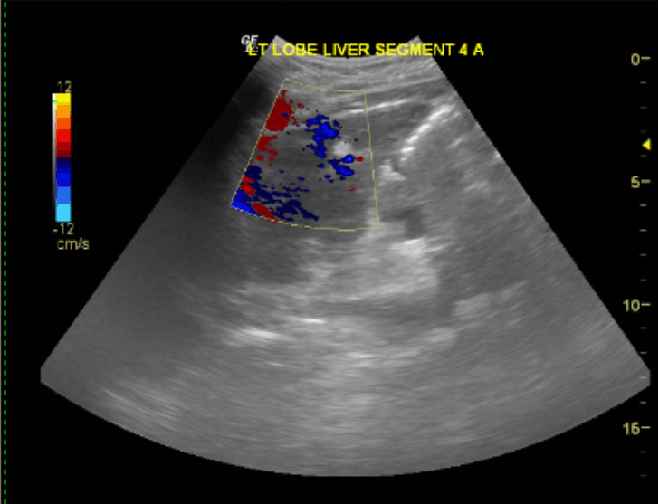
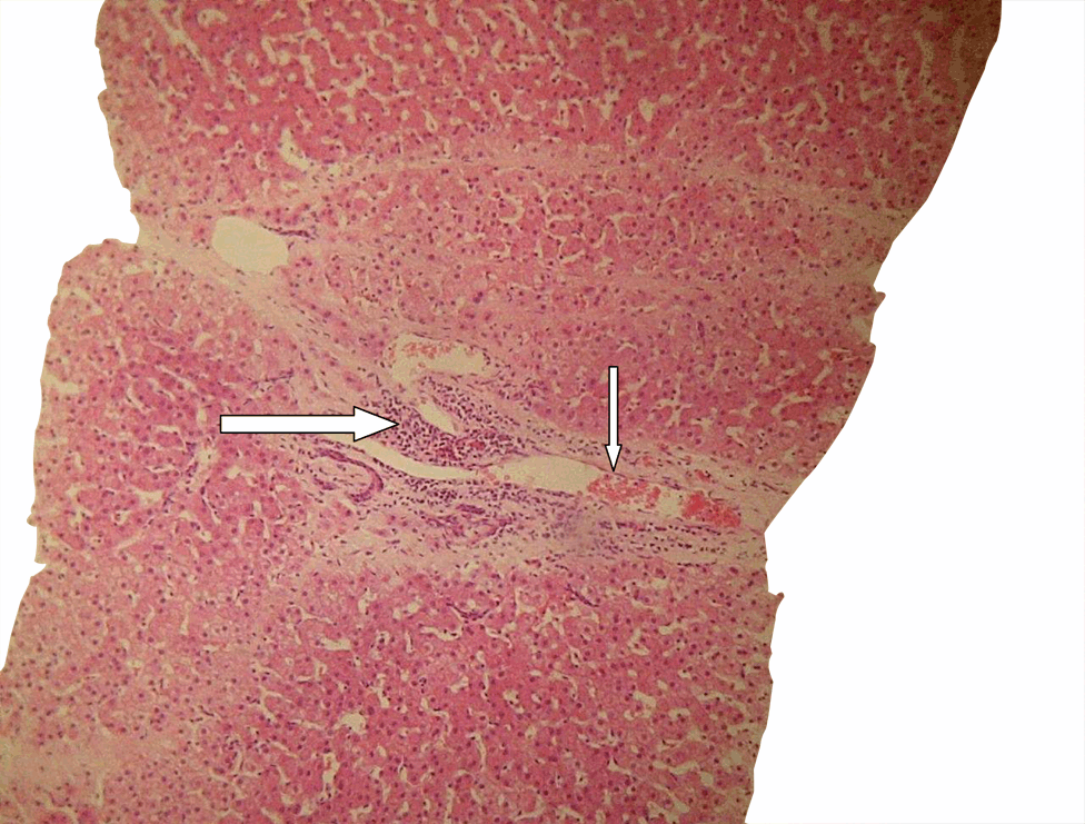
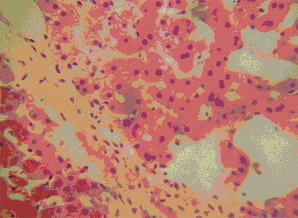
A 42-year-old male presented for evaluation as a routine pre-blood donation screen had shown that he was hepatitis B surface antigen (HBsAg) positive. He did not have a previous history of liver disease nor diabetes mellitus and was asymptomatic at presentation. His body mass index was 22.6 kg/m2 and other examination findings were unremarkable. Liver function tests including serum a-fetoprotein and carcinoembriogenic antigen levels were normal. The quantification of HBV deoxyribonucleic acid (DNA) levels from patient's serum was less than 20 IU/per mL (COBAS TaqMan kit) and a screen for hepatitis C virus was negative. Fasting samples were drawn for blood glucose and lipid profile and results were normal. An abdominal ultrasonography (USG) showed two distinct hyperechogenic intrahepatic masses. The first was an 8.7 mm in diameter nodule in segment VIII which on color flow Doppler ultrasound mode showed flow into and within the mass. These features were in keeping with a hemangioma. The second lesion seen in segment II was 12 mm in diameter and on color Doppler, demonstrated no flow within the mass (Figure 1). The differentials of the second mass included early hepatocellular carcinoma, a focal fatty infiltration, hepatic adenoma and focal nodular hyperplasia. The histologic assessment of tissue obtained from a ultrasound-guided liver biopsy of the second mass revealed nodules of proliferating hepatocytes arranged in two cell layers which were supported by well developed reticulin framework. The hepatocytes were pale but showed no evidence of fatty change. The hepatic nodules were separated by fibrous septa containing several vessels, including small arteries and veins as well as numerous ductules (Figures 2 and 3). Hence, a diagnosis was made of Hemangioma and Focal Nodular Hyperplasia in a patient with HBV infection was made. The patient was subsequently informed and appropriately counseled. He is presently on regular follow up at a hepatology outpatient unit. | ||||||
| ||||||
|
| ||||||
| ||||||
|
Discussion
| ||||||
|
Hepatic hemangioma and FNH lesions are the two most common liver cell-derived benign tumors and are estimated to be present in 0.4–20% and 8% of general adult population from the western world. [1] [2] [3] While each of these lesions are found commonly as isolated entities, an association between HH and FNH has also been well documented in literature. [1] [4] [5] While coexisting third lesions such as adenomas [6] and liver cysts [7] have been reported in association with these two pathologies, to the best of our knowledge this is the first documented case of such association from Nigeria and is the first time that these two lesions are being presented in association with HBV infection. The association between HH and FNH hinges around the presumed etiopathogenetic mechanisms that relates to local abnormalities in hepatic blood supply that somehow facilitate the hyperplastic development of these benign lesions. [4] Their shared property of development/growth with the intake of oral contraceptives has also been pointed out in support of this relationship. [6] [8] Literature is, however, void of a suggestion of explanation of a possible association between HBV infection and these tumors. | ||||||
|
Conclusion
| ||||||
|
We surmise that this relationship may simply represent a fortuitous one as Nigeria is a region of high endemicity for HBV infection (> 8%) with an estimated lifetime risk of infection being greater than 50%. [9] The probability such of coincidental coincidence is high. Granted that the presence of HBV infection may or may not be important from an etiopathogenetic standpoint, it is, however, pertinent to note that this relationship can exist in patients with HBV infection. A piece of information that could have various ramifications in the evaluation of hepatic lesions in HBV infection particularly in regions of high endemicity. | ||||||
|
Acknowledgements
| ||||||
|
The article has been read by the patient and written consent obtained for publication. We would like to thank the management of Foremost Radiology, and The Specialist Labs both in Surulere, Lagos State, Nigeria for carrying out the ultrasonography and histopathologic assessment respectively. | ||||||
|
References
| ||||||
| ||||||
|
[HTML Abstract]
[PDF Full Text]
|
|
Author Contributions:
Aderemi Oluyemi - Initial conception and design, Drafting the article, Critical revision of the article Adekule Adeyomoye - Contributed substantially in the design of the report. He provided the abdominal ultrasonography, guided the liver biopsy and Critical revision of this case report Nicholas Awolola - Contributed substantially in the design of the report. He provided the histology for the report and made critical revisions to the article content |
|
Guarantor of submission:
The corresponding author is the guarantor of submission. |
|
Source of support:
None |
|
Conflict of interest:
Authors declare no conflict of interest. |
|
Copyright:
© Aderemi Oluyemi et al. 2012; This article is distributed the terms of Creative Commons Attribution License which permits unrestricted use, distribution and reproduction in any means provided the original authors and original publisher are properly credited. (Please see Copyright Policy for more information.) |
|
|