| Table of Contents |  |
|
Case Report
|
| Pyoderma gangrenosum – rare manifestation of crohn's disease in 14-year-old child: Case report |
| Ajay Damor1, Varsha Shah1, Lalit Nainiwal1, Bhadra Trivedi2, Rohit Agrawal2 Vaibhav Patel3 |
|
1Assisstant Professor, Department of Pediatrics, SBKS MIRC, Pipariya, Vadodara, Gujarat, India.
2Third Year Pediatrics Resident, Department of Pediatrics, SBKS MIRC, Pipariya, Vadodara, Gujarat, India. 3Second Year Pediatrics Resident, Department of Pediatrics, SBKS MIRC, Pipariya, Vadodara, Gujarat, India. |
|
doi:10.5348/ijcri-2012-08-166-CR-13
|
|
Address correspondence to: Dr Ajay Damor Plot No 425/5, Sector 5A Gandhinagar - 382006 Gujarat India Phone: +91 9687625563 Email: drajdamor@yahoo.co.in |
|
[HTML Abstract]
[PDF Full Text]
|
| How to cite this article: |
| Damor A, Shah V, Nainiwal L, Trivedi B, Agrawal R, Patel V. Pyoderma gangrenosum – rare manifestation of crohn's disease in 14-year-old child: Case report. International Journal of Case Reports and Images 2012;3(8):46–50. |
|
Abstract
|
|
Introduction:
Pyoderma gangrenosum (PG) is an idiopathic, ulcerative, non-infective chronic inflammatory skin disorder of unknown etiology rarely affects children. It is associated with systemic medical illness in 50% of cases like inflammatory bowel disease, systemic arthritis, hematological diseases and malignancies. Characteristic lesions begin as pustule or vesiculopustule and progresses to an ulcer or deep erosion with violaceous undermined borders. The diagnosis of pyoderma gangrenosum is clinical and depends on exclusion of other causes of cutaneous ulceration. The management of PG is treatment of underlying systemic medical illness and judicious use of immunosuppressant.
Case Report: A 14-year-old female child who presented with multiple ulcerative lesions all over body especially on leg, foot, arm, hand, scalp and face, with largest one on right leg. Associated complaints were fever, abdominal pain, diarrhea, fatigue, anorexia and weight loss. On examination; along with multiple ulcers, she had pallor and clubbing. Systemic examination revealed no abnormality except mild tenderness in right lumber region. On investigation she was diagnosed as Crohn's disease (CD) and treated with corticosteroid. On subsequent follow up visit she had symptomatic improvement and healed ulcers. Conclusion: Pyoderma gangrenosum is rare manifestation of Crohn's disease in children. The diagnosis of PG is based on the typical clinical features and exclusion of other causes of ulcerative skin disorders and evaluation of systemic medical illness. The management is treatment of underlying systemic medical illness and judicious use of immunosuppressant. | |
|
Key Words:
Pyoderma gangrenosum, Crohn's disease
| |
|
Introduction
|
|
Pyoderma gangrenosum is an idiopathic, ulcerative chronic inflammatory skin disorder of uncertain etiology. The disease is more common in adults but children may also be affected on rare occasion. [1] Pathophysiology of PG is poorly understood; although the disease is idiopathic in 25–50% of patients, generally associated with other medical illnesses in 50% of case in adult and children. Diagnosis is clinical and depends on the exclusion of other causes of cutaneous ulceration and evaluation of associated secondary disease. We report a rare case of pyoderma gangrenosum with multiple ulcers associated with Crohn's disease in a 14-year-old child. |
|
Case Report
|
|
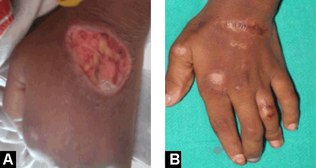
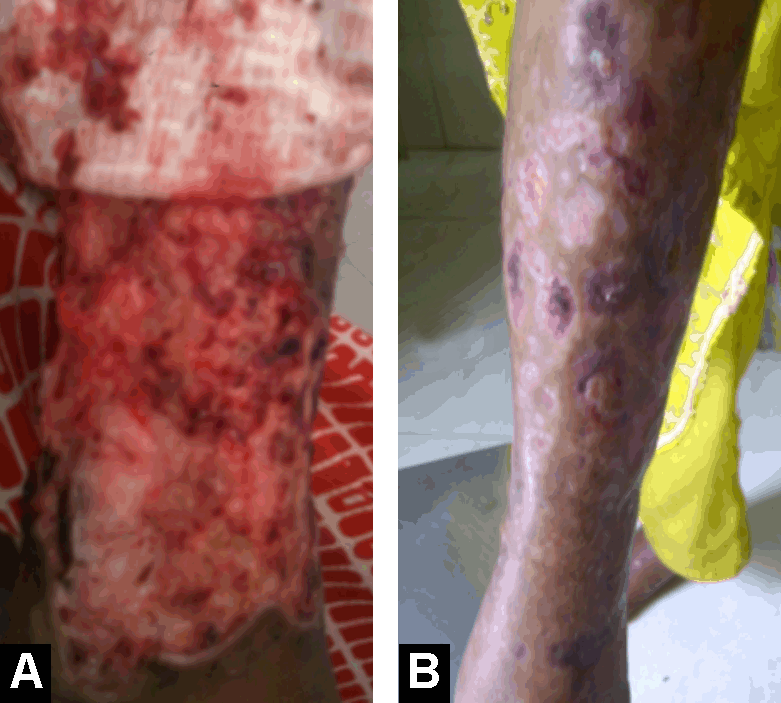
A 14-year-old female born to a non-consanguineous marriage presented with multiple ulcerative lesions all over body about 40 in number of different sizes, which began as small pustules progressed to large ulcers all over body especially on leg, foot, arm, hand, scalp and face, with the largest one on pretibial area of right leg for last six months. Associated complaints were intermittent moderate to high-grade fever, intermittent diffuse abdominal pain, and diarrhea without visible blood mucous or pus, fatigue, anorexia and weight loss for last one year. On examination, patient had multiple ulcers about 40 in number at different sites, with a mucopurulent base, violaceous undermined border and peripheral erythema (Figures 1A and 2A). Ulcers were varied in size. Largest ulcer was pretibial, about 16x10 cm size on right lower limb. She had pallor and clubbing. There was no lymphadenopathy. She was severely malnourished and her weight and height were less than 3rd centile for her age and she had delayed puberty. Systemic examination revealed no abnormality except mild tenderness at right iliac fossa. Family history was not significant. No history of tuberculosis contact in family members. On investigation, she had hypochromic microcytic anemia (Hb: 8 g/dL), complete blood count showed a leukocytosis (total count 16,000/cu mm with segmented neutrophils 71%, lymphocytes 24%, monocytes 3%, eosinophil 2%) with thrombocytosis (559,000/cu mm) with elevated erythrocyte sedimentation rate (ESR). Blood tests for urea, creatinine, electrolytes, liver function tests, sugar did not revealed any abnormality. The results for Venereal Disease Research Laboratory (VDRL), antinuclear antibody (ANA), enzyme-linked immunosorbent assay (ELISA) for HIV 1 and 2 were negative. Stool examination for occult blood was positive. A skin biopsy was taken from the edge of the lesion and histopathological examination reveal stratified squamous lining epithelium with areas of necrosis and ulceration. Underlying tissue shows granulation tissue, perivascular lymphocytic infiltrate with few neutrophils and histiocytes (Figure 3). These findings are consistent with clinical diagnosis of pyoderma gangrenosum. Pus culture from ulcer reveled S. aureus. Barium meal follows through showed edematous small bowel wall with coarse mucosal pattern, suggestive of Crohn's disease. Ileocolonoscopy revealed the skip lesions; cobble stoning of mucosa, apthous ulcers and also some deep irregular ulcers of varying sizes with normal intervening mucosa. Intestinal biopsy followed by histopathology showed the presence of noncaseating granuloma is diagnostic of CD. Anti-saccharomyces cerevisiae antibody (ASCA) was positive. Abdominal tuberculosis was ruled out as it is common entity in India and having similar clinical and histological finding. As per clinical finding and investigation she had pyoderma gangrenosum with Crohn's disease and she was treated with broad-spectrum antibiotics, pulse methyl prednisolone therapy for five days followed by oral prednisolone 2 mg/kg for four weeks, which was, tapered 2.5 mg every week. Supportive treatment with local ulcer dressing, anemia correction, and nutritional management was carried out simultaneously. After 8 weeks she had healed ulcers with scars all over body especially on leg, foot, arm, hand, scalp and face and symptomatic improvement for other associated complaints (Figures 1B and 2B). |
|
|
|
|
|
|
|
Discussion
|
|
Pyoderma gangrenosum was first described by Brocq in 1916 as 'phagédenisme géométrique'. [2] In 1930, at the Department of Dermatology, Mayo Clinic, Brunsting, Goeckerman and O'Leary coined the term Pyoderma Gangrenosum. [3] 'Pyoderma Gangrenosum' term was given because characteristic appearance of the lesion. The cutaneous lesions begin as pustule or vesiculopustule and progresses to an ulcer with undermined borders that evolves to an enlarging necrotic suppurative ulcer after gangrenous changes into the overlying skin. The lesions last from a few months to years, and heal with cribriform scarring. Lesions have been classified into: (A) Ulcerative, (B) Pustular, (C) Bullous, (D) Vegetative, (E) Periosteal, (F) Genital, (G) Infantile, and (H) Extra cutaneous. [4] The incidence of PG in adult estimated to be 3–10 patients per year occurring most commonly between 20–50 years of age. [5] The proportions of children less than 15 years of age in two larger series of patients with PG at Mayo Clinic were 4% and 4.6%. [6] [7] Although the disease is idiopathic in 25–50% of patients, an underlying immunologic abnormality may exist as suggested by its frequent association with systemic diseases with a suspected autoimmune pathogenesis. [5] The phenomenon of pathergy (development of skin lesion at the site of trivial trauma) would suggest altered, exaggerated, and uncontrolled inflammatory responses to non-specific stimuli. [3] Pathophysiology of PG is poorly understood, but dysregulation of immune system, especially altered neutrophil chemotaxis is the primary process. [8] The PG is known to be associated with a variety of systemic disorders. In adults with PG, associated systemic disorder found in 40–78% of cases. In children commonly associated systemic disease are inflammatory bowel disease, arthritis, leukemia, systemic lupus erythematous, immunodeficiency disorders and HIV, [9] etc. are the major underlying disorders in children [6, 7]. PG has been reported to occur in 2–12% of IBD in adult patients; in most of patient PG associated with ulcerative colitis, but prevalence of PG with Crohn's disease is much lower than ulcerative colitis. [10] Graham et al. reviewed 45 pediatric patient with PG; among them 12(26.6%) children had ulcerative colitis while only 7(9%) had Crohn's disease though IBD was major etiological factor for PG in children. Ulcers at certain anatomic sites are suggestive of pyoderma gangrenosum. Lesions are most commonly found on the lower legs, [11] but they may occur on the abdomen, thighs, buttocks, chest, head, neck, and anywhere on the skin. Graham et al. reported that anatomical distribution of ulcers among pediatric population were lower extremities (80%), head (26.1%), buttocks (15%), and children less than 2 years were notable for having perianal or genital region. [9] In our case report patient had multiple ulcers about 40 in number at different anatomical sites especially on leg, foot, arm, hand, scalp and face, with largest one on pretibial surface of right lower limb, PG is characterized by sterile ulcer; however several microorganisms cultured from PG had been reported in literature. [12] In our case report pus culture was positive for Staphylococcus aureus. Diagnosis is clinical and depends on the exclusion of other causes of cutaneous ulceration. No specific pathologic or laboratory findings exist. Histopathological findings in PG are not specific. However, a biopsy finding includes extensive sterile dermal neutrophilia. In early cases, mixed cell infiltrations may be present. Vascular inflammation in lesions of PG is not uncommon. Granuloma formation is generally believed to be incompatible with the diagnosis of PG evaluation of underlying systemic illness is of prime importance in PG. The diagnosis of PG in our case report was done by clinical examination of lesion, histopathology of biopsy and exclusion of other causes of cutaneous ulceration. The diagnosis of associated disease was Crohn's disease was made by clinical history and recurrence of symptoms. Diagnosis conformed by barium meal follow through, ileocolonoscopy followed by biopsy and histopathology suggestive of Crohn's disease. Currently, there are no general recommendations for the management of PG in children. (A) Local wound care includes dressings, limb elevation, etc. are helpful use of topical agent such as topical steroid, intralesional corticosteroid, benzoyl peroxide, tacrolimus 0.1%, 5-aminosalicylic acid may be helpful in adults but no data are available for pediatric population. (B) Systemic therapy includes corticosteroid, sulfa drugs, immunosuppressive agents such as cyclosporine, azathioprine, methotrexate, 6-mercaptopurine, mycophenolate mofetil, etc. but there is no guideline available for pediatric population. Corticosteroids usually are the first line therapy. Systemic steroids and cyclosporine A either alone or in combination are considered to be the first line of treatment. [11] Alternative to steroids include the sulfa drugs, clofazimine, and minocycline. In resistant cases immunosuppressive agents may be beneficial. Surgery has a limited role to play, however, local debridement and skin grafting may be attempted under immuno-suppression to hasten healing, decrease patient morbidity and hospital stay. Treatment done by broad spectrum antibiotics, pulse methyl prednisolone therapy for 5 days followed by oral prednisolone 2 mg/kg started for four weeks which was tapered 2.5 mg every week along with local wound dressing and nutritional therapy. The most frequently prescribed treatment for children was systemic corticosteroids, which generally very effective. [9] On follow up she had healed ulcers with symptomatic improvement of associated Crohn's disease. Effective management of the underlying disease often seems to result in improvement of Pyoderma gangrenosum. [5] Long-term outcome of PG is not exactly known, as long-term follow up data are not available in children. A high index of suspicion for diagnosis, continuous monitoring, and constant surveillance for associated disorders makes PG both interesting and challenging. |
|
Conclusion
|
|
As per our review we report a rare case of a 14 years old female presented with multiple ulcers (40 in number) all over body of varying sizes at different anatomical site, diagnosed as PG associated with Crohn's disease which was relatively rare association with PG as compare with ulcerative colitis. In our opinion, this is first case of PG presented with such large numbers of typical skin lesion of varying in size associated with Crohn's disease. She responded well to systemic followed by oral corticosteroid along with broad spectrum antibiotics with supportive care, and on 8th week follow up she had resolved ulcers with scar and significant symptomatic improvement of associated Crohn's disease. |
|
References
|
|


|
[HTML Abstract]
[PDF Full Text]
|
|
Author Contributions:
Ajay Damor – Conception and design, Drafting the article, Critical revision of the article, Final approval of the version to be published Varsha Shah – Critical revision of the article, Final approval of the version to be published Lalit Nainiwal – Drafting the article, Critical revision of the article, Final approval of the version to be published Bhadra Trivedi – Conception and design, Drafting the article, Final approval of the version to be published Rohit Agrawal – Conception and design, Drafting the article, Final approval of the version to be published Vaibhav Patel – Conception and design, Drafting the article, Final approval of the version to be published |
|
Guarantor of submission:
The corresponding author is the guarantor of submission. |
|
Source of support:
None |
|
Conflict of interest:
Authors declare no conflict of interest. |
|
Copyright:
© Ajay Damor et al. 2012; This article is distributed the terms of Creative Commons Attribution License which permits unrestricted use, distribution and reproduction in any means provided the original authors and original publisher are properly credited. (Please see Copyright Policy for more information.) |
|
|