Table of Contents    
CASE REPORT
 
Rare case of biclonal gammopathy
Mala Mahto1, Priya Balakrishnan2, Bidhan Chandra Koner3, Pramod Lali3, Tarun Kumar Mishra3, Alpana Saxena4
1Senior resident, Biochemistry, MAMC, New Delhi, India
2Junior resident, Radiotherapy, MAMC, New Delhi, India
3Professor, Biochemistry, MAMC, New Delhi, India
4Director Professor & Head, Biochemistry, MAMC, New Delhi, India

doi:10.5348/ijcri-2011-02-19-CR-3

Address correspondence to:
Dr. Mala Mahto
Room No. 203/204, 2nd floor
Department of Biochemistry
Pathology Block, Maulana Azad Medical College
New Delhi - 11002, India
Ph: +91-9891080560
Email: dr.malamahto@gmail.com

[HTML Abstract]   [PDF Full Text]

How to cite this article:
Mahto M, Balakrishnan P, Koner BC, Lali P, Mishra TK, Saxena A. Rare Case Of Biclonal Gammopathy. International Journal of Case Reports and Images 2011;2(2):11-14.


Abstract

Introduction: Multiple myeloma is characterized by plasma cell infiltration of the bone marrow and presence of a monoclonal protein in plasma and/or the urine. Multiple myeloma with biclonal gammopathy is rare (1%).
Case Report: In this report, we describe a case of biclonal multiple myeloma having two M bands on serum protein electrophoresis. He had elevated serum IgG and IgA levels. Ig G and IgA with Kappa and Lambda light chains respectively were detected by immunofixation. It is a very rare immunofixation pattern seen in any gammopathy. The patient presented with vertebral and pelvic bone involvement and neurosensory involvement of both the lower limbs. Patient has been lost for follow up at present.
Conclusion: It is a very rare form of multiple myeloma accounting for 1% of all cases. Clinical presentation and response to therapy in such cases appears to be like any other multiple myeloma cases. They should be treated like any other multiple myeloma case.

Key Words: Multiple myeloma, Biclonal gammopathy


Introduction

Multiple myeloma is a neoplastic clonal disease characterised morphologically by plasma cell infiltration of the medullary space and involvement of extraosseous tissues. [1] Multiple myeloma is an uncommon malignancy accounting for approximately 10% of all haematological malignancies. It is characterised by the production of M paraprotein. Biclonal gammopathies are characterised by simultaneous appearance of different M components. [2] The prevalence is approximately 1% of monoclonal gammopathies. [3] The most common combination is IgG and IgA (53%), followed by IgM and IgG combination (24%). [2] [3] While monoclonal gammopathy characterises a group of B cell disorders which result in the production of a specific and unique M component, biclonal gammopathy is characterised by the simultaneous appearance of two different M components. Although biclonal gammopathy is relatively rare in M proteinemia, the clinical features seem to be similar to monoclonal gammopathy. It is suggested that biclonal gammopathy results from either one monoclonal cell clone in monoclonal gammopathy or two different monoclonal cell clones. In our patient we report IgG (kappa) and IgA (lamba) type of biclonal gammopathy detected by appearance of two bands in the gamma region on serum protein electrophoresis whose classes were further confirmed by immunofixation.


Case Report

A 65-year-old male patient was referred to the radiotherapy department with numbness in bilateral lower limbs for one month. He was diagnosed as case of low grade multiple myeloma for the past one year when he had initially presented to orthopaedics department with complaints of backache and a midline swelling on the back. He had initially taken thalidomide as treatment for four months but then stopped it because of financial limitations and took herbal medications intermittently on his own till date when he was referred to radiotherapy department with neurosensory involvement of both legs due to radiculopathy.

Laboratory investigations
Complete blood count revealed leucocytosis with cell count of 10,500 cells/cmm, DLC showed polymorphs 53, lymphocytes 36, eosinophils 3, monocytes 9, basophils 1. ESR was 101mm/1st Hr. Routine biochemical parameters were normal. Peripheral smear examination revealed unremarkable WBCs and adequate platelets. Bone marrow biopsy done earlier at the time of diagnosis had revealed a cellular marrow with M:E ratio of 4:1. Erythroid series showed normoblastic maturation. Myelopoesis appeared normal. Megakaryocytes were adequate. Plasma cells were 18% of all nucleated cells including some binucelate forms. These features pointed towards multiple myeloma.

Radiological examination
X-ray skull had no osseous lesion. X ray pelvis showed lytic lesion in right ilium in supra-acetabular region. In X ray dorsal spine, lytic lesion in the body of D8 vertebra, with normal disc spaces was observed. X ray lumbar spine showed evidence of degenerative changes at L4 and L5 level.

Histopathological findings
Trucut biopsy from paravertebral soft tissue mass was taken. Microscopic examination revealed diffuse tumour made up of malignant plasma cells with uniform eccentric nuclei. Nucleoli were not prominent. Histopathological examination showed sheets of plasma cells (mature, binucleate, immature and typical) along with small trabeculae of bone. Features were compatible with plasma cell dyscrasia suggestive of multiple myeloma.

Serum protein electrophoresis and Immunofixation
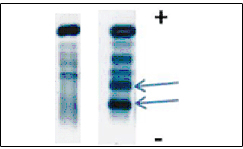
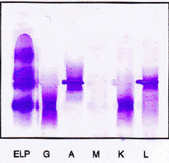
Serum electrophoresis on agarose (Sebia) revealed two sharp discrete bands in the gamma globulin region (Figure. 1). On being subjected to immunofixation (Sebia), serum revealed IgG- kappa and IgA- lambda gammopathy (Fig. 2).
Serum ß2 microglobulin measured by chemiluminescence was 4961ng/ml (Reference range: 1556-2286 ng/ml). Serum IgG level was 23.98 gm/l (Reference range: 6.53-13.51 gm/l), IgA level was 3.71 gm/l (Reference range: 0.86-3.20 gm/l). IgM level was <0.255 gm/l (Ref. range: 0.521-1.79 gm/l). All these parameters were assayed by nephelometry. Urine Bence-Jones protein was negative.
The patient was started on a combination of vincristine, adriamycin and dexamethasone. However, he was lost to follow up after two cycles of chemotherapy due to poor drug compliance by the fast degenerating general physical condition.


Click below to enlarge
Figure 1: Serum protein electrophoresis. (Arrow depicts two bands in the gamma region).


Click below to enlarge
Figure 2: Immunofixation depicting IgG Kappa& IgA Lamda


Discussion

The biclonal gammopathies are a group of disorders characterized by the production of two distinct monoclonal proteins, which may be due to proliferation of two clones of plasma cells, each producing an unrelated monoclonal immunoglobulin, or it may result from production of two monoclonal proteins by a single clone of plasma cells. During the normal development and differentiation of B lymphocytes, the surface immunoglobulins change from only IgM to both IgM and IgD, and the secreted antibody switches heavy-chain class (from IgM to IgG, for example). In spite of the switching of heavy-chain class, the antibody molecules retain the same antigenic specificity. These observations were among those that led to the hypothesis that the variable and constant regions of immunoglobulin chains were encoded in separate genes, and that during differentiation, a unique variable-region gene could be joined sequentially to different constant-region genes. Recent experiments using techniques of recombinant DNA and DNA cloning and sequencing have verified that the variable and constant region genes are encoded separately in the DNA and that genomic information is rearranged to result in joining of a variable-region gene to a constant-region gene . Although biclonal immunoglobulins may result from two independent transforming events yielding two unrelated plasma cell clones and monoclonal proteins, it has been postulated that some biclonal pairs may result from a transformation event in a cell undergoing a variable-region switch from one heavy-chain class or subclass to another. If this were the case, it would be predicted that the variable regions of the biclonal pair would be identical and that in addition, it might be possible to find some plasma cells producing both mono- clonal proteins. [3]

The existence of true biclonal myeloma based on the presence of more than 1 IgH subtype must be carefully reviewed because both serum M components may share the same clonal origin with an identical variable region rather than being 2 independent clones. Thus, at the RNA level clonotypic Cµ transcripts can be found in the BM of 68% of patients with IgG+ myeloma, consistent with the idea that these cells could be the clonogenic origin of the tumor clone. Similarly, in IgM+ myelomas C? transcripts with a clonogenic CDR3 region can be identified, consistent with the cell of origin being able to undergo the CSR process. Unfortunately, myelomas secreting 2 M components as detected by protein electrophoresis have not been studied in enough detail to allow us to comment on their clonal relationship. In this sense, they could also represent truly independent myeloma clones, with ongoing CSR detectable in subclones derived from the same myeloma progenitor, alternatively, lack of allelic exclusion within the same myeloma cell. [4]

In contrast to cases showing 2 different IgH subtypes, cases characterized by 2 different IgL peaks can be considered as truly biclonal, because no molecular mechanism for changing light chain expression within the same myeloma clone has been described so far. Biclonal myeloma cases expressing both kappa and lambda are not exceptional, although its frequency has been estimated to be only around 1% to 2%. This low frequency has led to dif?culty in interpreting the clinical characteristics of these patients; some authors having found no difference in the clinical characteristics and prognosis, while others suggest a poor response to therapy. Two cases expressing cytoplasmic Ig? and Ig? but secreting only IgG have been described, suggesting that it is possible to produce both light chain isotypes. This observation can be explained by the fact that the Ig? light chain was unable to assemble with the Ig heavy chain and illustrates how a functional IgH- IgL assembly may be one of the mechanisms leading to allelic exclusion and mono specificity. [5]

Occurrence of isotype switch and appearance of abnormal protein bands have, however been reported in myeloma patients after high dose therapy. This appears to be related to recovery of normal immunoglobulin production rather than alteration in disease biology. This change is also associated with increased survival. [6]

Mono- and biclonal gammopathy were also reported in two immunocompetent individuals caused by acute infection with Bartonella henselae resulting in cat-scratch disease (CSD). Bartonella henselae has been shown to induce host cell production of the vascular endothelial growth factor (VEGF) in vitro, resulting in proliferation of endothelial cells and thus representing a potential novel bacteria-dependent mechanism of tumor growth. Furthermore, it has been demonstrated that bacterial eradication by antibiotic treatment results in complete regression of the angiomatous tumors. Bartonella henselae is also capable of inducing the production of proinflammatory cytokines such as tumor necrosis factor alpha, interleukin (IL) 1b, IL-2, IL-6, IL-8, and IL-10. While IL-10 is an important differentiation factor for plasma cell formation and immunoglobulin secretion, IL-6 constitutes a major growth factor for myeloma and plasma cells, induces immunoglobulin production, and is an active factor in B-cell differentiation. The eradication of CSD following antibiotic treatment led to the termination of gammopathy. [7]


Conclusion

Biclonal multiple myeloma accounts for only 1% of all myelomas. This was the first case reported in our institution where so far 600 electrophoresis have been done. Clinical presentation and response to therapy is like other cases of multiple myeloma.


References
  1. Osterborg A, Hansson LO, Mellstedt H. Immunoglobulins in multiple myeloma. In: Garhton G ,Durie B(eds).Multiple myeloma.1st ed. London: Oxford University Press1996;36-50.    Back to citation no. 1
  2. Osterborg A, Mellstedt H. Monoclonal and biclonal immunoglobulin producing disorders. Eurp J 1989;43(Suppl 51):11-8.   [CrossRef]   [Pubmed]    Back to citation no. 2
  3. Kyle RA, Robinson R, Katzman J. The clinical aspects of biclonal gammopathies. Am J Med1981;71:999-1006.   [CrossRef]   [Pubmed]    Back to citation no. 3
  4. Kyle R A. Multiple Myeloma: A review of 869 cases. Mayo Clin Proc 1975;50:29-40.   [Pubmed]    Back to citation no. 4
  5. Gonzalez D, Burg M, Garcia-Sanz R, et. al. Immunoglobulin gene rearrangements and the pathogenesis of multiple myeloma. Blood 2007;110:3112- 3121.   [Pubmed]    Back to citation no. 5
  6. Vincent T Devita,Jr. Samuel Hellman, Stevens A Rosenberg;Cancer : Principles and practice of oncology: 7th edition.    Back to citation no. 6
  7. R. Krause , H. W. Auner , F. Daxbock ,A. Mulabecirovic , G. J. Krejs , C. Wenisch ,EC. Reisinger. Monoclonal and biclonal gammopathy in two patients infected with Bartonella henselae. Ann Hematol 2003;82:455-457.   [Pubmed]    Back to citation no. 7

[HTML Abstract]   [PDF Full Text]

Author Contributions:
Mala Mahto - Conception and design, Acquisition of data, Analysis and interpretation of data, Drafting the article, Critical revision of the article, Final approval of the version to be published
Priya Balakrishnan - Conception and design, Acquisition of data, Analysis and interpretation of data, Drafting the article, Critical revision of the article, Final approval of the version to be published
Bidhan Chandra Koner - Conception and design, Acquisition of data, Analysis and interpretation of data, Drafting the article, Critical revision of the article, Final approval of the version to be published
Pramod Lali - Conception and design, Acquisition of data, Analysis and interpretation of data, Drafting the article, Critical revision of the article, Final approval of the version to be published
Tarun Kumar Mishra - Conception and design, Acquisition of data, Analysis and interpretation of data, Drafting the article, Critical revision of the article, Final approval of the version to be published
Alpana Saxena - Conception and design, Acquisition of data, Analysis and interpretation of data, Drafting the article, Critical revision of the article, Final approval of the version to be published
Guarantor of submission:
The corresponding author is the guarantor of submission.
Source of support:
None
Conflict of interest:
The author(s) declare no conflict of interests.
Copyright:
© Mala Mahto et. al. 2011; This article is distributed the terms of Creative Commons Attribution License which permits unrestricted use, distribution and reproduction in any means provided the original authors and original publisher are properly credited. (Please see Copyright Policy for more information.)