
  |

|
 |
|
Case Report
| ||||||
| Late-onset thoracic aortic graft infection: A case report | ||||||
| Liran Shani1, Yuval Geffen1, Gil Bolotin1, Ayelet Raz-Pasteur1 | ||||||
|
1Rambam Health Care Campus, Haifa, Israel.
| ||||||
| ||||||
|
[HTML Abstract]
[PDF Full Text]
[Print This Article]
[Similar article in Pumed] [Similar article in Google Scholar] 
|


| How to cite this article |
| Shani L, Geffen Y, Bolotin G, Raz-Pasteur A. Late- onset thoracic aortic graft infection: A case report. Int J Case Rep Images 2015;6(6):356–360. |
|
Abstract
|
|
Introduction:
Thoracic aortic graft infection is a rare and devastating complication of aorta replacement surgery with an incidence range of 0.9–1.9%. Prosthetic aortic graft infections represent a major diagnostic and therapeutic challenge. Despite latest advancements in imaging and microbiological investigations there are still no agreed criteria to confirm the diagnosis.
Case Report: We present a case of late onset culture negative thoracic aortic graft infection in a Caucasian 65-year-old male, nine years after aortic replacement due to acute aortic dissection. Conclusion: Culture-negative vascular graft infection has not been described as a clinical entity so far. Review of current literature and Issues of diagnosis and management will be discussed. | |
|
Keywords:
Thoracic aortic graft infection, Culture negative vascular graft infection
| |
|
Introduction
| ||||||
|
Thoracic aortic graft infection is a rare and devastating complication of aorta replacement surgery with an incidence range of 0.9–1.9% and carries mortality rates ranging from 25–75%. Although most cases appear shortly after the surgery there have been descriptions of cases manifesting up to 15 years after the initial procedure [1] [2] [3] . Since sepsis is a common complication in these patients, early diagnosis and treatment are important. It is often difficult to identify the primary infection site, and there are no consensus criteria for diagnosing the condition. We present a case of late onset culture negative thoracic aortic graft infection in a male patient. Issues of diagnosis and management will be discussed. | ||||||
|
Case Report
| ||||||
|
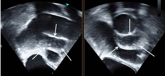
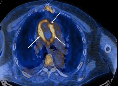
A 65-year old male with 10 days of fever and malaise was admitted for evaluation to the department of internal medicine. Nine years earlier the patient suffered from acute aortic dissection without aortic valve involvement. He had undergone emergency replacement of the ascending aorta with a GORE-TEX aortic graft. Fever started two days after an invasive dental procedure for which he was treated prophylactically with two doses of post-procedure amoxicillin. He continued to take his antibiotics under his dentist advice for 10 more days up until two days prior to his admission. His fever did not improve under the treatment. The patient had no history of contact with farm animals; he has not been traveling and did not consume out-of-routine food products including unpasteurized milk products. He had no respiratory, gastrointestinal or urinary symptoms. His other medical history included medically treated hypertension, prostatectomy following malignancy 15 years earlier treated with fesoterodine, smoking and venous insufficiency of the lower limbs. On admission the patient was in good condition. Vital signs showed a blood pressure of 118/81 mmHg, irregular heart rhythm of 88 bpm, oxygen saturation level of 98% at room air, and an oral temperature of 37.7°C. His stature was obese with a body mass index of 35.6 kg/m2. Physical examination of the heart and lungs was normal. His abdomen was soft with no tenderness. He had bilateral chronic lower legs edema due to venous insufficiency with no signs of cellulitis. Laboratory findings of the blood on admission disclosed a slightly elevated white blood cell count (10.96x10³/µL), normal hemoglobin (14.6 g/dl) and platelets level (162x103/µl), elevated CRP (103 mg/l) and erythrocyte sedimentation rate (70 mm/hr). BNP level was 130 Pico/ml. ECG of the patient revealed a normal sinus rhythm with atrial premature beats, his chest x-ray showed no signs of consolidation. Dental examination of the patient showed no abnormality, panoramic dental X-ray ruled out an infectious process. Further workup included computed tomography scan of the chest, abdomen and pelvis. While his abdomen and pelvis CT scan showed no specific finding, on chest CT, a small amount of fluid was demonstrated around his ascending aorta which did not exist five years earlier during workup for chest pain. Transesophageal echocardiography (TEE) demonstrated fluid surrounding the aortic graft, from the Sino tubular junction to the distal ascending segment of the graft. Suspected dehiscence of the proximal graft from its connection to ST junction was observed (Figure 1). Positron emission tomography- computed tomography (PET-CT) scan demonstrated pathological absorption around the ascending aorta and the nearby fluid which corresponded to an infectious process (Figure 2). Serologic panel taken included Bartonella, Brucella, Syphilis (RPR, TPHA) and Coxiella burnetii. Bartonella henselae IgG test was the only positive result. The patient had no contact with cats and did not meet the clinical criteria for Bartonella infection. PCR of blood and later aortic graft tissue excluded the diagnosis. Blood cultures, including long incubation periods and different culture mediums, were sterile. Ciprofloxacin treatment was started four days after admission when low grade fever did not resolve and a positive urine culture yielded a susceptible bacterium. Two days later ciprofloxacin was replaced with ceftriaxone when fever did not resolve. On the seventh day of his admission, under antibiotic treatment, the patient developed signs of sepsis with fever up to 390C , elevated white blood cell count (30x103 cells/µl) and acute renal failure manifested with anuria and creatinine level of up 7.5 mg/dl. The patient was treated with careful fluid administration, diuretics for volume control and went through several dialysis sessions until signs of renal recovery were shown. Broad spectrum antibiotic regimen that included piperacillin/tazobactam and vancomycin (for the possibility of graft infection with Methicillin-resistant Staphylococcus aureus) was given to the patient. With signs of improvement from sepsis and renal failure treatment was replaced to a combination of ciprofloxacin, metronidazole and vancomycin (treatment of culture negative vascular graft infection). This regimen was maintained until his discharge. Upon recovery, the patient was taken to the operating room. Surgical findings included an abscess between the superior vena cava and the proximal posterior part of the aortic graft. Structural integrity of the graft was maintained despite the infection. Under total circulatory arrest and hypothermia a new Dacron aortic graft was implanted after thorough debridement and rinsing. Samples were taken from the old graft and the abscess fluid. Postoperative recovery was slow. Remarkable complications included deep venous thrombosis of the lower extremity. Renal function did not return to normal and the patient was discharged with a stable creatinine level of 2 mg/dl. Pathologic examination of aortic graft segments extracted during surgery together with the fluid revealed inflammation, necrosis and calcifications. Cultures of the tissue samples including the abscess fluid were sterile. PCR analysis was negative for bacteria (including specific testing for Bartonella) and fungi. | ||||||
| ||||||
|
| ||||||
|
Discussion
| ||||||
|
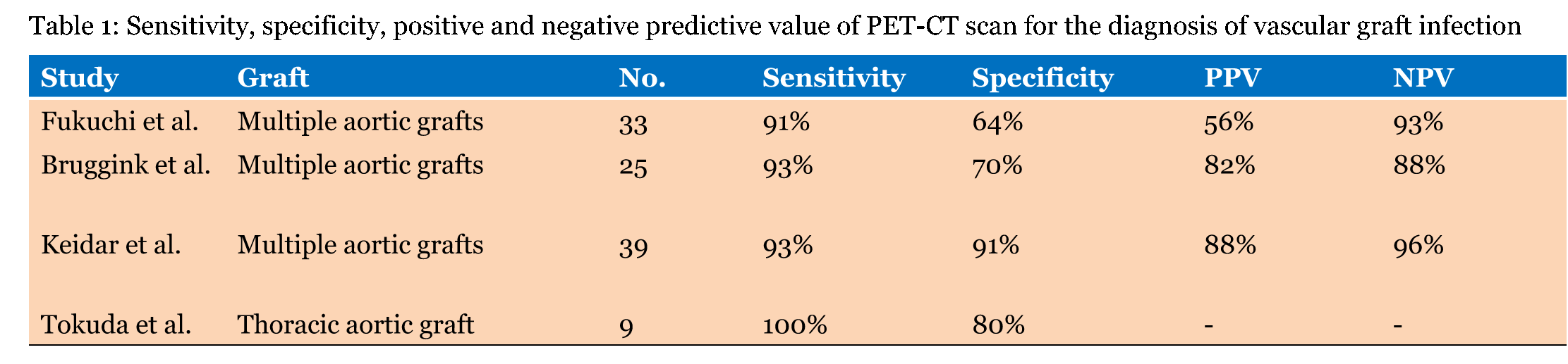
Four hundred and fifty thousand vascular grafts are being implanted annually in the United States with all graft types infection rate of 4% [4]. Diagnosis and management of thoracic aortic graft infections poses a major clinical challenge. Typical presentation is of a patient with no specific symptoms other than fever and malaise [5]. There is no consensus on diagnostic criteria or on the best management of aortic graft infection. Several imaging modalities can be used to establish a diagnosis of aortic graft infection. Computed tomography has a sensitivity of 94% and a specificity of 85% for diagnosing prosthetic graft infections in general [6]. MRI scan has been used on a limited scale to evaluate patients with suspected vascular graft infection. Shahidi et al. have demonstrated a sensitivity of 68% (95% CI 0.50–0.86) and specificity of 97% (95% CI 0.91–1.02)[7] [8]. Positron emission tomography-computed tomography (PET-CT) scan has been increasingly used to help in the diagnosis of vascular graft infection. Table 1 demonstrates the sensitivity, specificity, positive and negative predictive value for several clinical series. In general, the specificity of imaging for graft infection rises as time elapses form the surgical procedure [6]. Serological testing Tissue and blood PCR ruled out the presence of Bartonella infection in our patient. Microbiology of aortic graft infections Culture negative vascular graft infection Late-onset vascular graft infection Late-onset vascular graft infection is a rare event. In a series of 41 patients, jones et al. presented 50% of cases in the first 500 days after the surgical procedure. Only four patients suffered an infection more than seven years after the surgery, only one of them had culture negative infection [11]. Coselli et al. published in their clinical series a single case of very late-onset (15 years post-surgery) thoracic aortic graft infection [13]. The etiology of late vascular graft infection is said to be implantation of bacteria at the time of initial surgery, but in a few cases infection may result from seeding onto the graft during a bacteremia. Factors influencing late graft infection include type of graft material and the identity of the offending pathogen. Dacron grafts are more likely to develop a partial 'pseudointima', making it less susceptible to late bacteremic seeding compared to polytetrafluoroethylene. Infections with high-virulence organisms are unlikely to remain dormant for years after the initial procedure [13]. Thus, late onset infection usually represents infection of originally implanted (during the first surgery) bacteria of low virulence nature or bacteremic seeding of high virulence bacteria. Prevention of aortic graft infection There is no reference to antibiotic prophylaxis in people with vascular graft implantation. Much like endocarditis prevention strategies, secondary prevention of vascular graft infection has not been investigated in a viable research model. The low incidence and multifactorial pathophysiology makes it difficult generating such a model. Jones at el. Have reported a series of 41 patients with vascular graft infection over a course of 26 years, out of those only four experienced the complication over 10 years after the initial procedure and the data collected was limited. These four patients had isolates of high virulence bacteria indicating a mechanism of recent bacteremia [13]. In 2002, Lockhart conveyed a Survey of infectious disease experts regarding antibiotic prophylaxis for medical conditions. Sixty percent (477) of the members of the Infectious Diseases Society of America Emerging Infections Network responded. 35% recommend prophylaxis for patients with prosthetic vascular grafts [14]. Our patient, a 65-year-old male, described going through an invasive dental procedure two weeks prior to his admission. Although not indicated by the ESC or AHA guidelines his dentist prescribed him amoxicillin but only after the procedure itself. While the patient is not classified as high risk population (due to the presence of thoracic aortic graft) the nature of the dental procedure does make him a possible candidate for antibiotic prophylaxis (which he got but not in the appropriate manner). Surgical treatment | ||||||
| ||||||
|
| ||||||
|
Conclusion
| ||||||
|
Aortic graft infection remains to this day a catastrophic complication of aortic surgery. High suspicion and fast workup should be implemented for patients with an aortic graft presenting with signs of infectious disease at any period of time after the initial procedure. The term culture-negative vascular graft infection has not been coined yet and should be addressed as a clinical entity. Antibiotic prophylaxis that is given in the setting invasive medical procedures in patients with vascular grafts is largely unstudied. In the era of endovascular medicine and growing numbers of vascular grafts implanted, the current approach of withholding antibiotic prophylaxis for invasive medical procedure in this population may warrant reconsideration. | ||||||
|
References
| ||||||
| ||||||

|
[HTML Abstract]
[PDF Full Text]
|
|
Author Contributions
Liran Shani – Substantial contributions to conception and design, Acquisition of data, Drafting the article, Revising it critically for important intellectual content, Final approval of the version to be published Yuval Geffen – Substantial contributions to conception and design, Acquisition of data, Drafting the article, Revising it critically for important intellectual content, Final approval of the version to be published Gil Bolotin – Substantial contributions to conception and design, Acquisition of data, Drafting the article, revising it critically for important intellectual content, Final approval of the version to be published Ayelet Raz-Pasteur – Substantial contributions to conception and design, Acquisition of data, Drafting the article, Revising it critically for important intellectual content, Final approval of the version to be published |
|
Guarantor of submission
The corresponding author is the guarantor of submission. |
|
Source of support
None |
|
Conflict of interest
Authors declare no conflict of interest. |
|
Copyright
© 2015 Liran Shani et al. This article is distributed under the terms of Creative Commons Attribution License which permits unrestricted use, distribution and reproduction in any medium provided the original author(s) and original publisher are properly credited. Please see the copyright policy on the journal website for more information. |
|
|