
 |

|
 |
|
Case Report
| ||||||
| Cutaneous sarcoidosis presenting with diffuse panniculitis: A case report | ||||||
| Assane Diop1, Diallo Moussa2, Maodo Ndiaye2, Abbaspour Valiollah2, Pauline Dioussé2, Boubacar Diatta2, Fatimata Ly1, Suzanne Oumou Niang2, Mame Thierno Dieng2, Assane Kane2 | ||||||
|
1MD, Assistant Professor, Department of Dermatology, Institutd'HygiéneSociale, Faculty of Medicine, Cheikh Anta Diop University, Dakar, Senegal.
2MD, Assistant Professor, Department of Dermatology Hôpital Aristide le Dantec, Faculty of Medicine, Cheikh Anta Diop University, Dakar, Senegal. | ||||||
| ||||||
|
[HTML Abstract]
[PDF Full Text]
[Print This Article]
[Similar article in Pumed] [Similar article in Google Scholar] 
|


| How to cite this article |
| Diop A, Moussa D, Ndiaye M, Valiollah A, Dioussé P, Diatta B, Ly F, Niang SO, Dieng MT, Kane A. Cutaneous sarcoidosis presenting with diffuse panniculitis: A case report. Int J Case Rep Images 2015;6(6):328–331. |
|
Abstract
|
|
Introduction:
Cutaneous lesions in sarcoidosis are polymorphic. They can simulate most dermatological conditions. We report a case of cutaneous sarcoidosis presented as diffuse Panniculitis
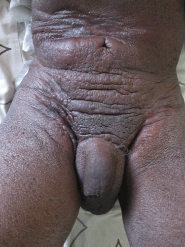
Case Report: A 62-year-old male with no significant past medical history consulted for infiltrated lesion on abdominal skin infiltration lasting for two years. On examination, there were multiple indurated plaques, topped with keratotic papules giving an orange peel view, extending on abdominal genitalia and thighs skin associated with lymph nodes enlargement in different sizes and elastic consistence in axillary and inguinal regions. The histopathological examination of the skin lesion and the lymph nodes revealed typical sarcoidosis granulomas. Thoracic abdominal CT scan was normal. After six months of treatment with methotrexate and prednisone, evolution was remarkable by a rapidly skin lesions des infiltration and disappearance of lymphadenopathy. Conclusion: Cutaneous sarcoidosis as disseminated panniculitis is rarely reported in literature. In our patient, methotrexate combined with prednisone was effective. Other studies have confirmed the efficacy of methotrexate, especially as a steroid sparing treatment. This efficiency would be the result of adenosine production and a decrease in TNF a secretion in granulomatous lesions. | |
|
Keywords:
Diffuse panniculitis, Methotrexate erythematous, Panniculitis, Sarcoidosis
| |
|
Introduction
| ||||||
|
Sarcoidosis is highly polymorphic in its clinical manifestations [1]. Clinically, sarcoidosis can mimic many skin diseases. However, Panniculitis as presenting feature in sarcoidosis is rarely reported [1] [2] [3]. We report a case of generalized granulomatous panniculitis in a patient with sarcoidosis. | ||||||
|
Case Report
| ||||||
|
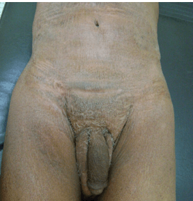
A 62-year-old male, without remarkable past medical history, was admitted for a large erythematous indurated plaque on abdominopelvic region lasted for two years. Physical examination showed an erythematous indurated plaque with an orange peel appearance in abdominal lower quadrant, pubic, external genitalia and thighs. The plaque was studded with hyperkeratotic papule especially on pubic region. A generalized ichthyosis sparing scalp and palmar-plantar region was noted (Figure 1). There were also consistent, painless, fixed lymphadenopathies of 2 to 7 cm in diameter, in auxiliary and inguinal regions. The skin and lymph node histopathologic studies revealed typical sarcoidosis granulomas. In skin, these granulomas were deeply situated in subcutaneous tissues and occupying fat lobules (Figure 2). The abdomen and thoracic CT scan was normal. Tuberculin test was anergic and HIV serological test was negative. CBC and blood smear were normal. After six months of treatment, combining methotrexate 25 mg intramuscularly per week and prednisone 0.5 mg / kg /day, the outcome was favorable, marked by fast skin lesions desinfiltration, disappearance of ichthyosis and lymphadenopathy regression (Figure 3). | ||||||
| ||||||
| ||||||
| ||||||
|
Discussion
| ||||||
|
We report a cutaneous and lymph node sarcoidosis in which skin involvement was confirmed by histological examination as panniculitis. Specific manifestations of cutaneous sarcoidosis are related with the location of granulomas in the dermis. In literature, the frequency varies between 9% and 30% in different studies [4] [5] . They are characterized by small and large nodules called sarcoid, infiltrating sarcoid and sarcoid on scars. Hypodermic sarcoid of Darier-Roussy represents 4–12% of the specific lesions of sarcoidosis [5] [6] and appear as cold nodules developing in a normal-looking skin. They are located more frequently on the limbs and rarely on the trunk. Cutaneous sarcoidosis with panniculitis as clinical presentation, it has rarely been reported [1] [7] [8]. To our knowledge, only 2 cases of sarcoidosis with extensive panniculitis have been reported. In one case, it was only a localized lesion on one limb [8]. In another case, the lesions were on supraclavicular, shoulder and upper back [7]. In our patient, the lesions were more generalized, on abdomen, pelvis, genitals and thighs. This very unusual clinical presentation can rise a problem of differential diagnosis with cutaneous lymphoma. However, the histopathologic features of sarcoidosis can make the difference with the skins as well as the lymph node lymphoma. The treatment of sarcoidosis remains is poorly codified [9]. Corticosteroids are the standard treatment, but with a purely suspensive effect [10] [11]. It is indicated in severe eye, neurological, cardiac, renal, laryngeal and lung involvement or progressive disabling lesions and lupus pernio with severe hypercalcemia. However, glucocorticoids cause many side effects at more than 50% of patients. Side effects are even more frequent in height doses and long duration treatment [12] thus, methotrexate is often proposed as an alternative, especially as a reserve when long-term treatment with prednisone is obligatory [12]. In our case, treatment with methotrexate and prednisone had achieved a complete remission of lesions after six months. Other studies [13] [14][15] have confirmed the effectiveness of methotrexate, especially as steroid sparing [16] treatment. This effectiveness would be the result of an increase in adenosine production and a decrease in the secretion and TNF a at granulomatous lesions [12]. | ||||||
|
Conclusion
| ||||||
|
Diffuse panniculitis as a presenting feature of sarcoidosis is rare. In such cases, only histopathology can confirm the diagnosis. Although treatment is not well classified, Methotrexate associated with prednisone seems to work well. | ||||||
|
References
| ||||||
| ||||||


|
[HTML Abstract]
[PDF Full Text]
|
|
Author Contributions
Assane Diop – Substantial contributions to conception and design, Acquisition of data, Analysis and interpretation of data, Drafting the article, Revising it critically for important intellectual content, Final approval of the version to be published Diallo Moussa – Analysis and interpretation of data, Revising it critically for important intellectual content, Final approval of the version to be published Maodo Ndiaye – Analysis and interpretation of data, Revising it critically for important intellectual content, Final approval of the version to be published Abbaspour Valiollah – Analysis and interpretation of data, Revising it critically for important intellectual content, Final approval of the version to be published Pauline Dioussé – Analysis and interpretation of data, Revising it critically for important intellectual content, Final approval of the version to be published Boubacar Diatta – Analysis and interpretation of data, Revising it critically for important intellectual content, Final approval of the version to be published Fatimata Ly – Analysis and interpretation of data, Revising it critically for important intellectual content, Final approval of the version to be published Suzanne Oumou Niang – Analysis and interpretation of data, Revising it critically for important intellectual content, Final approval of the version to be published Mame Thierno Dieng – Analysis and interpretation of data, Revising it critically for important intellectual content, Final approval of the version to be published Assane Kane – Analysis and interpretation of data, Revising it critically for important intellectual content, Final approval of the version to be published |
|
Guarantor of submission
The corresponding author is the guarantor of submission. |
|
Source of support
None |
|
Conflict of interest
Authors declare no conflict of interest. |
|
Copyright
© 2015 Assane Diop et al. This article is distributed under the terms of Creative Commons Attribution License which permits unrestricted use, distribution and reproduction in any medium provided the original author(s) and original publisher are properly credited. Please see the copyright policy on the journal website for more information. |
|
|