|
|
Case Report
| ||||||
| Drowning induced acute respiratory failure: A case report | ||||||
| Touzani Soumaya1, Houari Nawfal1, Elbouazzaoui Abderrahim2, Boukatta Brahim2, Kanjaa Nabil3 | ||||||
|
1Assistant Professor, Department of Anesthesiology and Intensive Care A4, University Hospital Hassan II, Fez, Morocco 2Associate Professor, Department of Anesthesiology and Intensive Care A4, University Hospital Hassan II, Fez, Morocco 3Head of Department, Department of Anesthesiology and Intensive Care A4, University Hospital Hassan II, Fez, Morocco | ||||||
| ||||||
|
[HTML Abstract]
[PDF Full Text]
[Print This Article] [Similar articles in PubMed] [Similar articles in Google Scholar] |
| How to cite this article |
| Touzani S, Houari N, Elbouazzaoui A, Boukatta B, Kanjaa N. Drowning induced acute respiratory failure: A case report. Int J Case Rep Images 2018;9:100939Z01ST2018. |
|
ABSTRACT
| ||||||
|
Introduction: Drowning is still a relevant medical challenge and a leading cause of accidental death worldwide. Hypoxia is the starting point for all morbidity and mortality and it must remain the focus of treatment. Case Report: We report the case of non-fatal drowning in an epileptic patient who presented to the emergency department with acute respiratory failure (ARF) and subsequent neurological status impairment. The patient’s condition was managed with early non-invasive ventilation (NIV). He was rapidly discharged from ICU despite severe hypoxemia on admission. Although the applications of NIV have been increased recently in the emergency settings, reports about its efficiency in drowning ARF are limited. Conclusion: Early administration of non-invasive ventilation would be interesting to manage drowning induced ARF through positive expiratory pressure while avoiding complications related to conventional ventilation. Keywords: Acute respiratory distress syndrome, Acute respiratory failure, Drowning, Non-invasive ventilation | ||||||
|
INTRODUCTION
| ||||||
|
Drowning is still one of the leading causes of accidental death worldwide. More than 500 000 deaths occur each year after unintentional drowning according to the World Health Organization (WHO) [1]. Acute respiratory failure (ARF) is the main component of drowning physiopathology. The end result of drowning is surfactant dysfunction, pulmonary edema, a decrease in pulmonary compliance and an increase in the ventilation/perfusion mismatch, frequently leading to profound hypoxia and acute respiratory distress syndrome (ARDS) and ultimately causing cardiovascular collapse and death [1]. This condition is still considered as a relevant medical challenge as clinical data are still lacking on the best medical strategy to use. The practice of non-invasive ventilation (NIV) has been increased recently in emergency settings [2], but its safety and efficiency in drowning related ARF remains unknown. This case report highlights the successful management of non-fatal drowning related ARF with early non-invasive ventilation. | ||||||
|
CASE REPORT
| ||||||
|
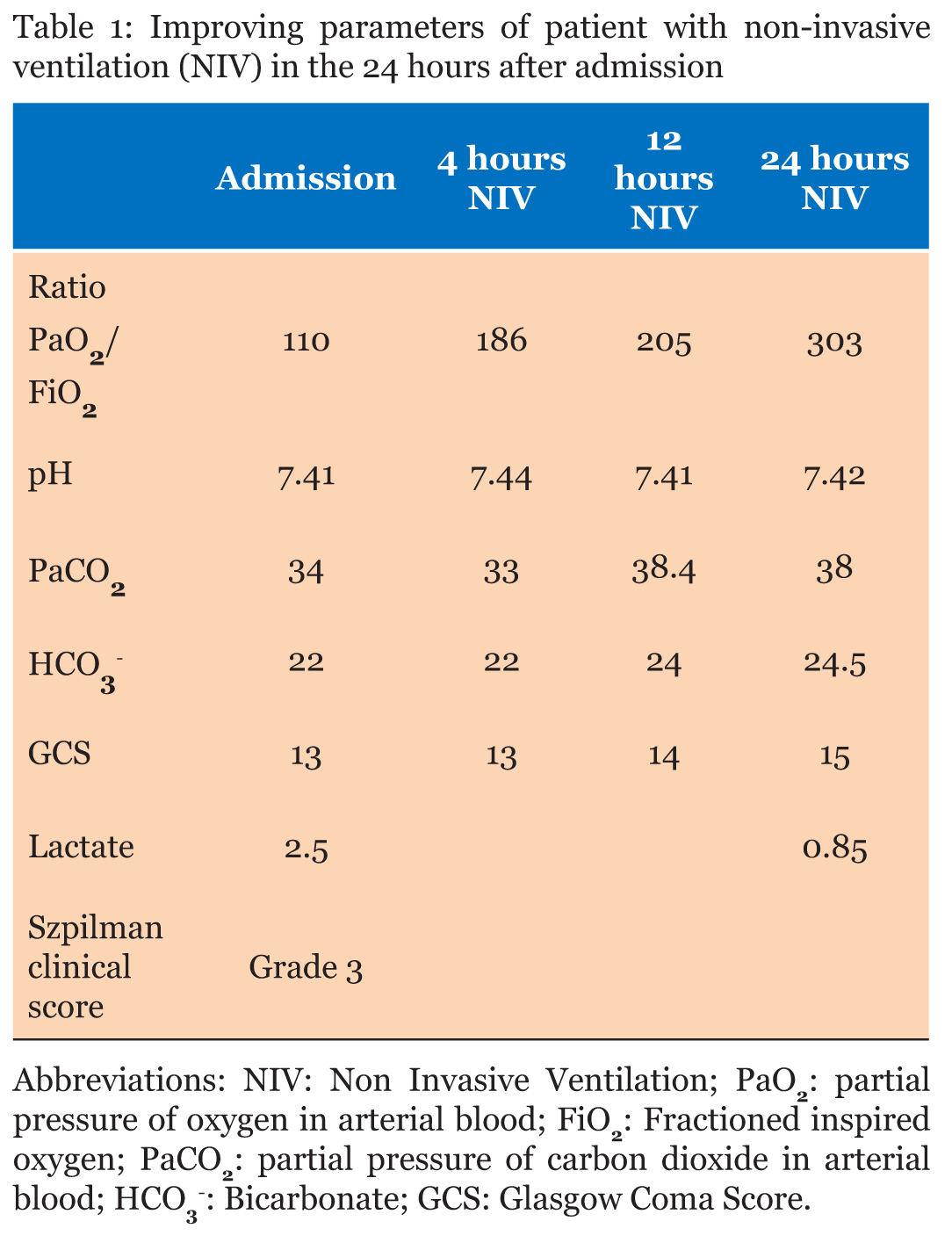
A 46-year-old man, with history of epilepsy and mental retardation, was brought to the emergency department (ED) after submersion in a thermal pool while sitting at the edge. No prior epileptic seizure was reported by his accompanying family member. Bystanders reported that the patient was submerged for approximately 1 minute. When rescued, he was unconscious and polypneic. The transport to hospital was immediate with no medical assistance. On admission to the ED, the physical examination revealed a Glasgow Coma Scale (GCS) score of 13/15 (E3, V4, M5), a pulse rate at 107 beats/min and blood pressure at 150/80 mmHg. Respiration was spontaneous at 29 breaths/min, with oxygen saturation (SpO2) on room air of 60%. He had bilateral crackles on auscultation of the lungs. He was hypothermic at 36.5° C. Electrocardiogram showed sinusal tachycardia. There was no evidence of trauma. Capillary blood glucose was 1.3 g/dl. Initial management included: a semi-sitting position, vascular access, warming and high flow of oxygen (10 l/min) through high concentration facial mask. The chest X-ray showed diffused alveolar opacities suggestive of pulmonary edema (Figure 1A). Chest CT scan disclosed multiple bilateral condensation areas and “ground glass” opacifications, consistent with respiratory distress syndrome (Figure 1B). An unenhanced computed tomography (CT) of the brain was unremarkable. His serum creatinine, electrolyte values, blood cell counts and rhabdomyolysis profile were normal. Arterial blood gas analysis (Table 1) revealed a severe hypoxemia (Ratio PaO2/FiO2 = 110) with compensated respiratory alkalosis (PaCO2= 34, HCO3- = 22, pH = 7.41) and hyperlactatemia at 2.5 mmol/l (Normal < 2 mmol/l). Non invasive ventilation (NIV) was immediately started in the ED, even before the CT chest and brain, and continued upon admission to the Intensive Care Unit (ICU).The head of the bed was elevated to 30° during ventilation to minimize the risk of aspiration. No sedation was needed. Non-invasive positive pressure ventilation on bi-level positive airway pressure (BiPAP) mode was applied via oronasal mask to the patient. Initial ventilator settings included: pressure support (PS) = 12 cmH2O; positive end-expiratory pressure (PEEP) = 5 cmH2O; FiO2 50%. The pressure support was adjusted to maintain a tidal volume of 6–8 ml/kg. The PEEP and FiO2 were adjusted (gradually increased or decreased in increments of respectively 2 cmH2O and 5%) to maintain blood oxygen saturation (SpO2) ≥90% or PaO2≥ 60 mmHg. The vital signs of the patients (neurological status, SpO2, hemodynamic parameters) were closely monitored at all times during the procedure. Invasive hemodynamic monitoring was not required and no remarkable echocardiographic findings were associated with either the respiratory failure or the assisted ventilation (especially no right ventricular dysfunction). Vital signs were stable over the successive 24 hours with progressive improvement of consciousness and progressive resolution of acute respiratory failure (Table 1). NIV was gradually stopped and replaced by low flow oxygen therapy through nasal cannula before complete weaning. The radiological infiltrate detected on admission gradually improved in the subsequent chest X-rays performed (Figure 2). The patient was discharged from ICU on day 3. | ||||||
| ||||||
| ||||||
| ||||||
DISCUSSION | ||||||
|
The WHO defined drowning as the process of experiencing respiratory impairment from submersion/immersion in liquid. Non-fatal drowning defines the rescued victims [3]. Drowning is a leading cause of death with over 90% of cases occurring in lower- and middle-income countries. Toddlers, elderly and those with medical conditions that cause consciousness loss, including epilepsy, are at greatest risk of death by drowning [4], [5]. Hypoxia is the starting point for all morbidity and mortality and it must remain the focus of treatment. Unexpected submersion triggers breath-holding and a struggle to surface. Reflex inspiratory efforts lead to hypoxemia by either laryngospasm or aspiration. The quantity of fluid aspirated, rather than the composition, determines subsequent pulmonary derangement. Aspiration of 1–3 ml/kg body weight of either salt or fresh water compromises the integrity of pulmonary surfactant leading to: alveolar collapse, atelectasis, noncardiogenic pulmonary edema, intrapulmonary shunting and ventilation-perfusion mismatch (V/Q), resulting in acute respiratory distress syndrome (ARDS). Profound hypoxia and metabolic and respiratory acidoses lead to cardiovascular collapse, neurologic damage and ultimately death [6]. Pulmonary compromise can develop insidiously or rapidly. Signs and symptoms usually include tachypnea, shortness of breath, hypoxia, crackles, which were present in our patient initially. The management and clinical severity of victims are adjusted according to Szpilman scoring system. High-flow O2 or early endotracheal intubation (ETI) are the recommended practices for patients with pulmonary edema (grade 3 or 4). In patients with severe ARDS, ETI and lung protective ventilation with positive end-expiratory pressure should be considered as first option ventilation [1]. The practice of non-invasive ventilation (NIV) has recently increased in the ED with the leading advantage of avoiding pneumonia and other complications associated with endotracheal intubation. The classical indications are acute bronchiolitis and cardiogenic pulmonary edema. The use of non-invasive positive pressure ventilation such as continuous positive airway pressure (CPAP) / bilevel positive airway pressure (BiPAP) in ARDS still controversial [2]. Many studies concluded that use of NIV may be safe in selected patients who are observed carefully [7]. Furthermore,pulmonary edema in drowning cases recovers faster and NIV could be a successful treatment [6]. Non-invasive ventilation can improve respiratory distress through: decreasing the work of breathing, reversing hypoventilation, increasing functional residual capacity, maintaining upper airway patency, recruitment of atelectatic regions and reducing V/Q mismatch. A beneficial effect of positive end expiratory pressure (PEEP) has indeed been reported in rare animal studies [8]. The favorable effect of early administration of PEEP has not been clinically demonstrated but is regularly discussed in few case reports and retrospective studies [9], [10], [11], [12], [13], [14]. Further prospective evaluation on ventilator supports (NIV, high-flow oxygen device) is needed. To our knowledge, Michelet and al. were the first to report, in a large cohort of drowning related ARF, the effectiveness of NIV. The positive results of NIV in their cases were associated with a stable and/or a rapid improvement of neurological status [13]. Thus, the neurological status and the risk of aspiration should be considered and ETI and mechanical ventilation must not be delayed when the NIV is insufficient. Pneumonia complicating drowning can be fatal. According to Van Berkel et al., intubation is a major risk factor (risk of pneumonia was 52% in intubated vs. 3% in non-intubated patients) and therefore the use of NIV appears even more interesting. Systematic antibiotic prophylaxis is reserved for cases of severe aspiration or drowning in heavily contaminated water [15]. In our case, ARF was characterized by a profound but reversal hypoxemia without relevant hypercapnia. Alteration of consciousness related to hypothermia was limited as the thermal water temperature was between 35 and 38°C. In our alert patient who was breathing spontaneously, NIV was beneficial resulting in recruitment of atelectatic regions and reducing shunt. The clinical and radiological improvement was observed in the first 24 hours despite the initial severe ARF. | ||||||
|
CONCLUSION
| ||||||
|
Drowning induced acute respiratory failure is frequent condition that can be fatal. Early administration of non-invasive ventilation, associated with a stable and/or a rapid improvement of neurological status, proved to be interesting on the basis of the use of positive end expiratory pressure (PEEP) while avoiding more severe complications related to conventional ventilation. | ||||||
|
REFERENCES
| ||||||
| ||||||
|
[HTML Abstract]
[PDF Full Text]
|
|
Author Contributions
Soumaya Touzani – Substantial contributions to conception and design, Acquisition of data, Analysis and interpretation of data, Drafting the article, Revising it critically for important intellectual content, Final approval of the version to be published Nawfal Houari – Substantial contributions to conception and design, Revising it critically for important intellectual content, Final approval of the version to be published Abderrahim Elbouazzaoui – Analysis and interpretation of data, Revising it critically for important intellectual content, Final approval of the version to be published Brahim Boukatta – Analysis and interpretation of data, Revising it critically for important intellectual content, Final approval of the version to be published Nabil Kanjaa – Analysis and interpretation of data, Revising it critically for important intellectual content, Final approval of the version to be published |
|
Guarantor of Submission
The corresponding author is the guarantor of submission. |
|
Source of Support
None |
|
Consent Statement
Written informed consent was obtained from the patient for publication of this case report. |
|
Conflict of Interest
Author declares no conflict of interest. |
|
Copyright
© 2018 Soumaya Touzani et al. This article is distributed under the terms of Creative Commons Attribution License which permits unrestricted use, distribution and reproduction in any medium provided the original author(s) and original publisher are properly credited. Please see the copyright policy on the journal website for more information. |
|
|