
 |
|
Case Report
| ||||||
| Diaphragmatic hernia following a liver resection: A rare cause of bowel obstruction | ||||||
| Nordjoe Yaotse Elikplim1, Outznit Mustapha1, Laamrani Fatima Zahra1, Jroundi Laila1 | ||||||
|
1Emergency Radiology Department, Centre Hospitalo-Universiataire Ibn Sina, Rabat, Morocco
| ||||||
| ||||||
|
[HTML Abstract]
[PDF Full Text]
[Print This Article] [Similar article in PubMed] [Similar article in Google Scholar] |


| How to cite this article |
| Elikplim NY, Mustapha O, Zahra LF, Laila J. Diaphragmatic hernia following a liver resection: A rare cause of bowel obstruction. Int J Case Rep Images 2018;9:100915Z01NE2018. |
|
ABSTRACT
| ||||||
|
Introduction: Delayed presentation of an iatrogenic diaphragmatic injury is rare. We report the case of a diaphragmatic hernia associated with a bowel obstruction occurring as a delayed complication of a right liver resection. This report emphasizes the rareness of such surgical outcome and the main CT-scan features of a diaphragmatic rent are discussed along with it. Case Report: A 30-year-old female was admitted to our emergency department with a clinical acute bowel obstruction syndrome. A chest and abdomen CT-scan revealed an ascent and strangulation by the entanglement of some jejunal loops through a right diaphragmatic hernia. A compression of the lung and the mediastinum were also observed. The patient was admitted to surgery. The non-viable ischemic loops were cut off, and the diaphragmatic defect is repaired after the reintegration of the abdominal structures and patient recovered uneventfully. Conclusion: Acquired diaphragmatic hernias are very rarely present on the right side. The CT scan findings suggestive of a diaphragmatic rent are highlighted and they should be carefully looked out for by the radiologist. Keywords: Diaphragmatic hernia, Liver surgery | ||||||
|
INTRODUCTION
| ||||||
|
Acquired diaphragmatic hernias are more commonly due to traumatic event, such as severe chest trauma [1]. Surgical diaphragmatic injuries are frequently an intra-operative findings and are immediately taken care of by the surgeon. However, in a lesser rate, undiagnosed iatrogenic diaphragmatic injuries are revealed by an acquired hernia with or without complications. Diaphragmatic hernia after liver resection is rare, with an incidence ranging from 0.6% to 6.2% of the series described in the literature [2]. We report a case of a right diaphragmatic hernia following the resection of the right hepatic lobe, and revealed by a bowel obstruction syndrome two years after the surgery. This report emphasizes the rareness of such surgical outcome and the main CT-scan features of a diaphragmatic rent are discussed along with it. | ||||||
|
CASE REPORT
| ||||||
|
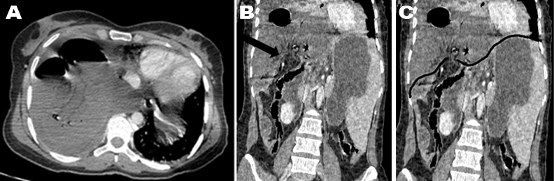
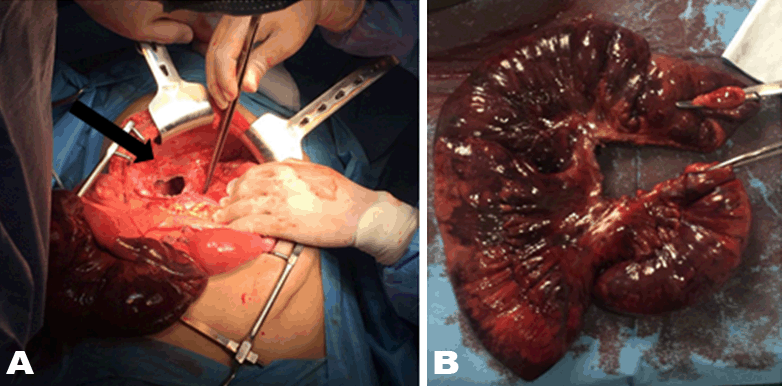
A 30-year-old female was admitted to our emergency department with abdominal pain, shortness of breath, vomiting and a cessation of the exhaust gas and stool since four days. Clinical examination revealed nothing more than a dehydration and an abdominal distension. No sign of vital distress was found. Laboratory findings revealed no major abnormalities: Leukocytes count 8.39x10³ cells/mm3; hemoglobin 13 g/dl. Blood urea nitrogen, creatinine and C-Reactive Protein were normal while levels of sodium 138 mmol/l, potassium 3,1mEq/l, chloride 95 mEq/l, bicarbonates 22mEq/l. Two year prior to these events, the patient underwent a surgical procedure, mainly, a right lob resection of the liver for a giant echinococcal cyst. An intestinal occlusion syndrome was diagnosed. A chest and abdomen CT-scan was performed, revealing an ascent and strangulation by the entanglement of some jejunal loops through a right diaphragmatic hernia (Figure 1). There was a thickening of the wall of the herniated intestinal structures. Right lung passive atelectasis by compression, a pleural effusion of moderate abundance and compression of the mediastinum were also present. The patient was admitted to surgery. Intra operative findings revealed the diaphragmatic defect and some ischemic intestinal loops Figure 2 (A and B). The non-viable ischemic loops were cut off, and the diaphragmatic defect was closed after the reintegration of the abdominal structures. The patient had an uneventful postoperative recovery. | ||||||
|
| ||||||
| ||||||
DISCUSSION | ||||||
|
A diaphragmatic hernia is a herniation of abdominal content into the thoracic cavity through normal and weakened apertures or a rent of the diaphragm. It can either be congenital or acquired. Acquired diaphragmatic hernias can either be iatrogenic or non-iatrogenic. Non-iatrogenic ones arise, in nearly all cases, from a diaphragmatic tear due a major trauma. Iatrogenic diaphragmatic hernias are a complication of any hospital management involving a harmful event on the diaphragm. Iatrogenic diaphragmatic injuries can occur in numerous situations, leading afterward to diaphragmatic hernia. The literature contains some examples: after surgery most of the times [2], [3] , [4], [5], [6], misplaced thoracic drain [7], and rarely from cardiopulmonary resuscitation [8]. Acquired diaphragmatic hernias are commonly due to traumatic disruptions of the diaphragm, following a severe trauma [1], [2]. They are mainly due to penetrating than blunt trauma with a 2:1 ratio [9], [10]. In these cases, the hernia is typically discovered in the course of the hospital stay but it can be delayed. Iatrogenic-related diaphragmatic hernias are less common. Surgical diaphragmatic injuries are nearly, in all cases, diagnosed intraoperatively and repaired by the surgeon who has to be thorough about the assessment of the diaphragm during the procedure. However, some of those injuries can be missed, leading to complications. Francesco Esposito [2] conducted a study, including a liver surgery performed for four years, to evaluate the prevalence of right diaphragmatic hernias following liver resection. The incidence in their study was 2.3 % (3/131) which is in accordance with the literature (< 6.2% after liver resection). Moreover, in their study, only one patient underwent an emergency surgery for bowel obstruction. Thus, the rareness of this surgical outcome could be illustrated. The rate of a late presentation of an iatrogenic diaphragmatic hernia is disparate ranging from 5% to 62% of the cases [3] , with delayed presentation occurring as late as many months or years after the supposedly harmful event [2], [3],[11], [12], [13]. Since the left diaphragm is more prone to injury than the right counterpart, acquired hernias are far more frequent on the left side. Aggravation of a small diaphragmatic rent is explained by the followings: a diaphragmatic wound, even small, cannot easily heal because of the diaphragm involvement in respiration, phonation, etc. A small rent could be stretched by any factor that can increase abdominal pressure such as cough or sport activities. Radiological diagnosis of diaphragmatic rupture is not simple because of the thinness of the diaphragm and associated lesions which may contribute to hide the main region of interest. In the acute settings, CT-scan is the best imaging method of exploration, because it is available and high volume can be quickly explored with new generation multi-slice machines. Frontal and sagittal reformatted images are very useful, notably in very small injuries. There are many CT findings suggestive of a diaphragmatic rupture described in the literature. These descriptions were made for traumatic diaphragmatic ruptures [14], [15], but they can be transposed to diaphragmatic hernias, may they be iatrogenic or not as listed below:
Chest X-ray is not precise but can provide some insights on the presence of a diaphragmatic hernia. MRI can be a useful tool in some cases where there is still doubts on the CT scan. However, this discussion is not that relevant in case of a herniation of abdominal contents because it became obvious and we don’t really need subtle signs to achieve the proper diagnosis. Surgery is the mainstay therapy of a diaphragmatic hernia even in asymptomatic patients. Two main approaches are described and being used. The surgical team can choose from either the thoracotomy approach or the abdominal approach. A third way of repairing the diaphragm is through laparoscopy, which is being used in left diaphragmatic hernias [16], [17]. Every one of these methods has their advantages and flaws. For example, the thoracotomy approach is considered by many authors to be the elective approach, and yet, it is proven to be associated with a longer hospital stay and a high risk of pulmonary embolism [18]. In our case, the surgical team chose the laparotomy approach, using a right subcostal incision. | ||||||
|
CONCLUSION
| ||||||
|
Acquired diaphragmatic hernias are very rarely present on the right side. There are many CT scan findings suggestive of the diagnosis as described in the literature and they should be carefully looked out for by the radiologist. Surgery is the mainstay therapy of diaphragmatic hernias. The originality of our case lies in the fact that late presentation of a diaphragmatic hernia in the right side,associated with a bowel obstruction is a rare outcome of whether a traumatic event or a surgical procedure. | ||||||
|
REFERENCES
| ||||||
| ||||||

|
[HTML Abstract]
[PDF Full Text]
|
|
Acknowledgements
This case report is not supported by any grants. |
|
Author Contributions
Nordjoe Yaotse Elikplim – Substantial contributions to conception and design, Analysis and interpretation of data, Drafting the article, Revising it critically for important intellectual content, Final approval of the version to be published Outznit Mustapha – Substantial contributions to conception and design, Analysis and interpretation of data, Drafting the article, Final approval of the version to be published Laamrani Fatima Zahra – Substantial contributions to conception and design, Analysis and interpretation of data, Drafting the article, Revising it critically for important intellectual content, Final approval of the version to be published Jroundi Laila – Substantial contributions to conception and design, Revising it critically for important intellectual content, Final approval of the version to be published |
|
Guarantor of Submission
The corresponding author is the guarantor of submission. |
|
Source of Support
None |
|
Consent Statement
Written informed consent was obtained from the patient for publication of this case report. |
|
Conflict of Interest
Author declares no conflict of interest. |
|
Copyright
© 2018 Nordjoe Yaotse Elikplim et al. This article is distributed under the terms of Creative Commons Attribution License which permits unrestricted use, distribution and reproduction in any medium provided the original author(s) and original publisher are properly credited. Please see the copyright policy on the journal website for more information. |
|
|