
 |
|
Clinical Image
| ||||||
| Dysphagia lusoria | ||||||
| Simon Phillpotts1, Alexander Azizi2, Matthew Train3, Steven Mann4 | ||||||
|
1Barnet General Hospital, Specialty Registrar, Gastroenterology, The Royal Free London NHS Foundation Trust, London, United Kingdom 2Barnet General Hospital, Foundation Doctor, Gastroenterology, The Royal Free London NHS Foundation Trust, London, United Kingdom 3Barnet General Hospital, Consultant, Radiology, The Royal Free London NHS Foundation Trust, London, United Kingdom 4Barnet General Hospital, Consultant, Gastroenterology, The Royal Free London NHS Foundation Trust, London, United Kingdom | ||||||
| ||||||
|
[HTML Abstract]
[PDF Full Text]
[Print This Article] [Similar article in PubMed] [Similar article in Google Scholar] |


| How to cite this article |
| Phillpotts S, Azizi A, Train M, Mann S. Dysphagia lusoria. Int J Case Rep Images 2018;9:100906Z01SP2018. |
|
CASE REPORT
| ||||||
|
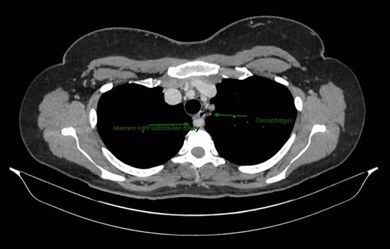
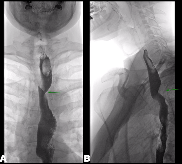
A 43-year-old lady with no past medical history was referred with five weeks of dysphagia and weight loss. She experienced food sticking retrosternally with associated pain radiating to her back. She was only able to tolerate a liquid diet and had subsequently lost nine kilograms of weight in five weeks. Physical examination was unremarkable. Her screening blood tests and abdominal ultrasound were normal. An oesophago-gastro-duodenoscopy (OGD) was performed which showed an entirely normal upper gastrointestinal tract. She subsequently underwent a computed tomography (CT) scan of her thorax, abdomen and pelvis and barium swallow. The CT demonstrated the aberrant right subclavian artery causing compression of the proximal oesophagus (Figure 1). This aberrant vessel was subsequently demonstrated causing a vascular impression of the mid oesophagus on barium swallow (Figure 2A and B). She has been managed conservatively, as definitive treatment would involve major vascular reconstructive surgery. Keywords: Barium, Dysphagia, Dysphagia lusoria, Subclavian | ||||||
| ||||||
|
| ||||||
|
DISCUSSION
| ||||||
|
Dysphagia lusoria is a rare cause of dysphagia. It is caused by an aberrant course of the right subclavian artery [1]. It is thought to occur due to abnormal involution or absence of the 4th right arch of the aorta during early embryological development. This anomaly causes the right subclavian artery to branch from the abdominal aorta distal to the left subclavian and therefore cross the midline, predominately adjacent to the oesophagus [2], [3]. The incidence of this aberrant right subclavian artery is reported as between 0.3 to 1.8% [4] ,[5]. Usually this anomaly does not cause symptoms. However, 20–40% of aberrant arteries are reported to cause trachea-oesophageal symptoms; the mean age of presentation is between 40–48 years old [1]. Manometric abnormalities may contribute to dysphagia in older patients [5]. Barium swallow classically shows a posterior, oblique indentation of the oesophagus. Contrast-enhanced computed tomography typically confirms the diagnosis. Symptomatic dysphagia lusoria can be treated with proton-pump inhibitors and dietary modification, or surgically with ligation and transposition to the right carotid artery [6]. | ||||||
|
CONCLUSION
| ||||||
|
Dysphagia lusoria is a rare cause of dysphagia. It is caused by an aberrant course of the right subclavian artery, which compresses the oesophagus and causes dysphagia. | ||||||
|
REFERENCES
| ||||||
| ||||||

|
[HTML Abstract]
[PDF Full Text]
|
|
Author Contributions
Simon Phillpotts – Substantial contributions to conception and design, Acquisition of data, Analysis and interpretation of data, Drafting the article, Revising it critically for important intellectual content, Final approval of the version to be published Alexander Azizi – Substantial contributions to conception and design, Acquisition of data, Analysis and interpretation of data, Drafting the article, Revising it critically for important intellectual content, Final approval of the version to be published Matthew Train – Substantial contributions to conception and design, Acquisition of data, Analysis and interpretation of data, Revising it critically for important intellectual content, Final approval of the version to be published Steven Mann – Substantial contributions to conception and design, Acquisition of data, Analysis and interpretation of data, Revising it critically for important intellectual content, Final approval of the version to be published |
|
Guarantor of Submission
The corresponding author is the guarantor of submission. |
|
Source of Support
None |
|
Consent Statement
Written informed consent was obtained from the patient for publication of this clinical image. |
|
Conflict of Interest
Author declares no conflict of interest. |
|
Copyright
© 2018 Simon Phillpotts et al. This article is distributed under the terms of Creative Commons Attribution License which permits unrestricted use, distribution and reproduction in any medium provided the original author(s) and original publisher are properly credited. Please see the copyright policy on the journal website for more information. |
|
|