|
|
Clinical Image
| ||||||
| Mycoplasma infection-induced leukocytoclastic vasculitis | ||||||
| Yasuyuki Taooka1, Yuka Ide2, Yusuke Higashi2, Gen Takezawa2 | ||||||
|
1MD, FACP, Department of General Medicine, Akiota Hospital, Hiroshima, Japan 2MD, Department of General Medicine, Akiota Hospital, Hiroshima, Japan | ||||||
| ||||||
|
[HTML Abstract]
[PDF Full Text]
[Print This Article] [Similar article in PubMed] [Similar article in Google Scholar] |
| How to cite this article |
| Taooka Y, Ide Y, Higashi Y, Takezawa G. Mycoplasma infection-induced leukocytoclastic vasculitis. Int J Case Rep Images 2018;9:100903Z01YT2018. |
|
CASE REPORT
| ||||||
|
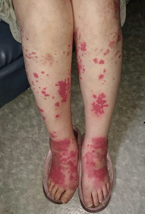
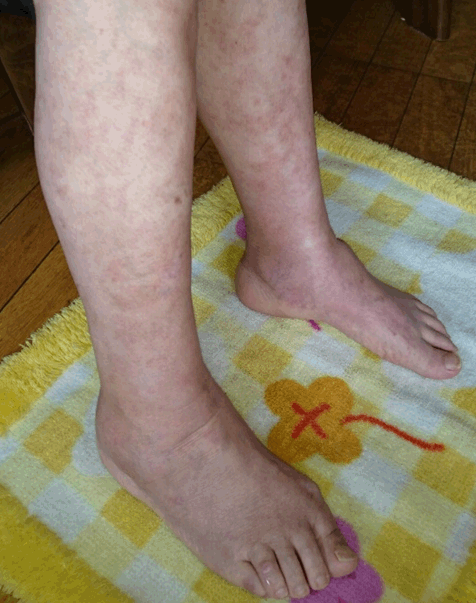
A 67-year-old woman on treatment for rheumatoid arthritis (RA) consulted our outpatient clinic complaining of a 3-day history of an itchy skin eruption of both lower extremities. For the treatment of RA, she was being treated with low-dose methotrexate (6 mg per week) for 24 months in our outpatient clinic. Nine days earlier, she noticed throat pain and dry cough, but these complaints were relieved several days later. Two weeks before her throat pain and dry cough had started, her grandson who lived with her also had throat pain, low-grade fever, and dry cough, but these symptoms disappeared spontaneously one week later. Therefore, she thought that she had a common cold from her grandson. On the day of consultation, diffuse erythema with an unclear margin with spotted purpura was seen on the lower extremities (Figure 1), and these skin lesions were subtly palpable. There was no abdominal pain or sensory disturbance. Her temperature was 36.6°C, and there were no other abnormal findings on physical examination. Urine examination did not show hematuria or proteinuria, and her stool occult blood examination was also negative. Blood laboratory examination showed: white blood cell count 9,570/ml; hemoglobin 13.0 g/dl; thrombocyte count 355,000/ml; total protein 8.0 g/dl; albumin 3.9 g/dl; AST 10 IU/ml; ALT 9 IU/ml; LDH 229 IU/L (normal rage: 106-211 IU/L); CPK 80 IU/l; BUN 11 mg/dl; Cr 0.63 mg/dl; and fasting blood sugar level 89 mg/dl. Serological examination showed: IgG 2,116 mg/dl; IgA 512 mg/dl; IgM 234 mg/dl; C3 149 mg/dl (normal range: 65-135 mg/dl); C4 31 mg/dl (normal range: 13–35 mg/dl); CH50 49 U/ml; C-reactive protein 7.5 mg/dl; MMP-3 685.4 ng/ml (normal range: 16.1-56.8 ng/ml); C-ANCA 1.4 U/ml (normal range: 0-3.4 U/ml); P-ANCA < 1.0 U/ml; and ASK x120 (normal range: 0-x2550). Serum cryoglobulin and anti-parvo B19 virus IgM were negative, but anti-mycoplasma IgM was positive, and serum anti-mycoplasma antigen was also positive. Suspecting cutaneous small vessel vascultis, skin biopsy of an erythematous lesion of her right lower leg was performed. Clarithromycin (400 mg per day) for 7 days and topical corticosteroid cream (diflucortolone valerate) were administrated. Pathological findings showed intradermal hemorrhage with neutrophil infiltration, swelling of vascular endothelial cells, and perivascular neutrophil infiltration. Fibrinoid degeneration of perivascular cell walls and cell nuclear debris (nuclear dust) were also observed in the upper layer of the dermis. Immunohistochemistry showed deposition of C3c component around vascular cell walls, but staining for IgA, IgM, and IgG was negative. A diagnosis of leukocytoclastic vasculitis was made. Three weeks later, the urticarial-like erythematous lesions and purpura of her lower extremities were improved (Figure 2), and her serum anti-mycoplasma IgM titer was four-fold increased. Finally, the diagnosis of mycoplasma infection-induced leukocytoclastic vasculitis was made. It was considered that the preceding infection transmitted by her grandson was a mycobacterial infection. However, her grandson’s serological examination could not be done because there was no informed consent for blood examination. Six months follow-up, there was no recurrence. Keywords: Leukocyteclastic vasculitis, Mycoplasma | ||||||
| ||||||
| ||||||
|
DISCUSSION
| ||||||
|
Cutaneous small vessel vasculitis with leukocytoclastic vasculitis is known as one of the immune complex-related allergic reactions. In addition, drug adverse effects, connective tissue diseases, inflammatory bowel diseases, infectious diseases, and hematological malignant diseases are also causes of vasculitis. Among infectious diseases, hepatitis C virus, streptococcus, and mycoplasma have been reported as causes [1], [2], [3] . In the differential diagnosis of this case, Henoch-Schönlein purpura and urticarial vasculitis were possible [1]. In this case, upper respiratory infection-like symptoms had preceded the skin eruption, but there were no episodes of abdominal pain and hematuria as in Henoch-Schönlein purpura, and IgA deposition around vascular cell walls was not confirmed on skin biopsy examination [1]. Although decreased serum levels of C3, C4, and CH50 are seen in urticarial vasculitis, these were not seen in this case. In this case, low-dose methotrexate therapy was continued to control RA disease activity. Previously, Halevy et al. [4] reported a case of methotrexate-induced leukocytoclastic vasculitis. Although methotrexate is a potential cause of cutaneous leukocytoclastic vasculitis, it was unlikely in the present case. | ||||||
|
CONCLUSION
| ||||||
|
A rare case of leukocytoclastic vasculitis was reported. When faced with a skin eruption as in the present case, the possibility of mycoplasma infection should be considered, and serological examination is useful for its diagnosis. | ||||||
|
REFERENCES
| ||||||
| ||||||
|
[HTML Abstract]
[PDF Full Text]
|
|
Author Contributions
Yasuyuki Taooka – Substantial contributions to conception and design, Acquisition of data, Analysis and interpretation of data, Drafting the article, Revising it critically for important intellectual content, Final approval of the version to be published Yuka Ide – Substantial contributions to conception and design, Acquisition of data, Analysis and interpretation of data, Drafting the article, Revising it critically for important intellectual content, Final approval of the version to be published Yusuke Higashi – Substantial contributions to conception and design, Acquisition of data, Analysis and interpretation of data, Drafting the article, Revising it critically for important intellectual content, Final approval of the version to be published Gen Takezawa – Substantial contributions to conception and design, Acquisition of data, Analysis and interpretation of data, Drafting the article, Revising it critically for important intellectual content, Final approval of the version to be published |
|
Guarantor of Submission
The corresponding author is the guarantor of submission. |
|
Source of Support
None |
|
Consent Statement
Written informed consent was obtained from the patient for publication of this clinical image. |
|
Conflict of Interest
Author declares no conflict of interest. |
|
Copyright
© 2018 Yasuyuki Taooka et al. This article is distributed under the terms of Creative Commons Attribution License which permits unrestricted use, distribution and reproduction in any medium provided the original author(s) and original publisher are properly credited. Please see the copyright policy on the journal website for more information. |
|
|