|
|
Case Report
| ||||||
| Fracture through a femoral stem prosthesis after trauma: A case report and review of the literature | ||||||
| R. F. Lyons1, C. G. Murphy1 | ||||||
|
1Galway University Hospital, Saolta Hospital Group, Galway, Ireland
| ||||||
| ||||||
|
[HTML Abstract]
[PDF Full Text]
[Print This Article] [Similar article in PubMed] [Similar article in Google Scholar] |
| How to cite this article |
| Lyons RF, Murphy CG. Fracture through a femoral stem prosthesis after trauma: A case report and review of the literature. Int J Case Rep Images 2018;9:100894Z01RL2018. |
|
ABSTRACT
| ||||||
|
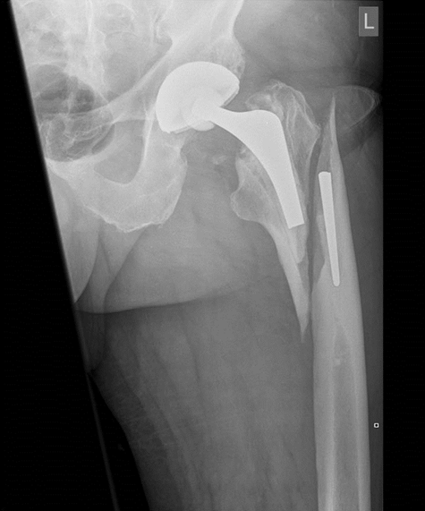
Introduction: Polished tapered collarless femoral implants are in common use in primary hip arthroplasty. Fracture of this implant construct is rare. We described a previously unreported Vancouver B1/2 fracture through the femoral stem implant post trauma with no signs of loosening. Case Report: A 67-year-old male presented with pain in the left hip after a fall. He had a total hip replacement five years previously. He denied preceding symptoms. A radiograph at admission revealed Vancouver B1/2 periprosthetic fracture with femoral stem fracture. He underwent a revision operation and did well post-operatively. Conclusion:Fracture of this implant construct is rare. This case highlights that a fracture through the femoral stem implant post trauma can occur with modern implants showing no fatigue. BMI should be considered when sizing total hip arthroplasty implants. Keywords: Exeter stem, Periprosthetic hip fracture, Revision arthoplasty, Stem fracture | ||||||
|
INTRODUCTION
| ||||||
|
Periprosthetic fractures are a major cause of morbidty and present operative challenges to those involved in trauma and revision arthroplasty. As the number of primary hip arthroplasties is over 700,000 in the UK, the incidence of periprosthetic fracture has been reported at over 6000 [1]. The estimated incidence of periprosthetic fractures has been estimated to be between 0.1–4.5 % in primary hip replacements, however after revision surgery the incidence can be up to 21% [2]. With increasing incidence of periprosthetic fractures, the associated mortality rates are high, being reported up to 18% within one year. When revision surgery is undertaken for a periprosthetic fracture compared to aseptic loosening, the mortality rates are significantly higher at 7.8% compared to 0.9% [3]. We describe a previously unreported Vancouver B1/2 fracture through the femoral stem implant post trauma with no signs of loosening. | ||||||
|
CASE REPORT
| ||||||
|
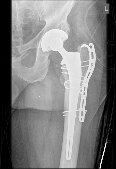
Five years after a primary hip arthroplasty, a sixty seven year old man fell 2ft from a construction excavator, onto his left side with direct impact onto the ground. No twisting motion was reported. The patient reported immediate pain in the left hip and leg and was unable to weight bear on the affected lower limb. He was referred to our trauma institution, where radiographs (Figures 1 and 2) were completed. Radiographs demonstrate a Vancouver B1/B2 type periprosthetic fracture with fracture through the femoral stem, with no obvious lysis around the stem. No previous fragility fractures were noted in the history. Radiographs showed no evidence of gross osteopenia or osteoporosis. A primary hip arthroplasty of the left hip had been performed in 2010 for severe degenerative osteoarthritis of that hip. A size0, offset44, collarless, polished, tapered, cemented femoral stem was inserted. A 58 mm outer diameter (OD) and 32 mm inner diameter (ID) acetabular component was inserted, using metal on polyethylene (MOP) articulating surfaces. Following his index surgery, the patient reported a normal post operative course, no interval symptoms and was back working in construction. A review of the medical notes confirmed an uncomplicated post-operative course with satisfactory examinations and radiographs at planned outpatient follow-up appointments. This man had a significant background history of rheumatoid arthritis (RA), taking fortnightly adalimumab injections and daily leflunomide. Of note he was 185cm and weighed 108kg with a BMI of 31. Routine haematological and biochemical blood investigations were carried out. White cell count (WCC) was 10x109xL, erythrocyte sedimentation rate (ESR) was 7mm/H and C reactive protein (CRP) was 0.1mg/L. Despite the background of RA, no lysis was noted around the stem at time of presentation. A posterior approach to the hip was undertaken for revision. Intraoperatively good bone stock was noted. The fracture site was identified with no further distal extension of the fracture noted. The bone-cement and prosthesis-cement interface were intact. The proximal components were well fixed in cement. No loosening of the prosthesis was appreciated. The proximal component was removed using drilled Kirchner (K-wires) wires, follwed by flexible and rigid osteotomes. An osteotomy was not utilised. The distal component was similarly well fixed in cement and both the prosthesis fragment and the cement were removed again using a combination of sequential K-wires, flexible and rigid osteotomes, and a metallic burr to bypass the cement restrictor and plug. The acetabular cup was assessed and found to be stable and compatible with the new implant configuration so was left in situ. Cerclage wires were used to reduce the femoral fracture. The distal femur was prepared with a reamer and a fully HA-coated dual offset collared uncemented stem was used, bypassing the fracture site. This was supplemented with a trochanteric grip plate. Figure 3 shows the fractured stem and Figure 4 is a radiograph of the post operative revision implant in place. The patient was mobilised on the day following surgery with protected weightbearing for six weeks following surgery. | ||||||
| ||||||
| ||||||
| ||||||
| ||||||
|
DISCUSSION
| ||||||
|
Periprosthetic fractures are a major cause of morbidty and present operative challenges to those involved in trauma and revision arthroplasty. As the number of primary hip arthroplasties is over 700,000 in the UK, the incidence of periprosthetic fracture has been reported at over 6000 [1]. The estimated incidence of periprosthetic fractures has been estimated to be between 0.1–4.5 % in primary hip replacements, however after revision surgery the incidence can be up to 21% [2]. With increasing incidence of periprosthetic fractures, the associated mortality rates are high, being reported up to 18% within one year. When revision surgery is undertaken for a periprosthetic fracture compared to aseptic loosening, the mortality rates are significantly higher at 7.8% compared to 0.9% [3]. In this case, both the proximal and distal segments were adequately cemented in place, hence eliminating cantilever bending forces as a mechanism for failure. There was no issue at the level of the bone-cement interface or the prothesis-cement interface, suggesting that the cementing technique was adequate. Poor cementing technique has been hypothesised as a risk factor for implant failure and fracture. However due to the intact cement mantels it would appear as an unlikely factor in the implant failure in this case [4]. To the best of our knowledge this is the first report of femoral stem fracture secondary to trauma with no compounding factors of loosening, infection or catilever forces causing fatigue. Fractures of the femoral stem in total hip arthroplasties were seen with the early implant designs, mainly due to the low fatigue strength and metallurgical defects. For example, the Exeter stem underwent a design change and since its production with Orthinox very few stem fractures have been reported. The estimated incidence of Exeter stem fracture up until 2003 was 3.9%. However this has been substantially decreased with the use of HNSS Orthinox [5]. Poor biomechanical factors for cemented stems including varus positioning, loosening, loss of proximal support, unfavorable geometry of the stem are all factors in implant failure [6]. The femoral stem used in this case, is a collarless, polished, double taper design. The clinical outcomes of these stems to date is excellent, making them a popular choice in primary hip arthroplasty worldwide. A fracture rate of 0.007% in primary Exeter hips has been quoted in the literature to date, this rate is increased in the revision group up to 0.21% [5]. However failures and complications have been reported in the literature with gross undersizing of the stem and defective cementing processes have been attributed as potential causative factors for failures [7]. Mechanical overload has been reported as a mechanism of failure of implants. Two cases described by Yates et al.[6] have described fracture of the neck of the implant in physically active patients who weighed in excess of 100kg. In these cases relatively large femoral stems similar to the stem in our case were implanted. In our case however the patient also weighed in excess of 100kg but a smaller stem (size 0) was implanted. In particular reference to the Exeter stem, no fractures have been reported in a stem size greater than a number 2. Some of the Exeter stem fractures reported, have occurred in revision cases. Manufactures of such implants have said that size 0 implants are not recommended in those above 70kg [5]. Harvie et al. reported a cohort of implant failures in the JRI Furlong implants. A common patient factor in all failures was a high BMI (above 25) [8]. With the worldwide prevalence of obesity having doubled, BMI should be considered when choosing an implant, especially the use of small stems and how failure has been reported in the literature [9] , [10]. Little has been published with regard to fracture of a femoral stem secondary to trauma where no cantilever forces appear. In 1995, a review of 27 femoral stem components over 10 years [7], identified three femoral stem fractures. No trauma was noted, but two of the cases identified pronounced lucencies between the cement and bone. The authors ascribe fatigue as the causative factor in these implant failures. Raj et al. [4] also reported on stem failure in a patient with an elevated BMI, again however these cases differ to the case we present as their patient reported no trauma, but did report significant preceding symptoms. A review of 14 stainless steel femoral stem fractures found that using smaller stems in particular in champagne glass canals can lead to cantilever bending and failure and this should be considered when treating patients with high body mass index [10]. To the best of our knowledge no case of a fractured implant secondary to trauma has been published. It must be considered however that this patient did have rheumatoid arthritis and published literature does suggest that bone stock is poorer in this cohort of patients, thus can increase the risk of periprosthetic fractures [11]. Classification and management in such cases has not been documented however revision surgery is the only viable option with a fractured implant. The Vancouver classification system is the most widely used system for classifying periprosthetic fractures and aids in treatment decisions [12], [13]. The fracture pattern in this case was classified as a Vancouver B1 with implant fracture. Huang et al. did consider a new fracture classification to incorporate a fractured stem however they only had three patients who had a concurrent femoral stem fracture [13]. Our case again highlights the difficulty to assign a classification type to every periprosthetic fracture. Although the implants were well fixed suggesting B1, the stem was in fact loose and thus required revision (B2). Intra-operative assessment is critical as up to 20% of B1 cases are upgraded to a B2 intra-operatively [14]. | ||||||
|
CONCLUSION
| ||||||
|
A femoral implant fracture associated with a periprosthetic fracure is extremely rare with little published literature available. In order to improve management of periprosthetic fractures, we must learn from previous experiences with implants, and be aware of the changing patient population. With the expected surge in the presentation of periprosthetic fractures, all operative plans and complications should be antipicated. | ||||||
|
REFERENCES
| ||||||
| ||||||
|
[HTML Abstract]
[PDF Full Text]
|
|
Author Contributions
R. F. Lyons – Substantial contributions to conception and design, Acquisition of data, Analysis and interpretation of data, Drafting the article, Revising it critically for important intellectual content, Final approval of the version to be published C. G. Murphy – Substantial contributions to conception and design, Acquisition of data, Analysis and interpretation of data, Drafting the article, Revising it critically for important intellectual content, Final approval of the version to be published |
|
Guarantor of Submission
The corresponding author is the guarantor of submission. |
|
Source of Support
None |
|
Consent Statement
Written informed consent was obtained from the patient for publication of this study. |
|
Conflict of Interest
Author declares no conflict of interest. |
|
Copyright
© 2018 R. F. Lyons et al. This article is distributed under the terms of Creative Commons Attribution License which permits unrestricted use, distribution and reproduction in any medium provided the original author(s) and original publisher are properly credited. Please see the copyright policy on the journal website for more information. |
|
|