

|
 |
|
Case Report
| ||||||
| Tracheal adenoid cystic carcinoma: A case report | ||||||
| Christine Costa1, Patrícia Dionísio1, Paula Monteiro2, José Rosal Gonçalves3, Salvato Feijó4 | ||||||
|
1Resident, Chest Department, Centro Hospitalar Lisboa Norte, Lisbon, Portugal
2Pulmonary Specialist, Chest Department, Centro Hospitalar Lisboa Norte, Lisbon, Portugal 3Chief of the Bronchology Unit, Chest Department, Centro Hospitalar Lisboa Norte, Lisbon, Portugal 4Chief of Department, Pulmonology Department, Cento Hospitalar de Leiria, Leiria, Portugal | ||||||
| ||||||
|
[HTML Abstract]
[PDF Full Text]
[Print This Article] [Similar article in Pumed] [Similar article in Google Scholar] |


| How to cite this article |
| Costa C, Dionísio P, Monteiro P, Gonçalves JR, Feijó S. Tracheal adenoid cystic carcinoma: A case report. Int J Case Rep Images 2018;9:100883Z01CC2018. |
|
ABSTRACT
| ||||||
|
Introduction: Adenoid cystic carcinoma is rare in the trachea and, due to slow growth, it can go unnoticed or be confused with other obstructive pathologies for months/years. Keywords: Adenoid cystic carcinoma, Trachea, Treatment | ||||||
|
INTRODUCTION
| ||||||
|
Primary tumors of the trachea are rare (<2% of tumors in the bronchial tree), and 40% are due to adenoid cystic carcinoma [1]. Adenoid cystic carcinoma (ACC) of the trachea affects men and women equally [2] from the 3rd to 9th decades of life with the mean in the 5th decade [1][3]. However, a case in a 14-year-old girl has been reported [4]. Predisposing factors are not known [1][3] and there is apparently no association between tobacco and adenoid cystic carcinoma [4]. Due to slow growth [1], initial symptoms can be subtle and go unnoticed or be confused with other obstructive pathologies, such as asthma or chronic obstructive pulmonary disease [1][2][3][5][6][7]. | ||||||
|
CASE REPORT
| ||||||
|
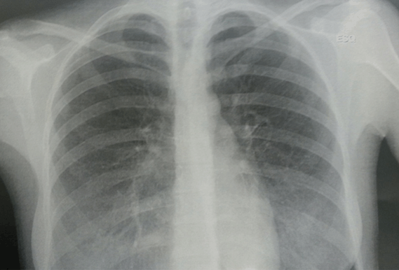
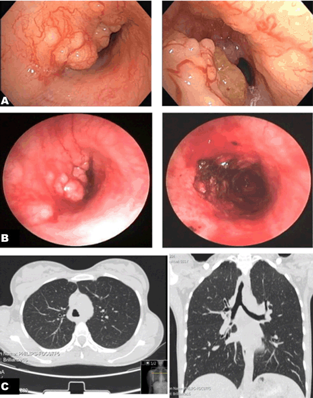
A 31-year-old woman, non-smoker, with no relevant personal history, complained of having productive cough, wheezing and dyspnea on exertion with a progressive evolution during her third trimester of pregnancy. She was observed at the emergency room, where a summary analytical study was conducted, revealing no alteration, as well as a chest radiography. Clinical presentation was considered to be linked to pregnancy and she was later discharged. One month after delivery, due to persistence of symptoms, she was observed in a pulmonology consultation where bronchial asthma was considered. For this reason, she started empirical treatment with inhaled corticoid plus bronchodilator, and supplementary diagnostic tests were ordered. In the reassessment, the patient denied a significant improvement, referring inspiratory stridor. The chest X-ray showed a reduction of tracheal diameter in its distal portion (Figure 1), so bronchofibroscopy (BFC) was performed which revealed an extensive mammillated mass on the anterior and left lateral walls of the thoracic trachea, reducing the lumen by 80% and infiltrating the carina and left main bronchus (Figure 2A). A rigid bronchoscopy with laser therapy for mass resection was immediately performed, leading to recovery of tracheal permeability (Figure 2B). After the procedure, a relief of symptoms was immediately noticed. Pathological examination of the resected specimen was consistent with ACC. After the anatomo-pathological result, computed tomography of the neck, chest and abdomen was performed which demonstrated asymmetric parietal thickening of the tracheal wall from the thoracic operculum to its bifurcation, with extension to the left main bronchus, contacting adjacent mediastinal structures, and no clear signs of invasion (Figure 2C). Due to its extension, surgical resection was not possible, so she was referred to our pulmonary oncology unit to further treatment and follow-up. | ||||||
| ||||||
|
| ||||||
|
DISCUSSION
| ||||||
|
Adenoid cystic carcinoma of the trachea is relatively rare and tends to occur at younger ages than other neoplasms of the bronchial tree [1][3] [4]. At the time of the diagnosis, patients usually present with inspiratory stridor, dyspnea, productive cough [1][3][4][6] or, less frequently, hemoptysis [3][5][6][7]. As dyspnea is generally the initial symptom, it is frequently confused with bronchial asthma, which can cause a delay in diagnosis. Further diagnostic examination consists of chest X-ray, which can either show latero-tracheal opacity or be normal, since tracheal tumors do not involve the lung parenchyma [1][7]. Computed tomography scan provides valuable information on resectability by identifying loco regional involvement and extension of the lesion. Bronchoscopy is mandatory, as it allows not only the collection of material for confirmation of diagnosis, but also enables localization of the lesion and evaluation of its extension [1]. There are three possibilities of treatment:
Surgery with tracheal resection and termino-terminal anastomosis followed by adjuvant radiotherapy is the treatment of choice and can be followed by adjuvant radiotherapy [1][3]. However, apart from requiring an experienced team, surgery requires the lesion to be smaller than half the tracheal length (< 6 cm) [1], and due to late diagnosis, with greater extension of the neoplastic lesion, this approach is often impossible. In unresectable cases, radiotherapy with a dose of 66–70 Gy should be considered [1]. Although there is not much information about late effects after definitive radiotherapy in ACC, high-dose thoracic radiotherapy has been associated with a risk of bronchial damage, such as narrowed airway [2]. Over the past few years, some cases of chemotherapy treatment with carboplatin and paclitaxel have been reported [8][9], but results are still controversial [2][6][7]. In patients with severe obstruction, endoscopic resection using the Nd-YAG laser allows quick disobliteration, a procedure that can be repeated as many times as necessary [5]. Adenoid cystic carcinoma spreads most commonly by direct extension, submucosal or perineural invasion, or hematogenous metastasis [2][3][5][6]. Pulmonary metastases are the most common [2][3] but metastases to the brain, bone, liver, kidney, skin, abdomen, and heart have also been reported [2]. Local recurrence of tracheal ACC is common and occurs at an average of 51 months after the primary treatment [2]. The 5 and 10 years overall survival rate is 52–91% and 29–76%, respectively [6]. | ||||||
|
CONCLUSION
| ||||||
|
This is one of the rare case reports of ACC whose symptoms began during the pregnancy. As we reported in this case, endoscopic laser resection should be performed in cases of severe tracheal obstruction, in order to immediately achieve total or partial recovery of tracheal lumen permeability. | ||||||
|
REFERENCES
| ||||||
| ||||||

|
[HTML Abstract]
[PDF Full Text]
|
|
Author Contributions
Christine Costa – Substantial contributions to conception and design, Acquisition of data, Acquisition of data, Analysis and interpretation of data, Drafting the article, Revising it critically for important intellectual content, Final approval of the version to be published Patrícia Dionísio – Substantial contributions to conception and design, Acquisition of data, Acquisition of data, Analysis and interpretation of data, Drafting the article, Revising it critically for important intellectual content, Final approval of the version to be published Paula Monteiro – Substantial contributions to conception and design, Revising it critically for important intellectual content, Final approval of the version to be published José Rosal Gonçalves – Substantial contributions to conception and design, Revising it critically for important intellectual content, Final approval of the version to be published Salvato Feijó – Substantial contributions to conception and design, Acquisition of data, Acquisition of data, Analysis and interpretation of data, Drafting the article, Revising it critically for important intellectual content, Final approval of the version to be published |
|
Guarantor of Submission
The corresponding author is the guarantor of submission. |
|
Source of Support
None |
|
Consent Statement
Written informed consent was obtained from the patient for publication of this case report. |
|
Conflict of Interest
Authors declare no conflict of interest. |
|
Copyright
© 2018 Christine Costa et al. This article is distributed under the terms of Creative Commons Attribution License which permits unrestricted use, distribution and reproduction in any medium provided the original author(s) and original publisher are properly credited. Please see the copyright policy on the journal website for more information. |
|
|