

|
 |
|
Case Report
| ||||||
| ST segment elevations and a pocket full of pus | ||||||
| Aniket S. Rali1, Tarun Dalia2, Aditya Rali3, Xiuxu Chen4, Ivan Damjanov4, Kamal Gupta1 | ||||||
|
1University of Kansas Medical Center, Division of Cardiovascular Diseases, Kansas City, USA
2University of Kansas Medical Center, Department of Internal Medicine, Kansas City, USA 3Emory University, School of Medicine 4University of Kansas Medical Center, Department of Pathology, Kansas City, USA | ||||||
| ||||||
|
[HTML Abstract]
[PDF Full Text]
[Print This Article] [Similar article in Pumed] [Similar article in Google Scholar] |


| How to cite this article |
| Rali AS, Dalia T, Rali A, Chen X, Damjanov I, Gupta K. ST segment elevations and a pocket full of pus. Int J Case Rep Images 2018;9:100880Z01AR2018. |
|
ABSTRACT
|
|
Introduction: Very few cases of myocardial abscess are reported in medical literature. Although a rare disease entity in the developed countries, it carries a very high mortality rate and hence requires prompt diagnoses and treatment. Here we report a case of myocardial abscess diagnosed on autopsy. Keywords: STEMI, Myocardial Abscess, Pericarditis |
|
INTRODUCTION
|
|
Myocardial abscess is predominantly seen in developing countries. Owing to early diagnoses and treatment of bacteremia, myocardial abscesses are very rare in developed countries with only a handful cases reported in medical literature. The incidence of myocardial abscess as determined in several autopsy studies ranges from 0.1–1.5% [1][2][3]. In the past, the most likely pathophysiology of bacterial myocardial abscess was believed to be preexisting bacteremia leading to spread of infection into the pericardium and myocardium [2]. However, recent reports suggest that most cases of myocardial abscess occur in conjunction with endocarditis of native or prosthetic valves [4]. Myocardial abscesses are often clinically silent and their presence is generally obscured by associated severe sepsis [3]. Herein, we describe a patient who presented with symptoms of chest and abdominal pain and was found to have myocardial abscess on autopsy. |
|
CASE REPORT
|
|
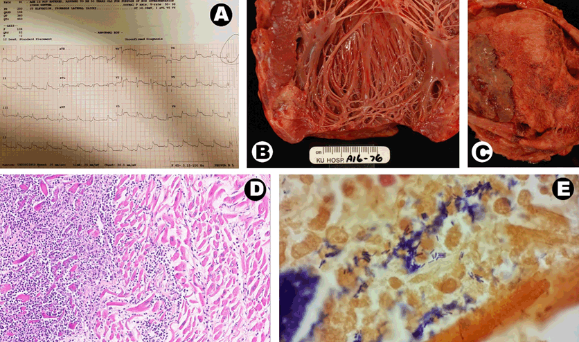
A 47-year-old Caucasian male was presented to the emergency room with complaints of progressively worsening shortness of breath, abdominal pain and chest pressure for three days. His past medical history was significant for uncontrolled diabetes mellitus, hypertension, dyslipidemia, morbid obesity and polymicrobial Fourneir’s gangrene with lower abdominal necrotizing fasciitis requiring several recent debridements. Wound cultures had grown Methicillin resistant Staphylococcus aureus (MRSA) and Enterococcus faecalis. Physical examination was notable for abdominal tenderness and multiple unhealed ulcers over left groin. He was tachypneic requiring 5 liters of supplemental oxygen but normotensive, non-tachycardic and afebrile. Laboratory studies showed significant leukocytosis (WBC 28 K/UL), elevated lactate (5 mmol/L) and creatinine (3.6 mg/dL), decreased serum bicarbonate (15 mmol/L), hyperkalemia (5.7 mmol/L) and mildly elevated troponin (0.48 ng/mL). Electrocardiography showed ST segment elevations in leads I, II, III, aVR, aVL, V2–V6 (Figure 1A). Emergency room team suspected ST segment elevation myocardial infarction (STEMI) and cardiology department was consulted for emergent cardiac catheterization. Due to high clinical suspicion of severe sepsis, the decision was made to hold off on cardiac catheterization. Patient was transferred for emergent CT scan to rule out intra-abdominal infection. While in the scanner, patient developed pulseless electrical activity (PEA) arrest and passed away despite attempted resuscitation. Intra-CPR echo was suggestive of a moderate-sized pericardial effusion. Autopsy was requested which showed bacterial myocarditis with abscess formation involving the left ventricle (Figure 1B–C) and fibrino-hemorrhagic pericarditis with 300 ml of purulent pericardial effusion. Histology section from myocardium showed dense inflammation suggestive of myocardial abscess. (Figure 1D) Gram stain of the autopsy specimen showed gram positive bacterial colonies (Figure 1E). |
|
|
|
DISCUSSION
|
|
Several aspects of this case are interesting including the presenting symptoms, ECG findings and the evolution of differential diagnoses. Initially patient’s symptoms of chest pain were attributed to acute coronary syndrome however laboratory and physical examination findings suggested an intra-abdominal pathology with possible extension of the inflammation into the pericardium and resultant pericarditis. Diffuse ST segment elevations on ECG and presence of pericardial effusion on bedside echo were also suggestive of this. We believe the etiology of PEA arrest to be multifactorial including acidosis, hypoxia and hyperkalemia. Cardiac tamponade could not be ruled out. This case highlights the importance of integration of all available clinical data and maintaining a broad differential diagnoses in reaching a rare diagnosis such as myocardial abscess. Myocardial abscess is a rare and lethal disease that requires prompt diagnoses and treatment. This should be included in the differential in any patient with known or suspected bacteremia and signs of cardiac involvement. In addition to treatment with intra-venous antibiotics, definitive treatment of myocardial abscess involves surgical drainage. In addition to bacteremia, and bacterial endocarditis, myocardial abscess can also occur as a complication of other clinical conditions such as acute myocardial infarction, trauma with penetrating injuries, following invasive cardiac procedures, left ventricular aneurysm infection, atrial myxoma infection and HIV-associated [5]. In our patient, there was no evidence of endocarditis on autopsy. |
|
CONCLUSION
|
|
Myocardial abscess should be considered in patients presenting with conduction defects and ST segment elevations on electrocardiography in the setting of suspected bacteremia or endocarditis. Transthoracic echocardiogram, transesophageal echocardiogram and nuclear medicine scans can all contribute to timely diagnoses. Definitive treatment of myocardial abscess involves surgical drainage. Due to high mortality associated with this disease, prompt diagnoses and treatment is of utmost importance. |
|
REFERENCES
|
|

|
[HTML Abstract]
[PDF Full Text]
|
|
Author Contributions
Aniket S. Rali – Substantial contributions to conception and design, Acquisition of data, Analysis and interpretation of data, Drafting the article, Revising it critically for important intellectual content, Final approval of the version to be published Tarun Dalia – Substantial contributions to conception and design, Acquisition of data, Analysis and interpretation of data, Drafting the article, Revising it critically for important intellectual content, Final approval of the version to be published Aditya Rali – Substantial contributions to conception and design, Acquisition of data, Analysis and interpretation of data, Drafting the article, Revising it critically for important intellectual content, Final approval of the version to be published Xiuxu Chen – Substantial contributions to conception and design, Acquisition of data, Analysis and interpretation of data, Drafting the article, Revising it critically for important intellectual content, Final approval of the version to be published Ivan Damjanov – Substantial contributions to conception and design, Acquisition of data, Analysis and interpretation of data, Drafting the article, Revising it critically for important intellectual content, Final approval of the version to be published Kamal Gupta – Substantial contributions to conception and design, Acquisition of data, Analysis and interpretation of data, Drafting the article, Revising it critically for important intellectual content, Final approval of the version to be published |
|
Guarantor of Submission
The corresponding author is the guarantor of submission. |
|
Consent Statement
Written informed consent was obtained from the patient for publication of this case report. |
|
Source of Support
None |
|
Conflict of Interest
Authors declare no conflict of interest. |
|
Copyright
© 2018 Aniket S. Rali et al. This article is distributed under the terms of Creative Commons Attribution License which permits unrestricted use, distribution and reproduction in any medium provided the original author(s) and original publisher are properly credited. Please see the copyright policy on the journal website for more information. |
|
|