|
|
|
|
Case Report
| ||||||
| A rare cause of obscure upper gastrointestinal bleeding: A challenging experience | ||||||
| Khalid Y. Nabrawi1, Saad S. Algahtani2, Ahmed Y. Shaheen1, Abdullah M. Alshehri1, Mushabab Algahtani1, Mohamed-Elbagir K. Ahmed3 | ||||||
|
1Department of Medicine, Gastroenterology Unit, Aseer Central Hospital, P O Box 34, Abha, Saudi Arabia
2Department of Interventional Radiology, Armed Forces Hospital, Southern Region, P O Box 101, Khamis Mushait, Saudi Arabia 3Department of Medicine, College of Medicine, King Khalid University, P O box 641, Abha, Saudi Arabia | ||||||
| ||||||
|
[Abstract HTML]
[Full Text HTML]
[Full Text PDF]
[Print This Article] [Similar article in Pumed] [Similar article in Google Scholar] |
| How to cite this article |
| Nabrawi KY, Algahtani SS, Shaheen AY, Alshehri AM, Algahtani M, Mohamed-Elbagir KA. A rare cause of obscure upper gastrointestinal bleeding: A challenging experience. Int J Case Rep Images 2018;9(1):16–19. |
|
ABSTRACT
| ||||||
|
Introduction: Liver cirrhosis leads to a serious complication mainly the development of portal hypertension which causes catastrophic consequences namely variceal bleeding, encephalopathy, and hepatocellular carcinoma. Bleeding is commonly caused by ruptured esophageal, gastric, and rarely ectopic varices. We report here a patient with liver cirrhosis who presented with obscure upper gastrointestinal bleeding proved to be due to an ectopic varix. Keywords: Ectopic varix, Gastrointestinal bleeding, Interventional radiology | ||||||
|
INTRODUCTION
| ||||||
|
Liver cirrhosis is a serious complication of chronic liver disease which is caused by both viral diseases (hepatitis B virus and hepatitis C virus) and non-viral conditions such as alcohol related, autoimmune liver disease, metabolic, and inherited condition. It constitutes the fifth-leading cause of adult deaths and ranks eighth in economic cost among the major illnesses. Its most serious complication is the development of portal hypertension which leads to serious complications namely variceal bleeding, encephalopathy, and hepatocellular carcinoma [1][2][3][4]. Bleeding is commonly caused by ruptured esophageal, gastric, and ectopic varices. Ectopic varices are defined as dilated portosystemic collateral veins in locations other than the gastroesophageal region. They account for 1–5% of all variceal bleeds in patients with intrahepatic portal hypertension and 20–30% of those with extrahepatic portal hypertension (PHT) [5][6] . Numerous sites have been identified for the occurrence of ectopic varices including 18% in the jejunum or ileum, 17% in the duodenum, 14% in the colon, 8% in the rectum, and 9% in the peritoneum, in addition, to surgical stomas and retroperitoneal sites [7]. Although PHT is the main cause of ectopic varices, other conditions such as surgical procedures involving abdominal organs and vessels, anomalies in venous outflow, abdominal vascular thrombosis, hepatocellular carcinoma, and familial origin are also documented causes [8][9]. Ectopic variceal bleeding is considered potentially fatal with a high mortality rate reaching 40% in duodenal variceal bleeding [10]. Many therapeutic modalities have been developed, including endoscopic treatments, endoscopic variceal obturation (EVO), endoscopic variceal band ligation (EVL)], radiologic interventions such as balloon-occluded retrograde transvenous occlusion (BRTO), transjugular intrahepatic portosystemic shunt (TIPS), and surgery. We report here a patient with a rare cause of obscure variceal bleeding successfully managed by the interventional radiologist. | ||||||
|
CASE REPORT
| ||||||
|
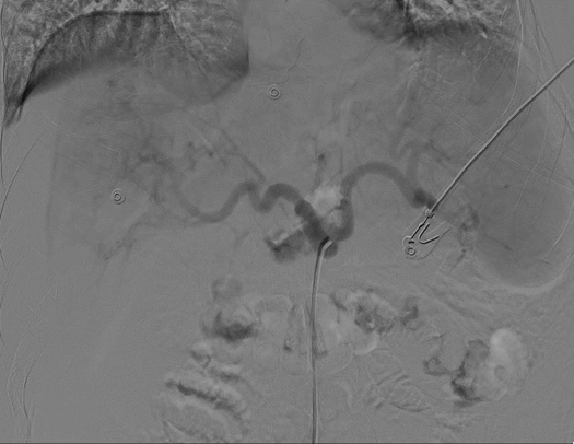
Our patient was a 55-year-old male known to have hepatitis C virus induced liver cirrhosis complicated by hepatocellular carcinoma with portal vein invasion. The patient has history of colonic injury with colectomy and Hartmann’s procedure which has been done two years back before presentation. He reported to the emergency unit at Aseer central hospital in Abha, Saudi Arabia complaining of recurrent melena and hematochezia for the past 10 days. There was no abdominal pain, heartburn, or vomiting. Prior to admission to our hospital, he reported to a local hospital where he was resuscitated with 13 unit packed red blood cells and then referred to our hospital. Upon admission, he was restless but fully conscious. The vital signs were as follows: blood pressure of 118/57 mmHg lying, heart rate of 116/min with small regular pulse, respiratory rate of 20\min, and body temperature of 36.6°C . Head and neck examinations of the patient were unremarkable except for pale conjunctivae and yellowish discoloration of the sclera. His abdomen was soft, distended with positive shifting dullness, and the liver span was 9 cm. Examination of other systems was unremarkable, but the lower limbs showed pitting edema up to his knees. Initial laboratory data were as follow: WBC 11.6x10⁹ ml, hemoglobin 8 g/dl, platelet count 69,000 ml, total bilirubin 14.9 mg/dl (direct 8.6) and albumen 2.1 g/dl. INR was 1.7 with negative hepatitis B markers and positive anti hepatitis C antibodies. The patient was treated by intravenous fluids, pantoprazole, intravenous octreotide and intravenous ceftriaxone. He was also transfused packed red blood cells. Emergency esophagogastroduodenoscopy revealed the presence of grade 2 esophageal varices but without stigmata of recent bleeding. No bleeding lesion was seen in the stomach or duodenum. Also, no bleeding focus was found at colonoscopy up to the terminal ileum. Computed tomography (CT) angiography revealed the presence of sub - mucosal varix at level of a small bowel jejunal loop (Figure 1). The decision was to refer the patient to intervention radiologist, because of continuous active bleeding. The patient was transfused 4 units of fresh frozen plasma prior to procedure for correction of coagulopathy. Selective angiography of celiac trunk, superior mesenteric artery, and the inferior mesenteric artery did not reveal any bleeding or A-V shunt. Then CT angiography of the abdomen confirmed the presence of a large varix draining upwards in the superior mesenteric vein. This was embolized under ultrasound guided using coil without any immediate complication (Figure 2). Following the procedure, the patient remained stable and maintained his hemoglobin with no recurrence of bleeding. He was discharged from hospital in a stable condition and offered regular follow-up in the gastroenterology clinic when seen 2 weeks later in the clinic, the patient was stable without recurrence of bleeding. | ||||||
| ||||||
| ||||||
|
DISCUSSION
| ||||||
|
Available information concerning ectopic variceal bleeding control is mostly derived from case reports or small case series because of the lack of randomized controlled trials. The largest series ever of ectopic variceal bleeding was reported by Norton et al who reviewed 169 cases [10]. Furthermore, the confirmation of the site and management of ectopic varices require a proper study of the vascular supply and local hemodynamics of the varices using a multidisciplinary approach which involves gastroenterologist, hepatologists, interventional radiologists, and surgeons. However, there is no proven, superior treatment modality for the management of ectopic variceal bleeding [9][10]. A previous report by Sharma et al. described the experience with a bleeding of duodenal ectopic varices [11]. Moreover, Park et al. published the most recent large series of eight patients with ectopic variceal bleeding treated by different modalities but without radiological intervention [12]. One of the very few reports of ectopic jejunal bleeding varices which is similar to ours was that published by Park et al. However, their patient had liver cirrhosis, but unlike the present one, without any previous abdominal surgery, and was managed by embolization of the jejunal varices via the portal vein [13]. Our patient was finally managed by the interventional radiologist. Hence, the role of interventional radiology becomes vital in patients whose gastrointestinal bleeding remains resistant to medical and endoscopic treatment. Radiology offers diagnostic imaging studies and endovascular therapeutic interventions that can be performed promptly and effectively with successful outcomes. In particular, ultrasound guided intervention provides safe and inexpensive imaging evidence of morphological abnormalities associated with cirrhosis and PH [14]. Also, the presence of portocollateral circulation on ultrasound, computed tomography (CT), or magnetic resonance imaging scan such as recanalized paraumbilical vein, spontaneous splenorenal circulation, and dilated left and short gastric veins, or the finding of a reversal of flow within the portal system, is very specific for clinically significant portal hypertension. Furthermore, computed tomography angiography and nuclear scintigraphy can also help localize the source of bleeding and provide essential information for the interventional radiologist to guide therapeutic management with endovascular angiography and transcatheter embolization [15]. Another area where the interventional radiologist can offer help to control acute variceal hemorrhage refractory to endoscopic and pharmacological treatments is the performance of Transjugular Intrahepatic Portosystemic Stenting (TIPSS). However, being a highly specialized procedure, it requires adequate training, experience, and knowledge of the relevant equipment, anatomy, and how to deal with any complications. It should, therefore, be performed in centers with adequate personnel, multidisciplinary support and equipment required to optimize management and minimize risks [16][17][18][19]. | ||||||
|
CONCLUSION
| ||||||
|
In conclusion, this report refreshes the minds about the current methods of diagnosis and management of ectopic varices and emphasizes the important role of the interventional radiologist in this matter. | ||||||
|
REFERENCES
| ||||||
| ||||||
|
[Abstract HTML]
[Full Text HTML]
[Full Text PDF]
|
|
Author Contributions
Khalid Y. Nabrawi – Substantial contributions to conception and design, Acquisition of data, Analysis and interpretation of data, Drafting the article, Revising it critically for important intellectual content, Final approval of the version to be published Saad S. Algahtani – Substantial contributions to conception and design, Acquisition of data, Analysis and interpretation of data, Drafting the article, Revising it critically for important intellectual content, Final approval of the version to be published Ahmed Y. Shaheen – Substantial contributions to conception and design, Acquisition of data, Analysis and interpretation of data, Drafting the article, Revising it critically for important intellectual content, Final approval of the version to be published Abdullah M. Alshehri – Substantial contributions to conception and design, Acquisition of data, Analysis and interpretation of data, Drafting the article, Revising it critically for important intellectual content, Final approval of the version to be published Mushabab Algahtani – Substantial contributions to conception and design, Acquisition of data, Analysis and interpretation of data, Drafting the article, Revising it critically for important intellectual content, Final approval of the version to be published Mohamed-Elbagir K. Ahmed – Substantial contributions to conception and design, Acquisition of data, Analysis and interpretation of data, Drafting the article, Revising it critically for important intellectual content, Final approval of the version to be published |
|
Guarantor of Submission
The corresponding author is the guarantor of submission. |
|
Source of Support
None |
|
Conflict of Interest
Authors declare no conflict of interest. |
|
Copyright
© 2018 Khalid Y. Nabrawi et al. This article is distributed under the terms of Creative Commons Attribution License which permits unrestricted use, distribution and reproduction in any medium provided the original author(s) and original publisher are properly credited. Please see the copyright policy on the journal website for more information. |
|
|