
|
 |
|
Case Report
| ||||||
| A case of isolated ST segment elevation in augmented vector right secondary to occluded or under perfused STOUP-collateral circulation | ||||||
| Edward Rojas1, Molly Malone2, George Stoupakis3 | ||||||
|
1MD, Department of Medicine, Rutgers, New Jersey Medical School, Newark, New Jersey 2MD, Department of Emergency Medicine, Hackensack University Medical Center, Hackensack, New Jersey 3MD, FACC, Department of Cardiology, Hackensack University Medical Center, Hackensack, New Jersey | ||||||
| ||||||
|
[HTML Abstract]
[PDF Full Text]
[Print This Article] [Similar article in Pumed] [Similar article in Google Scholar] 
|


| How to cite this article |
| Rojas E, Malone M, Stoupakis G. A case of isolated ST segment elevation in augmented vector right secondary to occluded or under perfused STOUP-collateral circulation. Int J Case Rep Images 2017;8(8):519–522. |
|
ABSTRACT
|
|
Introduction:
Lead augmented vector right (aVR) has been historically underestimated in the evaluation of acute coronary syndromes in the emergency department and other clinical settings. ST-elevation in aVR has shown to be a predictive marker of critical stenosis of the left main coronary and is associated with increased 30 day mortality. Keywords: Augmented vector right (aVR) lead, Collateral circulation, Left main coronary artery, ST-elevation myocardial infarction (STEMI) |
|
INTRODUCTION
|
|
The utility of lead augmented vector right (aVR) in the evaluation of acute coronary syndrome has been historically underestimated in clinical practice [1]. Nonetheless, ST elevation isolated to lead aVR and especially in combination with other ischemic changes, has been shown to be a predictive marker of critical stenosis of the left main coronary artery. Augmented vector right ST-elevation has been associated with a higher 30-day mortality that was independent of concomitant ST segment changes in other leads [2]. The increased mortality of this condition is demonstrated in this case report; our patient succumbed to his disease eleven days after presentation to the emergency department. We report an unusual case of acute ST-elevation myocardial infarction consistent with left main disease presenting with aVR ST elevation in the setting of hypotensive collateral insufficiency in a patient with ischemic stroke. This case highlights the importance of recognizing an unusual pattern on electrocardiography (ECG) for early detection of left main disease. Also demonstrates, a case of ST elevations due an occluded or under perfused collateral circulation (STOUP- collateral circulation). |
|
CASE REPORT
|
|
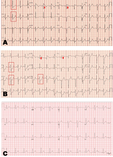
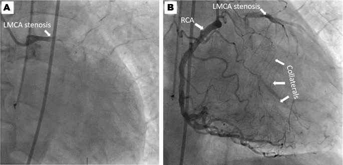
A 75-year-old Asian male with prior history of cerebrovascular accident, type 2 diabetes, hypertension and dementia was brought to the emergency department with a chief complaint of aphasia, left sided facial droop and right sided hemiparesis. Symptoms began acutely at 09:00 a.m. on the day of admission when returning home from church. In the emergency department, blood pressure was initially 137/72 mmHg, heart rate of 86bpm, and electrocardiogram (ECG) showed 0.1 mV ST elevations in aVR and V1, and ST-depression in lead I and II (Figure 1A). Computed tomography scan of head without contrast showed no acute intracranial hemorrhage and the patient was deemed an appropriate candidate for t-PA. After initial t-PA bolus, patient was started on a t-PA infusion. Due to worsening hypertension reaching 200/88 mmHg, he was started on a titratable nicardipine infusion. Transiently, the patient experienced an episode of nausea and vomiting and became hypotensive to 70/45 mmHg leading to the discontinuation of nicardipine. Shortly thereafter, the patient was noted to have new ST changes on telemetry monitor. A second ECG The patient was taken directly to the catheterization laboratory where he was found to have a total occlusion of the distal left main coronary artery (LMCA) which was collateralized from a large, dominant right coronary artery (RCA) that had no obstructive lesions. To confirm that left circulation was chronically occluded, we attempted to wire into the left anterior descending (LAD) artery, but the wire could not cross through the left main stenosis, confirming a chronic occlusion (Figure 2A). |
| Treatment, outcome and follow-up |
|
This patient developed a STEMI due to a totally occluded LMCA and hypotensive collateral insufficiency of the RCA in the setting of treatment of an acute ischemic stroke. His coronary angiogram showed a chronically occluded distal LMCA with a dominant RCA and significant collateral circulation (Figure 2B). The patient did not require any immediate revascularization in the catheterization laboratory due to improved mean arterial pressure and improved collateral perfusion. Given recent administration of t-PA, intravenous heparin was not recommended due to increased risk of bleeding. Post-cardiac catheterization and after clearance by neurosurgery and neurology, the patient was started on aspirin and metoprolol succinate. He was managed in the intensive care unit for 48 hours, extubated and transferred to stroke unit. Echocardiogram was performed showing a left ventricular ejection fraction of 40% correlating with moderate systolic dysfunction, and mild concentric left ventricular hypertrophy. Also, the entire anterior, basal and mid anterolateral wall, mid anteroseptal segment, apical lateral segment, and apex were hypokinetic. Grade II diastolic dysfunction was also seen. During his hospital study, the patient was found to have multiple episodes of atrial fibrillation that was rate controlled with digoxin and nicardipine hydrochloride with appropriate response. The patient was eventually planned for staged coronary bypass surgery. However, one week after presentation developed septic shock secondary to aspiration pneumonia and ultimately expired. |
|
|
|
|
|
DISCUSSION
|
|
Interpretation of the ECG in the appropriate clinical setting remains the cornerstone in diagnosis of STEMI [3][4][5] and helps in localizing stenotic lesions [6]. Nonetheless, lead aVR has been historically ignored in clinical practice partially because of its relative isolation (aVR has no other adjacent lead) [1][2][7]. Analysis of this particular lead is of great importance mainly due to its anatomical position. Yamaji et al. [8] were able to illustrate that ST-elevation in aVR especially when greater than that in V1, predicts with high sensitivity and specificity LMCA occlusion. Acute occlusion of the LMCA is generally associated with high mortality. Without collateral circulation, patients are at risk of cardiogenic shock. ST-segment elevation in lead aVR was also strongly associated with increased rates of in-hospital recurrent ischemic events and heart failure. Wong et al. [2] demonstrated a higher mortality in patients with aVR ST-elevation compared to those with ST elevation in any other leads. This might be partially explained by its association with severe LMCA disease [9]. Our patient initially presented with a 0.1 mV ST- elevation in aVR that progressed into frank STEMI. We hypothesize that hypotension-related hypoperfusion of RCA collateral circulation, in the setting of a chronically occluded LMCA was the cause of STEMI. Similar cases have been described in literature associating aVR ST-elevations with significant left main coronary artery or ostial LAD stenosis [10][11]. The role of collateral circulation in coronary artery disease is a growing and promising field [12] and our case is an example of the enormous therapeutic potential of inducing arteriogenesis and angiogenesis to establish collateral circulations able supply ischemic areas of the myocardium. Prompt recognition of isolated aVR ST changes due to critical LMCA and LAD occlusions would allow for identification of high risk patients and appropriate activation of the catheterization laboratory [13]. An early invasive approach might be especially beneficial in patients presenting with this finding [14]. |
|
CONCLUSION
|
|
This case of isolated ST segment elevation in augmented vector right (aVR) secondary to an occluded or under perfused (STOUP) collateral circulation demonstrates the importance of early recognition of changes in aVR, as well as the therapeutic potential of an efficient collateral circulation. |
|
REFERENCES
|
|

|
[HTML Abstract]
[PDF Full Text]
|
|
Author Contributions
Edward Rojas – Substantial contributions to conception and design, Acquisition of data, Analysis and interpretation of data, Drafting the article, Revising it critically for important intellectual content, Final approval of the version to be published Molly Malone – Analysis and interpretation of data, Revising it critically for important intellectual content, Final approval of the version to be published George Stoupakis – Analysis and interpretation of data, Revising it critically for important intellectual content, Final approval of the version to be published |
|
Guarantor
The corresponding author is the guarantor of submission. |
|
Source of support
None |
|
Conflict of interest
Authors declare no conflict of interest. |
|
Copyright
© 2017 Edward Rojas et al. This article is distributed under the terms of Creative Commons Attribution License which permits unrestricted use, distribution and reproduction in any medium provided the original author(s) and original publisher are properly credited. Please see the copyright policy on the journal website for more information. |
|
|