|
|
|
|
Case Report
| ||||||
| Temporary exacerbation of primary epiploic appendagitis: A case report | ||||||
| Masahide Hara1, Yumiko Ando2, Kazuhide Tohara3 | ||||||
|
1MD, Deputy Director, Nishibeppu National Hospital, Beppu City, Oita, Japan
2MD, Director of Department of Radiology, Nishibeppu National Hospital, Beppu City, Oita, Japan 3MD, Director of Department of Surgery, Nishibeppu National Hospital, Beppu City, Oita, Japan | ||||||
| ||||||
|
[HTML Abstract]
[PDF Full Text]
[Print This Article] [Similar article in Pumed] [Similar article in Google Scholar] 
|
| How to cite this article |
| Hara M, Ando Y, Tohara K. Temporary exacerbation of primary epiploic appendagitis: A case report. Int J Case Rep Images 2017;8(7):462–465. |
|
ABSTRACT
|
|
Introduction:
Primary epiploic appendagitis (PEA) is a disease with a good prognosis, and numerous reports have indicated that it can be cured without surgical treatment. However, there have been few reports on the necessity of anti-microbial therapy. There is also a paucity of information regarding what kind of management should be provided if the symptoms do not improve. Keywords: Antibiotics, Non-steroidal anti-inflammatory drugs, Primary epiploic appendagitis, Self-limiting disease |
|
INTRODUCTION
|
|
It is important to distinguish primary epiploic appendagitis (PEA) from other diseases in the clinical evaluation of patients with acute abdomen. The reported treatment modalities for PEA have primarily focused on surgical techniques [1]. In recent years, many reports have stated that if the diagnosis is correct, most patients have a good prognosis without surgical treatment [2][3]. However, there are few reports on how to manage PEA in patients with deteriorated symptoms. In addition, there is limited information regarding whether antibiotics are useful as conservative treatment. We describe a patient with PEA of the sigmoid colon, and we discuss available literature on the topic. |
|
CASE REPORT
|
|
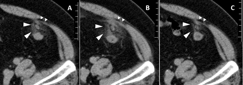
A 69-year-old male with myocardial infarction had been treated conservatively. He reported smoking 30 cigarettes per day for 52 years, and drinking one bottle of beer and a glass of shochu with hot water per day. He had no significant family medical history. He was receiving outpatient treatment for high blood pressure and ventricular extrasystole. The presented to the hospital with left lower abdominal pain beginning the previous evening. He was admitted to the hospital on the same day with a suspected diagnosis of colonic diverticulitis. He was 164.8 cm tall, weighed 96.0 kg, had a body mass index (BMI) of 35.3 kg/m2, and a temperature of 36.0°C. His blood pressure was 112/87 mmHg, and his pulse was regular at 86 beats per minute. On physical examination, his abdomen was bulging, and bowel sounds were normal. Tenderness and rebound were present in the left lower quadrant. There were no palpable masses in the abdomen. The venous blood examination showed a C-reactive protein level of 1.43 mg/dL (normal range 0–0.14 mg/dL) and white blood cell count of 8,800/mm3 (normal range 3,300–8,600/mm3). The leukocytes comprised 56% neutrophils (normal range 38–72%), 29% lymphocytes (normal range 19–50%), 5% monocytes (normal range 4–11%), 8.4% eosinophils (normal range 0.5–9.6%), and 0.9% basophils (normal range 0.2–3.6%). No other laboratory abnormalities were present. The abdominal radiograph showed no abnormality. The abdominal computed tomography (CT) scan showed no intraperitoneal free air or signs of ileus. A hyper-attenuated nodular soft tissue mass surrounded by a fatty rim and localized peritoneal thickening was identified. The mass was located anterior to the sigmoid-descending colon junction. It was not continuous with the sigmoid colon, and he was diagnosed as having PEA (Figure 1A). Since secondary epiploic appendagitis could not be completely ruled out, he began fasting. He received fluid replacement therapy and 500 mg of levofloxacin per day intravenously. On the third day of hospitalization, his abdominal pain exacerbated and abdominal tenderness and rebound were observed. Venous blood analysis revealed a WBC count of 7,300/mm3, and the C-reactive protein level was 1.60 mg/dl. To reconfirm the diagnosis, CT scan was repeated. Computed tomography scan showed exacerbation of the density of the fat rim and peritoneal thickening (Figure 1B). There were no inflammatory findings on the intestinal wall, and abscess formation was not observed. Therefore, conservative treatment was continued accompanied by the oral administration of 180 mg daily of loxoprofen sodium. On the seventh day of hospitalization, the patient’s abdominal pain and tenderness gradually improved. Laboratory test results revealed a white blood cell count of 5,600/mm3 and a C-reactive protein level of 0.53 mg/dL. The CT scan showed alleviation of the hyper-attenuated adipose tissue and peritoneal hyperplasia. Levofloxacin and loxoprofen sodium administration were completed, and he was discharged on the eighth day of hospitalization. On outpatient follow-up on the eighth day after discharge, he no longer reported abdominal pain, and the white blood cell count and C-reactive protein level were normalized at 8,300/mm3 and 0.11 mg/dL, respectively. Abdominal CT scan revealed further improvement (Figure 1C). |
|
|
|
DISCUSSION
|
|
Epiploic appendages are pedunculated, leaf-like fat structures wrapped by serosa. The colon has approximately 50 to 100 and 2–5 cm long epiploic appendages. They protrude from the anterior and posterior portions of the large intestine along the tenia libera. Although their role is not precisely understood, they are believed to play a protective role as cushions, localized prevention against infection, and as energy reservoirs during starvation [4]. Epiploic appendages are nourished by small arteries and veins. Since they are pedunculated and rich in mobility, they are prone to ischemic infarction due to torsion. This is the reported pathomechanism of epiploic appendagitis [5]. However, secondary epiploic appendagitis is caused by the inflammation of adjacent organs, such as the colon diverticulum, gallbladder, and appendix [6]. Epiploic appendages exist along the entire length of the colon. Therefore, it is necessary to differentiate epiploic appendagitis from many other diseases. Diverticulitis, appendicitis, and omental infarction are often difficult to differentiate from epiploic appendagitis. Other potential diagnoses include tuberculous peritonitis, neoplasm, urachal cyst, mesenteric panniculitis, and trauma [7]. Symptoms of epiploic appendagitis include localized abdominal pain and rebound tenderness, sometimes with mild fever. In many cases, the white blood cell count remains normal or mildly increased. Choi et al. [8] examined 31 patients with epiploic appendagitis. They reported that abdominal tenderness was found in all cases, and rebound was found in 8 cases. The average body mass index was 25.9 kg/m2. The diagnosis before the imaging examination was diverticulitis in 13 patients. On diagnostic CT scan, the epiploic appendage with inflammation is observed as an ovoid mass close to the colon. Intraperitoneal adipose tissue with hyperdensity due to inflammation surrounds the mass [6][9][10]. Symptoms are often relieved within a week with analgesic therapy alone [6]. There have been many reports that conservative treatment can be continued if typical CT findings are recognized [11][12]. Schnedl et al. reported that 1,000 mg of ciprofloxacin daily was often used for epiploic appendagitis in their hospitals [13]. The Infectious Diseases Society of America guidelines for intra-abdominal infection provided recommendations for antibiotic therapy in 2010 [14]. According to the guidelines, adult patients with mild-to-moderate severity community-acquired infection should be treated with ticarcillin-clavulanate, cefoxitin, ertapenem, moxifloxacin, or tigecycline as single use. Cefazolin, cefuroxime, ceftriaxone, cefotaxime, levofloxacin, or ciprofloxacin are also preferable in combination with metronidazole. However, for adults with high-risk community-acquired infection, doripenem, meropenem, imipenem-cilastatin, piperacillin-tazobactam, ciprofloxacin, or levofloxacin accompanied by metronidazole should be chosen. In our case, a single intravenous infusion of levofloxacin was administered because secondary epiploic appendagitis due to diverticulitis could not be ruled out. Metronidazole was not available. Abadir et al. investigated 15 patients with omental infarction or epiploic appendagitis and reported that nine patients received antibiotics [15]. Six of 15 patients had peritoneal signs, and 12 patients were treated conservatively. Thus, there is no consensus on the role of antibiotics in the treatment of epiploic appendagitis. Many studies have reported that CT scan is useful to rule out the need for surgical treatment [8][16]. In our patient, antibacterial therapy and fasting were initiated on the first day of hospitalization, but his symptoms worsened on the third day of hospitalization. Secondary epiploic appendagitis and abscess formation were ruled out through subsequent CT scan. Mild diverticulitis was considered difficult to distinguish from PEA. Computed tomography scan showed that the lesion was a solid, lobular-like soft-tissue density mass, and it was not adjacent to the colon wall. On the basis of these characteristic findings, we diagnosed the lesion as PEA, not diverticulitis. In the case of diverticulitis, the interior of the lesion is not solid, and it has a three-layer structure. That is, there is a hyper-attenuated mucosal layer from the inside, thickened submucosa with a low density, and a large amount of serosa. Oral administration of non-steroidal anti-inflammatory drugs (NSAIDs) was initiated, and his symptoms subsequently improved. This indicates that NSAIDs may be more effective for the treatment of PEA than antibiotics. One study reported that diagnostic laparoscopy is required for patients without clinical improvement of diverticulitis [17]. However, we re-examined the CT scan instead of laparoscopy, and we confirmed that the diagnosis was PEA. Therefore, we decided to continue conservative management. |
|
CONCLUSION
|
|
Primary epiploic appendagitis (PEA) is a self-limiting disease. To our knowledge, there are no other reports of patients with PEA with symptoms worsening after the administration of antibiotics alone. We consider this case valuable in terms of determining appropriate treatment for PEA. To alleviate the symptoms conservatively, a confirmed diagnosis by computed tomography scan and the administration of non-steroidal anti-inflammatory drugs from an early stage are necessary. |
|
REFERENCES
|
|
|
[HTML Abstract]
[PDF Full Text]
|
|
Author Contributions
Masahide Hara – Substantial contributions to conception and design, Acquisition of data, Analysis and interpretation of data, Drafting the article, Revising it critically for important intellectual content, Final approval of the version to be published Yumiko Ando – Analysis and interpretation of data, Critical revision of the article, Final approval of the version to be published Kazuhide Tohara – Analysis and interpretation of data, Revising it critically for important intellectual content, Final approval of the version to be published |
|
Guarantor
The corresponding author is the guarantor of submission. |
|
Source of support
None |
|
Conflict of interest
Authors declare no conflict of interest. |
|
Copyright
© 2017 Masahide Hara et al. This article is distributed under the terms of Creative Commons Attribution License which permits unrestricted use, distribution and reproduction in any medium provided the original author(s) and original publisher are properly credited. Please see the copyright policy on the journal website for more information. |
|
|