
|
 |
|
Case Report
| ||||||
| Sigmoid colon fistula with tubo-ovarian pelvic abscess: A case report | ||||||
| Pyong Wha Choi | ||||||
|
Department of Surgery, Ilsan Paik Hospital, Inje University College of Medicine, Goyang-si, Gyeonggi-do, South Korea
| ||||||
| ||||||
|
[HTML Abstract]
[PDF Full Text]
[Print This Article] [Similar article in Pumed] [Similar article in Google Scholar] 
|


| How to cite this article |
| Choi PW. Sigmoid colon fistula with tubo-ovarian pelvic abscess: A case report. Int J Case Rep Images 2017;8(7):444–447. |
|
ABSTRACT
| ||||||
|
Introduction:
A tubo-ovarian abscess reflects an inflammatory adhesion of pelvic organs including the fallopian tube and ovary forming a palpable complex, which represents the ultimate process of pelvic inflammatory disease (PID). Fistula formation between the sigmoid colon and other pelvic organs such as the bladder, uterus, and ovary is mainly caused by colorectal cancer or diverticulitis. However, cases in which a tubo-ovarian abscess leads to sigmoidal fistula are extremely rare. Keywords: Colon, Fistula, Pelvic inflammatory disease, Tubo-ovarian abscess | ||||||
|
INTRODUCTION
| ||||||
|
Tubo-ovarian abscess is a severe inflammatory condition in the pelvis caused by the aggravation of pelvic inflammatory disease (PID) or local extension of an infection such as appendicitis [1]. Medical treatment using combination antibiotics is the mainstay of treatment. Failure to obtain medical treatment may lead to rupture into the abdominal cavity, which requires emergency surgery [2]. However, fistula formation with nearby organs in the pelvis is an extremely rare complication of tubo-ovarian abscess. The bladder, rectum, and sigmoid colon have been reported as fistula-forming organs with tubo-ovarian abscess [3] [4][5][6]. Here we present a case of sigmoidal fistula secondary to a tubo-ovarian abscess. | ||||||
|
CASE REPORT
| ||||||
|
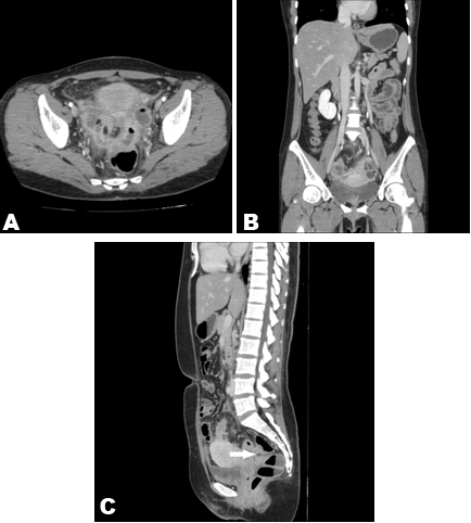
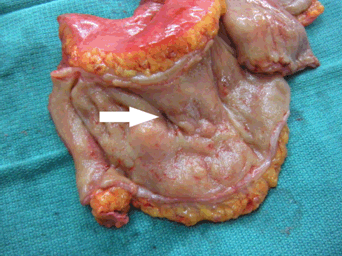
A 46-year-old premenopausal woman gravida 2 para 2 presented to the emergency department with lower abdominal pain. She had no specific past medical history including surgery and reported that the pain had started two weeks prior. The patient previously visited a local clinic. With the presumptive impression of PID, she received conservative management including analgesics for pain control and antibiotics for one week. The pain was transiently relieved. No improvement was seen during her local hospital stay, so she was referred to the department of obstetrics and gynecology. Her vital signs at admission were as follows: temperature 37.5°C, blood pressure 120/85 mmHg, pulse 92 beats/minute, and respiratory rate 18 breaths/minute. The lower abdomen was soft and tender without signs of peritoneal irritation. A pelvic examination showed severe tenderness in both adnexal regions, but a digital rectal examination was negative. Laboratory results were within the normal range except for the white blood cell count of 19,500/µL. There were no specific findings on chest or abdominal X-ray. Abdominopelvic computed tomography (CT) scan showed PID with a tubo-ovarian abscess and an air-containing abscess in the rectovaginal pouch with suspicious fistula in the sigmoid colon (Figure 1). Colonoscopy indicated an elevated hard mucosal change 7–20 cm from the anal verge. Although no definite fistula opening was detected, a pus-like substance was continuously draining from the rectosigmoid colon (Figure 2). With the diagnosis of sigmoid fistula and a tubo-ovarian pelvic abscess, elective surgery was performed. During the operation, the tubo-ovarian inflammatory complex and abscess cavity in the rectovaginal pouch abutting the sigmoid colon were revealed, while omental and small bowel adhesions were noted in the abscess cavity. After the colon, omentum, and small bowel were dissected from the abscess cavity and the pus was drained, total abdominal hysterectomy, bilateral salpingo-oophorectomy, and resection of the sigmoid colon including the affected segment with the primary anastomosis were performed. After the surgery, the fistula opening was identified in the surgical specimen that was not preoperatively detectable (Figure 3). The patient’s postoperative course was uneventful and she was discharged on the 10th postoperative day. | ||||||
|
| ||||||
| ||||||
| ||||||
|
DISCUSSION
| ||||||
|
Pelvic inflammatory disease is common, but the differential diagnosis of a surgical abdomen such as appendicitis is crucial because the optimal treatment can vary among disease conditions. Pelvic inflammatory disease is caused by an infection that ascends from the lower genital tract into the fallopian tube and peritoneal cavity [1]. Its treatment of choice is medical, but resistance to medical treatment or the spread of a local inflammatory condition like appendicitis, adnexal surgery, and cesarean section may lead to tubo-ovarian abscess [1][2]. Although antibiotics for gram-negative organisms combined with clindamycin or metronidazole are recommended in the treatment of tubo-ovarian abscess, medical treatment failure may result in rupture of the abscess into the peritoneal cavity, one of the most serious complications leading to sepsis and mortality. Emergency surgery is required in such cases [7] . Fistula formation around the organs is another rare complication of tubo-ovarian abscess, but it develops chronically and does not require emergency surgery. The mechanism of fistula formation has not been well established, but the defense mechanism of the spread of inflammation in the abdominal cavity may be a clue to its pathogenesis. Since redundant organs such as the small bowel, sigmoid colon, and omentum play an important role in preventing the spread of inflammation into the abdominal cavity by adhesion to the pelvis, local tubo-ovarian and pelvic abscess may be confined in the pelvis without rupturing into the abdominal cavity. Thus, in such cases, when antibiotic therapy is not effective and an abscess does not rupture into the peritoneal cavity, it may invade other spaces such as the preperitoneal space and form an abscess in the abdominal wall or a nearby organ such as the bladder, rectum, or sigmoid colon and form a fistula as in the present case [3][4][5][6][8]. Therefore, sealing off the inflamed pelvis using the small bowel, sigmoid colon, and omentum is a sort of defense mechanism but also may be a triggering factor of fistula formation in the pelvic organs. Abdominal pain, the main symptom of fistula secondary to tubo-ovarian abscess, may be relieved by pus drainage into the sigmoid colon or bladder. Depending on the fistula-forming organ, pyuria or purulent diarrhea may also be present, and these symptoms may be pathognomonic in patients with tubo-ovarian abscess and fistula formation [3] [4][5][6] . The diagnosis of tubo-ovarian abscess is made based on imaging studies. Transvaginal ultrasonography is the first-line imaging study because it provides high-resolution images and avoids radiation, but its interpretation may vary among practitioners [1][9]. If the US result is equivocal or there is a suspicious lesion such as malignancy, CT scan can be a preferred modality [10]. However, the detection of a fistula tract or opening on a CT image may be limited. Thus, to detect a fistula tract or opening, a contrast study, magnetic resonance imaging (MRI), and colonoscopy can be useful [4][6]. In the present case, ultrasound was performed in the local clinic, but the report was not available at admission. Although the CT image indicated a suspicious lesion of a fistula tract with the sigmoid colon, colonoscopy findings made it possible to consider fistula formation with the sigmoid colon. A tubo-ovarian abscess secondary to sigmoid colonic diverticulitis could be considered in the differential diagnosis because diverticulitis may be one of the etiologies for tubo-ovarian abscess [1]. However, there was no evidence of diverticulitis on the CT image; finally, the operative findings and gross findings of the surgical specimen led us to the confirmatory diagnosis. Surgical treatment for tubo-ovarian abscess varies from drainage and unilateral salpingo-oophorectomy to total abdominal hysterectomy and bilateral salpingo-oophorectomy, but in cases of fistula formation, surgical options have not been well established [1]. Fistula opening sealing with a primary suture but no colon resection may be one option, while involved colon resection with or without diversion may be another [5][6]. Surgery accompanied by colon resection requires longer operative time than that with fistula opening sealing only, but when the fistula opening sealing is not available due to severe inflammation, colon resection may be a suitable option as in the present case, and if the patient’s condition is unstable, Hartmann’s operation may be performed. However, secondary operations to reverse the colostomy might lead to general anesthesia–associated risks and postoperative morbidity, particularly in older patients. Thus, the optimal surgical option for fistula tract should be individualized to a patient’s condition. | ||||||
|
CONCLUSION
| ||||||
|
Sigmoidal fistula secondary to tubo-ovarian abscess is an extremely rare complication for which the optimal surgical treatment modality has not been established. Prompt preoperative diagnosis and individualized surgical treatment should be provided to avoid morbidity and mortality. | ||||||
|
REFERENCES
| ||||||
| ||||||


|
[HTML Abstract]
[PDF Full Text]
|
|
Author Contributions
Pyong Wha Choi – Substantial contributions to conception and design, Acquisition of data, Analysis and interpretation of data, Drafting the article, Revising it critically for important intellectual content, Final approval of the version to be published |
|
Guarantor
The corresponding author is the guarantor of submission. |
|
Source of support
None |
|
Conflict of interest
Authors declare no conflict of interest. |
|
Copyright
© 2017 Pyong Wha Choi. This article is distributed under the terms of Creative Commons Attribution License which permits unrestricted use, distribution and reproduction in any medium provided the original author(s) and original publisher are properly credited. Please see the copyright policy on the journal website for more information. |
|
|