|
|
|
|
Case Report
| ||||||
| A possible new risk factor causing pneumomediastinum in a middle-aged patient: Anatomical weakness in the bronchial wall | ||||||
| Hirofumi Namiki1, Kazuhiko Matsuno2, Tadashi Kobayashi3 | ||||||
|
1MD, Yonaguni Municipal Clinic, Japan Association for Development of Community Medicine, 125-1 Yonaguni,
Yonaguni-cho, Yaeyama-gun, Okinawa, Japan
2MD, PhD, Department of Pulmonary medicine, Naha City Hospital, 2-31-1 Furujima Naha, Okinawa, Japan 3MD, PhD, Department of General Medicine, Hirosaki University School of Medicine & Hospital, 53 Hon-cho, Hirosaki-shi, Aomori-ken, Japan | ||||||
| ||||||
|
[HTML Abstract]
[PDF Full Text]
[Print This Article] [Similar article in Pumed] [Similar article in Google Scholar] 
|
| How to cite this article |
| Namiki H, Matsuno K, Kobayashi T. A possible new risk factor causing pneumomediastinum in a middle-aged patient: Anatomical weakness in the bronchial wall. Int J Case Rep Images 2017;8(7):429–432. |
|
ABSTRACT
| ||||||
|
Introduction:
Pneumomediastinum is a rare condition that occurs after physical trauma or other situations that lead to air leakage into the mediastinum. The present report describes a middle-aged patient with pneumomediastinum caused by breath-holding at work. Keywords: Conservative treatment, Cough, Thorax, Japan, Pneumothorax | ||||||
|
INTRODUCTION
| ||||||
|
Previous reports have shown that pneumomediastinum rarely occurs in young male especially children [1]. However, many authors understand that the occurrence of pneumomediastinum may be more frequent than initially believed because many patients refrain from medical visits [2]. In addition, almost patients without any risk factors are diagnosed to spontaneous pneumomediastinum [3] [4]. This report describes a middle-aged patient who developed pneumomediastinum after breath-holding at work. Bronchoscopy revealed a new possible risk factor for pneumomediastinum, an anatomical weakness in the bronchial wall. | ||||||
|
CASE REPORT
| ||||||
|
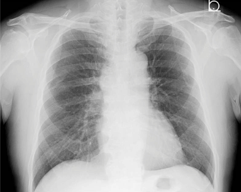
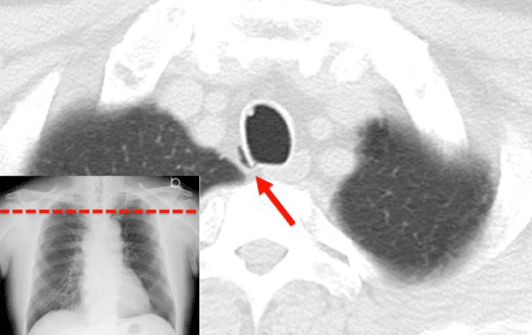
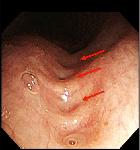
A 59-year-old male developed sudden-onset coughing at working the evening before his presentation at a clinic. His symptoms appeared immediately after a few seconds when breath-holding for heavy lifting started, and had persisted for approximately 12 hours. He had no other symptom except for dry cough. His medical history included only hypertension. He was undergoing regular medication (Indapamide 1 mg/day) for hypertension. There was no family history of respiratory disease. He was a not-smoker. Physical examination revealed normal vital signs, and no tenderness or crackles on his neck and chest. All other findings were normal. Blood test and X-ray findings were also normal (Figure 1). For differential diagnosis of pneumothorax, a computed tomography scan of his chest at the clinic was obtained, and pneumomediastinum was revealed (Figure 2). Thus, the patient was diagnosed with pneumomediastinum and received bronchoscopy at a general hospital in another island, which found an anatomical weakness in the bronchial wall (Figure 3). The patient had a conservative treatment with cough medicine (codeine phosphate, 2 g every 8 hours) and daily follow-up. After two months of treatment, his symptoms gradually disappeared. | ||||||
| ||||||
|
| ||||||
| ||||||
|
DISCUSSION
| ||||||
|
Investigation of the epidemiology of pneumomediastinum has indicated that pneumomediastinum is a rare event in middle-aged and older patients, but that it occasionally occurs in natural births and patients aged 5–34 years [1]. Most affected patients (76%) are male [5]. In addition, several reports of pneumomediastinum in middle-aged patients reported that affected patients had some risk factors [6]. The development of spontaneous pneumomediastinum in a middle-aged man is rare. We considered that increased pressure on the bronchus secondary to a bronchial anomaly might have caused the pneumomediastinum in our patient. The mediastinal tissues in older patients are fibrosed, making air movement more difficult [7]. However, the increased pressure in the bronchus upon the breath-hold could have caused the air to rupture the bronchial wall that had a cratered surface [8][9]. Our patient had no other predisposing factors for pneumomediastinum, such as chronic obstructive pulmonary disease or airway infection, post- tuberculosis. Some reports of spontaneous pneumomediastinum have revealed that no inducing factor may be found [3] [4]; however, bronchoscopy was not always performed in these studies. Therefore, in our patient, the potential causes of the pneumomediastinum were incidentally revealed by bronchoscopy and may have been related to the cratered surface of the bronchus. With respect to therapy, conservative management was chosen in our patient and provided a good outcome. However, the optimal management of pneumomediastinum depends on its severity and cause. Some patients might require hospitalization for over 24 hours of observation, and others might require drugs for pain, anxiety, or infection. Conservative treatment might be sufficient for patients with pneumomediastinum that causes only mild discomfort. In the present case, we strongly discouraged physical activity and instructed the patient to remain on bed rest and treatment with cough medicine. Spontaneous pneumomediastinum is generally considered to be a benign disease with a good prognosis. Both recurrence of spontaneous pneumomediastinum and prolonged cases (over two months) have been reported [4]. In general, however, few cases of recurrent pneumomediastinum have been reported, highlighting its benign nature [10]. Additional diagnostic evaluation should be conducted in patients with recurrence to detect underlying pathologies such as pulmonary or esophageal pathology. As our patient in middle-aged who had no other predisposing factors underwent bronchoscopy, we had detected anatomical weakness in the bronchus. To the best of our knowledge, no cases of pneumomediastinum associated with bronchial anomalies in middle-aged patients have been reported. Anatomical weakness in the bronchial wall should be considered as possible risk factor for pneumomediastinum in a middle-aged patient, if patient had no common predisposing factors. | ||||||
|
CONCLUSION
| ||||||
|
Pneumomediastinum should be considered in middle-aged patients who develop sudden coughing immediately after breath-holding. Anatomical weakness in the bronchial wall should be considered as a possible new risk factor. | ||||||
|
REFERENCES
| ||||||
| ||||||
|
[HTML Abstract]
[PDF Full Text]
|
|
Author Contributions
Hirofumi Namiki – Substantial contributions to Conception and design, Acquisition of data, Analysis and interpretation of data, Drafting the article, Revising it critically for important intellectual content, Final approval of the version to be published Kazuhiko Matsuno – Acquisition of data, Analysis and interpretation of data, Revising it critically for important intellectual content, Final approval of the version to be published Tadashi Kobayashi – Substantial contributions to Conception and design, Analysis and interpretation of data, Drafting the article, revising it critically for important intellectual content, Final approval of the version to be published |
|
Guarantor
The corresponding author is the guarantor of submission. |
|
Source of support
None |
|
Conflict of interest
Authors declare no conflict of interest. |
|
Copyright
© 2017 Hirofumi Namiki et al. This article is distributed under the terms of Creative Commons Attribution License which permits unrestricted use, distribution and reproduction in any medium provided the original author(s) and original publisher are properly credited. Please see the copyright policy on the journal website for more information. |
|
|
|
ABOUT THE AUTHORS
| |||||||||
| |||||||||
|
|