|
|
|
|
Clinical Image
| ||||||
| Intramyocardial calcification in a 37-year-old patient with severe aortic stenosis | ||||||
| Aoife M. Granahan1, Katie E. O’ Sullivan2, Sarah A. Early3 | ||||||
|
1MB, BCh, BAO, MRCS SHO, Surgery, St. James’s University Hospital, Dublin, Ireland 2MD, MRCS, SPR, Cardiothoracic Surgery, St. James’s Hospital, Dublin, Ireland 3MD, FRCS, Consultant, Cardiothoracic Surgery, St. James’s Hospital, Dublin, Ireland | ||||||
| ||||||
|
[HTML Abstract]
[PDF Full Text]
[Print This Article]
[Similar article in Pumed] [Similar article in Google Scholar] 
|
| How to cite this article |
| Granahan AM, O’ Sullivan KE, Early SA. Intramyocardial calcification in a 37-year-old patient with severe aortic stenosis. Int J Case Rep Images 2017;8(7):481–483. |
|
CASE REPORT
| ||||||
|
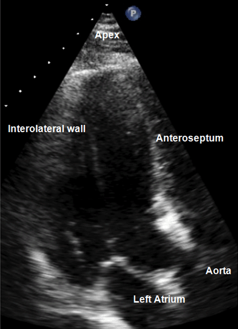
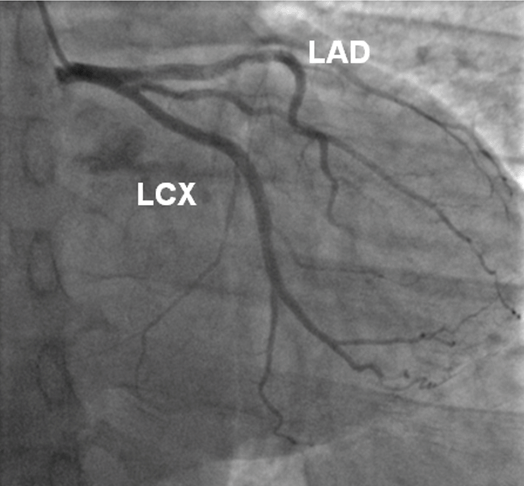
A 37-year-old male with symptoms of progressive dyspnea and intermittent chest pain over a one-year period with a longstanding history of aortic stenosis was referred for aortic valve replacement. His personal cardiovascular history was unremarkable, most notably for tuberculosis, rheumatic heart disease, pericarditis or myocardial ischemia. Family history was non-contributory. Examination revealed an ejection systolic murmur consistent with aortic stenosis. Vital signs revealed an early warning score of zero, laboratory investigations were found within normal parameters and electrocardiogram (ECG) was in sinus rhythm. Echocardiography (ECHO) demonstrated an ejection fraction of 55% and confirmed severe aortic stenosis and moderate aortic incompetence with peak and mean gradients of 90 and 50 mmHg respectively. Extensive myocardial calcification on ECHO (Figure 1) and coronary angiography (Figure 2) prompted further investigation with computed tomography scan. Computed tomography scan was notable for a thick band of intramyocardial calcification extending from beneath the left coronary cusp to half way down the interventricular septum (Figure 3). Calcification was not noted in any other organ system and there was no evidence of malignancy. Serum calcium levels were within normal range. Cardiac catheterization revealed no evidence of coronary artery disease. Aortic valve replacement was performed. Intraoperative findings revealed a heavily calcified aortic valve. The native valve was meticulously excised and replaced using a 25-mm carbomedics mechanical prosthesis with interrupted sutures. Postoperative echocardiogram was satisfactory and postoperative recovery was uneventful. | ||||||
| ||||||
| ||||||
| ||||||
|
DISCUSSION
| ||||||
|
Epidemiological studies reveal that aortic stenosis increases with age, 25% of people aged over 65 years have aortic sclerosis, and 3% over 75 years have severe stenosis. Approximately, 16% of patients with sclerosis progress to stenosis in seven years [1]. The overwhelming cause of aortic stenosis is calcific degeneration, 2% of the population account for congenital bicuspid valves, rheumatic disease is now extremely rare [2]. Cases of myocardial calcification which are not associated with myocardial infarction or metastatic deposition are extremely rare [3][4]. The calcification of myocardium can occur secondary to two mechanisms. Firstly, dystrophic calcification occurs in dead or degenerative tissue in the presence of normal calcium/phosphate balance such as previous myocardial infarction. Other potential causes of dystrophic calcification include myocarditis, ventricular aneurysms, tuberculosis, sarcoidosis and hemorrhage [5][6][7][8][9]. Secondly, metastatic calcification, which occurs when a derangement of calcium phosphate metabolism results in calcium deposition in normal tissue, such as chronic renal failure or hyperparathyroidism [3]. Our patient had no known history or aberrant biochemical analyses to suggest that any of the differentials mentioned were the causative pathology. Extensive calcification and porcelain heart due to endomyocardial fibrosis has also been reported in literature [10]. Endomyocardial fibrosis is characterized by the presence of fibrous tissue in the endocardium eventually extending into the myocardium, a finding not identified in our patient. Mitral annular and leaflet calcification occurs frequently with degenerative aortic stenosis and is known to reduce leaflet opening and result in significant mitral stenosis in 25% of patients with calcific aortic stenosis [11]. In our patient, however, no involvement of the mitral valve was noted. | ||||||
|
CONCLUSION
| ||||||
|
This is an unusual case as significant calcification of the interventricular septum developed in the absence of any known etiologic factor or evidence of pre-existing myocardial injury. Valvular heart disease, is an underappreciated yet serious and growing public health problem. Keywords: Aortic stenosis, Aortic valve replacement, Cardiothoracic surgery, Intramyocardial calcification, Symptomatic valvular disease | ||||||
|
REFERENCES
| ||||||
| ||||||
|
[HTML Abstract]
[PDF Full Text]
|
|
Author Contributions
Aoife M. Granahan – Substantial contributions to conception and design, Acquisition of data, Analysis and interpretation of data, Drafting the article, Revising it critically for important intellectual content, Final approval of the version to be published Katie E. O’ Sullivan – Analysis and interpretation of data, Revising it critically for important intellectual content, Final approval of the version to be published Sarah A. Early – Analysis and interpretation of data, Revising it critically for important intellectual content, Final approval of the version to be published |
|
Guarantor
The corresponding author is the guarantor of submission. |
|
Source of support
None |
|
Conflict of interest
Authors declare no conflict of interest. |
|
Copyright
© 2017 Aoife M. Granahan et al. This article is distributed under the terms of Creative Commons Attribution License which permits unrestricted use, distribution and reproduction in any medium provided the original author(s) and original publisher are properly credited. Please see the copyright policy on the journal website for more information. |
|
|